Article
Printed in Brazil - ©2016 Sociedade Brasileira de Química0103 - 5053 $6.00+0.00*e-mail: [email protected]
In memorian of Prof Gentil José Vidotti.
Bioactive Indole Alkaloids from
Croton echioides
Claudio R. Novello,*,a Luís C. Marques,b Murilo E. Pires,c Ana P. Kutschenco,d Celso V. Nakamura,e Samara Nocchi,e Maria H. Sarragiottoc and João C. P. Mellod
aDepartamento de Química e Biologia, Universidade Tecnológica Federal do Paraná, 85601-970 Francisco Beltrão-PR, Brazil
bUniversidade Bandeirante de São Paulo, 04710-000 São Paulo-SP, Brazil
cDepartamento de Química, dDepartamento de Farmácia and eDepartamento de Ciências Básicas da Saúde, Universidade Estadual de Maringá, 87020-900 Maringá-PR, Brazil
Bioguided fractionation of a hydroethanolic extract from the stem bark of Croton echioides Baill. led to isolation of the new indole alkaloid N-trans-feruloyl-3,5-dihydroxyindolin-2-one as a stereoisomeric mixture and the known alkaloids N-trans-p-coumaroyl-tryptamine, N-trans-p -coumaroyl-5-hydroxytryptamine, N-trans-4-methoxy-cinnamoyl-5-hydroxytryptamine, N-trans -feruloyl-5-hydroxytryptamine (moschamine), from the ethyl acetate fraction. The flavonoids 3-o-methyl kaempferol, 3-o-methyl quercetin, 3,7-di-o-methyl quercetin and 3,3’-di-o-methyl quercetin, together with the benzoic acid derivatives 4-hydroxybenzoic acid, 4-hydroxy-3-methoxybenzoic acid and 4-hydroxy-3,5-di4-hydroxy-3-methoxybenzoic acid were also isolated. Alkaloids and the flavonoids showed strong antioxidant properties in vitro radical scavenging assay (2,2-diphenyl-1-picrylhydrazyl, DPPH), with IC50 values ranging from 9.2 to 17.5 µmol L-1, lower than those of the positive control Trolox (IC50 = 17.9 µmol L-1). Alkaloids showed cytotoxic activity against the HCT-116 human cancer cell line, with IC50 valuesranging from 86.8 to 210.7 µmol L-1. Compound N-trans-4-methoxy-cinnamoyl-5-hydroxytryptamine was the most active,with an IC50 value of 86.8 µmol L-1.
Keywords:Croton echioides,Euphorbiaceae, indole alkaloid, cytotoxic activity, antioxidant activity
Introduction
Plants of the genus Croton are well known for their
many therapeutic uses, including anti-inflammatory,
antiulcer, antitumor, antioxidant and cytotoxic activity.1-3
This genus is a rich source of special metabolites, mainly clerodane diterpenes, flavonoids, and different classes
of alkaloids.4 Alkaloids related to benzylisoquinolines,
such as morphinandienones and tetrahydroprotoberberine alkaloids are the most frequent class found in members of
the genus Croton.5 Glutarimide alkaloids and a new class
of sesquiterpene guaiane-type alkaloids have recently been
reported from Croton species.6
Croton L. is the second-largest genus in the family Euphorbiaceae, with about 1,200 species, 350 of which
occur in Brazil.5 Croton echioides Baill., popularly
known as “quebra-faca”, “caatinga branca”, “velame” and “canela-de-velho”, is a small native tree found in the
North-eastern region of Brazil.7,8 The stem bark of this
plant has been widely marketed as an aphrodisiac and tonic, as a substitute for the roots of Amazon Marapuama,
Ptychopetalum olacoides Benth. (Olacaceae).9 To our
knowledge, no chemical or biological study of this species has been reported.
In this paper, we describe the isolation and structure identification of a new indole alkaloid identified as
N-trans-feruloyl-3,5-dihydroxyindolin-2-one (1) together
with the known alkaloids N-trans-p-coumaroyl-tryptamine
(2),10 N-trans-p-coumaroyl-5-hydroxytryptamine (3),11
N-trans-4-methoxy-cinnamoyl-5-hydroxytryptamine (4)12
and N-trans-feruloyl-5-hydroxytryptamine (moschamine)
(5),13 the flavonoids 3-o-methyl kaempferol (6),14
3-o-methyl quercetin (7),14 3,7-di-o-methyl quercetin
(8)15 and 3,3’-di-o-methyl quercetin (9),16 and benzoic
4-hydroxy-3-methoxybenzoic acid (11) and
4-hydroxy-3,5-dimethoxybenzoic acid (12)17 from the stem bark of
Croton echioides. The in vitro cytotoxicity against the HCT-116 human cancer cell line was determined for the
isolated alkaloids 1-5 and the free radical
(2,2-diphenyl-1-picrylhydrazyl, DPPH) scavenging activities were also
determined for the compounds 1-12.
Experimental
General experimental procedures
Infrared (IR) spectra were recorded on a Bomem MB-Series spectrometer (Bomem, Quebec, CA, USA) in a 400
to 4000 cm-1 spectral region using KBr matrix. Shimadzu
UV-1650-PC spectrometer (Shimadzu, Kyoto, Japan) was used for UV-Vis determinations. Optical rotation was measured on a JASCO P-2000 polarimeter (Jasco, Tokyo, Japan) in MeOH. High-resolution electrospray ionization mass spectrometry (HR-ESIMS) were recorded on a Bruker-Daltonics MicroTof (Bruker-Bruker-Daltonics, Bremen, Germany), and the ESIMS were recorded on a Thermo Scientific Orbitrap LTQ XL spectrometer (Thermo Fisher System,
San Jose, CA, USA) performed at Universityof Münster,
Germany. Nuclear magnetic resonance (NMR) spectra were recorded on a Varian Mercury Plus 300 MHz spectrometer (7.02 T) (Varian, Palo Alto, CA, USA) operated at 75 MHz
for 13C and 300 MHz for 1H and 2D NMR, using deuterated
MeOH as solvent and tetramethylsilane (TMS) as internal standard. Medium pressure liquid chromatography (MPLC) separation was performed using a Waters 6000A (Waters, Milford, Massachusetts, USA) pump with a manual sample injection valve 10 mL loop coupled with different columns (450 × 10 mm internal diameter (i.d.); 340 × 30 mm i.d.; 300 × 50 mm i.d.) loaded with Sepra C18-E 50 µm (Phenomenex, Torrance, CA, USA) as the stationary phase. The MPLC system is equipped with a Pharmacia-Biotech FRAC-200 (Amersham Biosciences KK, Tokyo, Japan) fraction collector. Preparative counter-current chromatography was performed using a high-speed counter-current chromatography (HSCCC) (P. C. Inc., Potomac, USA) chromatograph equipped with a polytetrafluoroethylene (PTFE) column (2.5 mm i.d.; 200 mL total capacity) coupled to a Waters Mod. 6000A (Waters, Milford, Massachusetts, USA) pump and a manual sample injection valve with a 10 mL loop. Column chromatography was carried out on Sephadex
LH 20 (PharmaciaFine Chemicals Inc., New Market, NJ,
USA). Pharmacia-Biotech FRAC-200 fraction collector (Amersham Biosciences KK, Tokyo, Japan) was used for isolations procedures. Thin-layer chromatography (TLC)
was performed on precoated silica gel aluminum sheets (Kieselgel 60 F254, 0.20 mm, Merck, Darmstadt, Germany). Fractions and pure compounds were monitored
by TLC, and spots were revealed by heating (110 °C) the
plates sprayed with a 2% vanillin in chloride acid/EtOH (1:2) solution.
HSCCC separations
The elution systems used in HSCCC separations were optimized by determination of the partition coefficient (Kp) of target compounds in a series of biphasic liquid systems by UV-Vis determinations. Sets of biphasic solvent systems composed of hexane, petroleum ether, ethyl acetate, methanol, ethanol and water was examined at different arrangements and volume ratios. The biphasic liquid system was prepared by adding portions of the solvents in a separatory funnel and, after the equilibrium was reached, 1 mL of each phase (upper and lower) were added in a test tube. After that, 1 mg of the solute was introduced. The test tube was shaken manually until equilibrium was reached. The system was equilibrated for at least 1 h and the concentration of the solute in the upper and the lower phase was analyzed by UV-Vis. The Kp was calculated as the ratio of the solute concentration in the upper and the lower phase. For the samples purification process, the
most suitable solvent system (H2O/EtOH/EtOAc/hexane;
2.5:1:1.5:1.5 v/v) was prepared and thoroughly equilibrated in a separatory funnel at room temperature and the two phases were separated shortly before use. The sample solution was prepared by dissolving the crude sample in 10 mL of the stationary lower phase of the solvent system. The multilayer coiled column was first entirely filled with the lower phase. The upper mobile phase was then pumped into the inlet of the column on tail to head option at the flow rate
of 1.0 mL min-1, while the apparatus was run at 800 rpm.
After hydrodynamic equilibrium was reached, a sample was injected. The eluent was continuously monitored by TLC and the fractions were collected according to the chromatographic profile. After target compounds were eluted, the HSCCC apparatus was stopped and the column contents were fractionated.
Plant material
T h e a e r i a l p a r t s o f f l o w e r i n g p l a n t s o f
Extraction and isolation
Air-dried and ground stem bark (7.6 kg) of C. echioides
were extracted with EtOH/H2O (70%, 76 L) through
an Ultra-Turrax Ika Works UTC 115/KT (Ika Works, Wilmington, NC, USA) at room temperature for 48 h, subdivided into 5 min of Ultra-Turrax turbo extraction followed by 2 h of rest, successively. The crude extract was filtered, the solvent was evaporated under reduced pressure, and the residue was lyophilized to give the crude extract (383 g, yield 5.04%). Part of the crude extract (300 g) was suspended in water (2.5 L) and partitioned
with hexane, CH2Cl2, EtOAc and BuOH (2.5 L) to give
the fractions respectively termed HF (62.6 g, yield 20.9%), DF (44.2 g, yield 14.7%), ethyl acetate fraction (EAF) (23.5 g, yield 7.8%) and BF (53.6 g, yield 17.9%). The EAF was subjected to chromatography column over
Sephadex LH-20 (460 × 55 mm i.d.) eluting with a gradient
of EtOH/H2O, MeOH, Me2CO/H2O and AcOH/H2O
to give 26 fractions (EAF-1 to EAF-26). Fraction EAF-9 (2.43 g, yield 10.3%) was separated by preparative
MPLC (column 300 × 50 mm i.d., MeOH/H2O 1:1 v/v,
3 mL min-1) to give four subfractions (EAF-9.1 to EAF-9.4).
Subfraction AF-9.2 (190 mg, yield 7.8%) was purified by
HSCCC using H2O/EtOH/EtOAc/hexane (2.5:1:1.5:1.5 v/v,
1 mL min-1), to yield 1 (47.2 mg, yield 24.8%),
4-hydroxy-3-methoxybenzoic acid (35.0 mg, yield 18.4%) and 4-hydroxy-3,5-dimethoxybenzoic acid (15.0 mg, yield 7.9%). Subfraction EAF-9.3 (241 mg, yield 9.9%) was purified by HSCCC in the same conditions as AF-9.2 to provide 4-hydroxybenzoic acid (5.0 mg, yield 2.1%). The EAF-17 (1.04 g, yield 4.4%) fraction was subjected to
MPLC in a C-18 column (340 × 30 mm i.d., MeOH/H2O,
3:7 v/v, 2 mL min-1), to give two subfractions (AF-17.1
and AF-17.2). Subfractions EAF-17.1 (106 mg, yield 10.2%) and AF-17.2 (60 mg, yield 5.8%) were purified by
MPLC in a C-18 column (450 × 10 mm i.d., MeOH/H2O,
1:1 v/v, 1.5 mL min-1) to yield 4 (5.0 mg, yield 8.3%)
and 5 (13.4 mg, yield 22.3%). Purification of the EAF-18
fraction (398.8 mg, yield 1.7%) by MPLC in a C-18 column
(340 × 30 mm i.d., MeOH/H2O, 7:3 v/v, 2 mL min-1),
afforded 8 (28.4 mg, yield 7.1%) and 9 (25.2 mg, yield
6.3%). Purification of EAF-19 (283 mg, yield 1.20%)
by MPLC (340 × 30 mm i.d., EtOH/H2O, 3:7 to 9:1 v/v,
2 mL min-1) provided 6 (10.5 mg, yield 3.7%). Subfraction
EAF-19.1 (65.0 mg, yield 23.0%) was purified by MPLC
(450 × 10 mm i.d., MeOH/H2O, 5:5 v/v, 1.5 mL min-1) to
provide 3 (51.6 mg, yield 79.4%). Purification of EAF-20
(220.6 mg, yield 0.9%) by MPLC (340 × 30 mm i.d.,
MeOH/H2O, 7:3 v/v, 2 mL min-1) afforded compounds 7
(78.2 mg, yield 35.4%) and 2 (16.2 mg, yield 7.3%).
N-trans-Feruloyl-3,5-dihydroxyindolin-2-one (1)
Amorphous gray solid; mp: 156-158 °C; [α]D25 = 0°
(c 0.25, MeOH); IR (KBr) νmax / cm-1 3390, 1711, 1650,
1600, 1208; NMR data see Table 1; HR-ESIMS, positive
ion mode, m/z 407.1220 [M + Na]+; calcd. for C
20H20N2O6:
407.1219.
N-trans-p-Coumaroyl-tryptamine(2)
Amorphous yellow solid; NMR data were in agreement
with literature values;10 HR-ESIMS, positive ion mode,
m/z 329.1257 [M + Na]+; calcd. for C
19H18N2O2: 329.1266.
N-trans-p-Coumaroyl-5-hydroxytryptamine (3)
Amorphous gray solid; NMR data were in agreement
with literature values;11 HR-ESIMS, positive ion mode,
m/z 345.1212[M + Na]+;calcd. for C
19H18N2O3: 345.1215.
N-trans-4-Methoxy-cinnamoyl-5-hydroxytryptamine (4)
Amorphous gray solid; mp 160-162 °C; 1H NMR
(300 MHz, CD3OD) d 7.47 (d, 1H, J 16.2 Hz, H-15), 7.45
(d, 2H, J 8.4 Hz, H-17, H-21), 7.15 (d, 1H, J 8.7Hz, H-7),
7.02 (s, 1H, H-2), 6.95 (d, 1 H, J 2.4 Hz, H-4), 6.93 (d,
2H, J 9.0 Hz, H-18, H-20), 6.65 (dd, 1H, J 8.7, 2.4 Hz,
H-6), 6.44 (d, 1H, J 15.6 Hz, H-14), 3.81 (s, 3H, OCH3),
3.56 (t, 2H, J 7.2 Hz, H-11), 2.92 (t, 2H, J 7.2 Hz, H-10);
13C NMR (75 MHz, CD
3OD) 168.0 (C, C-13), 161.3 (C,
C-19), 149.8 (C, C-5), 140.3 (CH, C-15), 131.9 (C, C-8), 129.3 (CH, C-17, C-21), 128.2 (C, C-9), 127.7 (C, C-16), 123.2 (C, C-2), 118.2 (CH, C-14), 114.1 (CH, C-18, C-20), 111.6 (CH, C-7), 111.3 (C, C-3), 111.2 (CH, C-6), 102.4
(CH, C-4), 54.7 (CH3, OCH3), 40.3 (CH2, C-11), 25.2 (CH2,
C-10); ESIMS, positive ion mode, m/z 359.5 [M + H]+;
calcd. for C20H19N2O3: 337.5.
N-trans-Feruloyl-5-hydroxytryptamine (5)
Amorphous gray solid; NMR data were in agreement
with literature values;11 HR-ESIMS, positive ion mode,
m/z 375.1310 [M + Na]+; calcd. for C
20H20N2O4:375.1321.
Cytotoxic assays
The cytotoxicity activity against HCT-116 cells was
determined by in vitro sulforhodamine B test (SRB-test).18
HCT-116 cell lines, originated from a human colorectal carcinoma, were cultured and maintained in Dulbecco’s modified Eagle’s medium (DMEM, Gibco, Grand Island, NY, USA) supplemented with 10% heat-inactivated fetal
bovine serum (FBS, Gibco) and 50 µg mL-1 gentamycin, in
incubator set at 37 °C, 5% CO2 and 95% relative humidity.
were digested by using 0.25% trypsin (Gibco) containing 1 mmol EDTA (ethylenediamine tetraacetic acid). The cells were seeded in 96-well tissue plates (TPP-Techno Plastic Products, Trasadingen, Switzerland) at a density
of 8 × 10⁵ cell mL-1 in 100 µL of medium for 24 h in
the CO2 incubator. The sample solutions with different
concentrations (3.12, 6.25, 12.5, 25, 50 and 100 µg mL-1)
were prepared and added to the culture media. After incubation for 48 h, the cell monolayers were washed with 100 µL phosphate buffered saline (PBS) sterile, fixed with trichloroacetic acid and stained for 30 min with SRB
(7.16 mmol L-1) (Sigma Chemical Co., St. Louis, MO, USA)
in acetic acid solution. The dye was removed by four washes
with acetic acid solution (175 mmol L-1), and protein-bound
dye was extracted with 10 mmol L-1 unbuffered tris base
[tris(hydroxymethyl)aminomethane] for determination of optical density in a computer-interfaced, 96-well microtiter plate reader (Power Wave XS, BIO-TEK, Winooski, VT,
USA). The IC50 values were determined from a percentage
of cells relative to untreated control and the half-life of the HTC cells in the presence and absence of each tested sample was estimated as the time point at which the cell viability decreased to 50% in relation to the beginning of the test. All the tests were performed in triplicate.
DPPH assay
The free radical scavenging activity was determined by in vitro DPPH (2,2-diphenyl-1-picrylhydrazyl) assay.19
Sample stock solutions (1.0 mg mL-1) of analyte and Trolox
(positive control) were diluted to final concentrations of
250, 125, 50, 25, 10 and 5 µmol L-1, in methanol. 1 mL
of a 0.3 mmol L-1 DPPH solution in methanol was added
to 2.5 mL of sample solutions of different concentrations, and allowed to react at room temperature. After 30 min the absorbance values were measured at 518 nm and converted into the percentage antioxidant activity. 1 mL of methanol
DPPH solution 0.3 mmol L-1 plus methanol (2.5 mL) was
used as a negative control. The IC50 values were calculated by
linear regression. All the tests were performed in triplicate.
Results and Discussion
The ethanol-aqueous extract of stem bark of C. echioides
was successively partitioned into hexane, dichloromethane,
ethyl acetate and butanol. The resulting fractions were
subjected to free radical DPPH scavenging assay, which demonstrated that the ethyl acetate fraction was the
most active, with IC50 = 48.9 ± 0.2 µg mL-1. In order to
isolate bioactive compounds, this fraction was submitted to fractionation on a chromatographic column and the subfractions were also assayed in the DPPH. From that results, the most active subfractions were purified by chromatographic methods affording indolinone and
N-substituted tryptamine alkaloid type compounds (1-5)
which chemical structures are shown in Figure 1. In addition, were isolated flavonoids derived from kaempferol
and quercetin (6-9), as well was benzoic acid derivatives
(10-12). The structures of the isolated compounds were elucidated by analysis of their NMR spectroscopic data (including NMR 2D) and HR-ESIMS, and by comparison with those reported.
Compound 1 was isolated as a stereoisomeric mixture
at C-3 position due to the absence of optical rotation
deviation. The IR spectrum showed mainly two strong
bands at 1711 and 1650 cm-1 corresponding to the carbonyl
stretching vibrations of (γ) lactam and NHCO, respectively.
A broad band was found at 3390 cm-1 due to N−H and OH
stretching. The positive HR-ESIMS spectrum gave the ion
peak at m/z 407.1220 [M + Na]+ (calculated 407.1219),
corresponding to the molecular formula C20H20N2O6.
The NMR spectrum showed signals for oxindole and
trans-feruloyl moieties which were confirmed by 1H and
13C NMR data assignments (Table 1). Specifically, from
the 1H NMR spectra three aromatic hydrogens at d 6.87
(d, J 2.1 Hz, H-4), 6.68 (dd, J 8.4 and 2.1 Hz, H-6) and
6.72 (d, J 8.4 Hz, H-7), which evidenced the presence of
the 1,3-dihydro-3,5-dihydroxy-2H-indol-2-one moiety
substituted at C-3 position by one aminoethyl unit at
3.26 (m, H-11) and d 2.13 (t, J 7.8 Hz, H-10). This was
confirmed by the signals for a carbonyl group at d 181.7
(C-2), a quaternary carbon at d 77.0 (C-3), together with
the signals for the aromatic ring at 154.8 (C-5), 134.6 (C-9), 133.7 (C-8), 116.7 (C-6), 113.0 (C-4) and 112.0 (C-7), and
for an aminoethyl group at d 38.2 (C-10) and 35.7 (C-11),
in the 13C NMR spectra. The presence of a trans-feruloyl
group was evidenced by the signals for the aromatic system
at dH/dC 7.09 (d, J 1.8 Hz, H-17)/ 111.6 (C-17), 6.78 (d,
J 8.4 Hz, H-20)/116.5 (C-20) and dH/dC 6.99 (dd, J 8.4
and 1.8 Hz, H-21)/123.2 (C-21), a trans-olefin system at
dH/dC 7.36 (d, J 15.6 Hz, H-15)/142.0 (C-15) and 6.32 (d,
J 15.6 Hz, H-14)/118.7 (C-14), and for a methoxy group
at dH/dC 3.87 (3H, s)/56.4. The signals of the carbonyl
group and aromatic carbons of the trans-feruloyl moiety
were observed at dC 169.2 (C-13), and at dC 149.8 (C-19),
149.3 (C-18) and 128.3 (C-16), respectively. The linkage of
the moieties trans-feruloyl and
1,3-dihydro-3,5-dihydroxy-2H-indol-2-one was confirmed by the 3J correlation of the
H-15 (d 7.36) and H-11 (d 3.26) signals with the carbon
carbonyl group at d 169.2 (C-13) in the HMBC spectra. In
addition, it was observed an HMBC correlation between
the methylene hydrogens at d 2.13 (H-10) and the carbonyl
and quaternary carbons at C-2 and C-3, respectively. These
data supported the structure determination of 1 as N-
trans-feruloyl-3,5-dihydroxyindolin-2-one.
Compound 4 has previously been synthesized, but no
complete NMR assignments were reported.12 Here, we report
its isolation and characterization from a natural source. The alkaloids (1-5),flavonoids (6-9) and benzoic acid
derivatives (10-12) were evaluated for their antioxidant
activity by the free scavenging DPPH method. The
results for compounds 1-5, expressed by IC50, are shown
in Table 2. Compounds 3-5 and the flavonoids 7 and 8
showed prominent antioxidant properties, with IC50 values
ranging from 9.2 to 17.5 µmol L-1, lower than those of the
positive control Trolox (IC50 =17.9 µmol L-1). Comparison
of the IC50 value of compounds 2 (203.7 µmol L-1) and 3
(10.7 µmol L-1) showed that the hydroxy group at the C-5
of the tryptamine moiety is fundamental for the antioxidant activity of these indole alkaloids.
The cytotoxic activity of isolated indole alkaloids (1-5)
was tested in vitro against the HCT-116 human cancer cell
line, and the results are expressed in Table 2, with IC50
valuesranging from 86.8 to 210.7 µmol L-1. Compound 4
was the most bioactive, with an IC50 value of 86.8 µmol L-1.
According to the texted protocol, the alkaloid 1 was not
bioactive (IC50 > 250µmol L-1), which suggests that the
indole unit present in alkaloids (2-5) is important for the
cytotoxic activity.
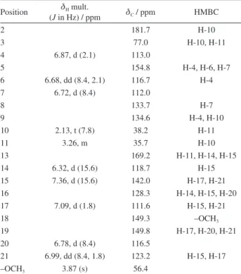
Table 1. 1H, 13CNMR and HMBC data of the compound 1 in CD 3OD
Position dH mult.
(J in Hz) / ppm dC / ppm HMBC
2 181.7 H-10
3 77.0 H-10, H-11
4 6.87, d (2.1) 113.0
5 154.8 H-4, H-6, H-7
6 6.68, dd (8.4, 2.1) 116.7 H-4
7 6.72, d (8.4) 112.0
8 133.7 H-7
9 134.6 H-4, H-10
10 2.13, t (7.8) 38.2 H-11
11 3.26, m 35.7 H-10
13 169.2 H-11, H-14, H-15
14 6.32, d (15.6) 118.7 H-15
15 7.36, d (15.6) 142.0 H-17, H-21
16 128.3 H-14, H-15, H-20
17 7.09, d (1.8) 111.6 H-15, H-21
18 149.3 –OCH3
19 149.8 H-17, H-20, H-21
20 6.78, d (8.4) 116.5
21 6.99, dd (8.4, 1.8) 123.2 H-15, H-17
–OCH3 3.87 (s) 56.4
HMBC: heteronuclear multiple bond correlation.
Table 2. DPPH antioxidant activity and in vitro cytotoxicity against HCT-116 cells of compounds 1-5
Compound Antioxidant activity IC50 / (µmol L-1)
Cytotoxicity IC50 / (µmol L-1)
1 30.0 ± 0.7 n.a.
2 203.7 ± 25.3 144.3 ± 28.6
3 10.7 ± 0.4 210.7 ± 1.4
4 17.5 ± 0.4 86.8 ± 9.7
5 14.5 ± 0.4 142.1 ± 60.5
Trolox 17.9 ± 1.1 –
To our knowledge, this is the first report of indolinone
alkaloid from a species of Croton, which is an important
contribution to chemotaxonomic knowledge of this genus. Concerning the reported activities for the known compounds, the alkaloids have mainly antioxidant and
tyronase inhibitory activities.20-22 Also, anti-inflammatory
activity (compounds 2 and 3),23 growth-promoting
activity for fibroblasts (compound 3)24,25 and in vitro
cytotoxic activity against the CaCo2 colon cancer cells
(compound 5)21 were previously reported. This is the first
report on the cytotoxicity of compounds 1-5 against the
HCT-116 human cancer cell line.
Conclusions
The present report is the first chemical study of
C. echioides Baill., despite this plant species being widely marketed as a substitution to the traditional
Amazon Marapuama, Ptychopetalum olacoides. Five
indole alkaloids were isolated and characterized
by NMR data. One of them (N-trans-
feruloyl-3,5-dihydroxyindolin-2-one) is a new natural product from
Croton species. Their antioxidant and cytotoxicity activity were reported together with other phenolic compound types. These results represent a relevant contribution to the chemical and biological knowledge of this vegetal species and can support its correct and safe medicinal uses.
Supplementary Information
The NMR spectra of compounds 1 to 5 are available
free of charge at http://jbcs.sbq.org.br as a PDF file.
Acknowledgments
This work was supported by the Brazilian granting agencies CNPq, CAPES/Proap, INCT_if, FINEP and Fundação Araucária. The authors are grateful to Prof Janet W. Reid, JWR Associates, Trumansburg, New York, for the English language revision.
References
1. Monteiro, P.; Vendramini-Costa, D.; Ruiz, A. L.; Foglio, M. A.; Carvalho, J.; Planta Med. 2014, 80, 16.
2. Furlan, C. M.; Santos, K. P.; Sedano-Partida, M. D.; Motta, L. B.; Santos, D. Y. A. C.; Salatino, M. L. F.; Negri, G.; Berry, P. E.; Ee, B. W.; Salatino, A.; Braz. J. Bot. 2015, 38, 702. 3. Qiu, M.; Cao, D.; Gao, Y.; Li, S.; Zhu, J.; Yang, B.; Zhou, L.;
Zhou, Y.; Jin, J.; Zhao, Z.; Fitoterapia2016, 108, 86.
4. Ndunda, B.; Langat, M. K.; Wanjohi, J. M.; Midiwo, J. O.; Kerubo, L. O.; Planta Med. 2013, 79, 1766.
5. Salatino, A.; Salatino, M. L. F.; Negri, G.; J. Braz. Chem. Soc. 2007, 18, 11.
6. Jin-Tong, S.; Yang, H.; Xiao-Ling, W.; Tao, S.; Hong-Xiang, L.; Xiao-Ning, W.; Fitoterapia2015, 107, 54.
7. Barbosa, M. R. V.; Lima, I. B.; Lima, J. R.; Cunha, J. P.; Agra, M. F.; Thomas, W. W.; Oecol. Bras. 2007, 11, 313.
8. Silva, J. S.; Sales, M. F.; Carneiro-Torres, D. S.;Rodriguesia 2009, 60, 879.
9. Novello, C. R.; Marques, L. C.; Miyazaki, C. R.; Milaneze-Gutierre, M. A.; Carneiro-Torres, D. S.; Sarragiotto, M. H.; Mello, J. C. P.; Braz. J. Pharmacog.2012, 22, 946.
10. Andrianaivoravelona, J. O.; Terreaux, C.; Sahpaz, S.; Rasolondramanitra, J.; Hostettmann, K.; Phytochemistry1999,
52, 1145.
11. Zhang, H. L.; Nagatsu, A.; Watanabe, T.; Sakakibara, J.; Okuyama, H.; Chem. Pharm. Bull. 1997, 45, 1910.
12. Jenett-Siems, K.; Weigl, R.; Kaloga, M.; Schulz, J.; Eich, E.;
Phytochemistry 2003,62, 1257.
13. Queiroz, M. M. F.; Queiroz, E. F.; Zeraik, M. L.; Marti, G.; Favre-Godal, Q.; Simões-Pires, C.; Marcout, L.; Carrupt, P. A.; Cuendet, M.; Paulo, M. Q.; Bolzani, V. S.; Wolfender, J. L.; Phytochem. Lett. 2014, 10, 88.
14. Lee, H. L.; Kim, H. J.; Song, Y. S.; Jin, C.; Lee, K. T.; Cho, J.; Lee, Y. S.; Arch. Pharm. Sci. Res.2003, 26, 1018.
15. Grayer, R. J.; Eckert, M. R.; Lever, A.; Veitch, N. C.; Kite, G. C.; Paton, A. J.; Biochem. Syst. Ecol. 2010,38, 335.
16. Wang, Y.; Hamburger, M.; Gueho, J.; Hostettmann, K.;
Phytochemistry1989, 28, 2323.
17. Tan, J.; Bednarek, P.; Liu, J.; Schneider, B.; Svatos, A.; Hahlbrock, K.; Phytochemistry2004, 65, 691.
18. Skehan, P.; Storeng, R.; Scudeiro, D.; Monks, A.; McMahon, J.; Vistica, D.; Warren, J. T.; Bokesch, H.; Kenney, S.; Boyd, M. R.; J. Natl. Cancer Inst.1990, 82, 1107.
19. Mensor, L. L.; Menezes, F. S.; Leitão, G. G.; Reis, A. S.; Santos, T. C.; Coube, C. S.; Leitão, S. G.; Phytother. Res. 2001, 15, 127. 20. Maciel, M. A. M.; Dantas, T. N. C.; Camara, J. K. P.; Pinto, A. C.; Veiga Jr., V. F.; Kaise, C. R.; Pereira, N. A.; Carneiro, C. M. T. S.; Vanderlinde, F. A.; Lapa A. J.; Agner, A. R.; Colus, I. M. S.; Echevarria-Lima, J.; Grynberg, N. F.; Esteves-Souza, A.; Pissinate, K.; Echevarria, A.; Adv. Phytomed.2006, 2, 225. 21. Shoeb, M.; MacManus, S. M.; Jaspars, M.; Trevidu, J.; Nahar,
L.; Kong-Thoo-Lin, P.; Sarker, S. D.; Tetrahedron2006, 62, 1172.
22. Takahashi, T.; Miyazawa, M.; Bioorg. Med. Chem. Lett. 2011,
21, 1983.
24. Takii, T.; Hayashi, M.; Hiroma, H.; Chiba, T.; Kawashima, S.; Zhang, H. L.; Nagatsu, A.; Sakakibara, J.; Onozaki, K.; J. Biochem. 1999, 125, 910.
25. Nasim, S.; Lee, N. H.; Bull. Korean Chem. Soc. 2009, 30, 1729.
Submitted: November 18, 2015 Published online: April 13, 2016