Ar
ti
cl
e
0103 - 5053 $6.00+0.00
*e-mail: [email protected]
Effects of Temperature and Chromium (III) Ion on the
Structure of Bovine
β
-Lactoglobulin-A
Adeleh Divsalar,*,a,b Ali Akbar Saboury,a Faizan Ahmadc and Ali Akbar Moosavi-Movahedia
aInstitute of Biochemistry and Biophysics, University of Tehran, Tehran, Iran
bDepartment of Biological Sciences, Tarbiat Moallem University, Tehran, Iran
cDepartment of Bioscience, Jamia Millia Islamia, New Delhi, India
Estudos termodinâmicos relativos ao efeito da temperatura (de 27 a 47 ºC) sobre a estrutura da b-lactoglobulina-A bovina (BLG-A), em solução de cloreto de sódio 50 mmol L-1, na presença
e ausência de íons Cr(III), usando espectroscopia de absorção no UV-Visível, dicroísmo circular (DC), próximo e distante e, espectroscopia de luorescência, foram realizados. Os estudos com CD UV-distante, a diferentes temperaturas e na presença e ausência de íons Cr(III) não mostraram alteração signiicativa na estrutura secundária da proteína. Ao contrário, os estudos empregando DC UV-próximo evidenciaram alterações na estrutura terciária da BLG natural com o aumento da temperatura, indicada pela exposição de fragmentos de tirosina (Tyr). Estudos de espectroscopia de luorescência em BLG-A natural, na ausência e presença de íons Cr(III) apresentam considerável alteração na estrutura terciária da proteína, devido ao aumento da temperatura. Com a estabilização da proteína na presença de íons Cr(III), a estrutura terciária da BLG revela considerável alterações a 37 e 47°C as quais são concordantes com o aumento nos valores de Tm nestas temperaturas.
Thermodynamic studies of the effect of temperature (27-47 ºC) on the structure of bovine
β-lactoglobulin-A (BLG-A) in the absence and presence of Cr(III) containing 50 mmol L-1
sodium chloride have been carried out using UV-Visible absorption spectroscopy, far and near circular dichroism (CD) and luorescence spectroscopy. The far-UV CD studies do not show any signiicant change in the secondary structure of the protein at different temperatures in the absence and presence of Cr(III). On the contrary, the near-UV CD studies show change in the tertiary structure of the native BLG on increasing the temperature, indicating the exposure of Tyr residues. Fluorescence spectroscopic studies on the native BLG-A in the absence and presence of Cr(III)represent considerable change in the tertiary structure of the protein due to the increase in temperature. Due to protein stabilization in the presence of Cr(III) ions, the tertiary structure of BLG represents considerable alterations at 37 and 47 οC that is in agreement with increasing of
Tm values at these temperatures.
Keywords:β-lactoglobulin, temperature, Cr(III) ions, tertiary structure, Tm value
Introduction
Whey protein products are important food ingredients because of their desirable functional properties such as gelation and emulsiication. The major proteins in these whey protein products are α-lactalbumin (α-La; 20%) and β-lactoglobulin (BLG; 50%).1β-Lactoglobulin is a small
water-soluble protein that forms the major component of ruminant milk whey.2 Seven different genetic variants
have been identiied but in industrial preparations A and B variants are the most prevalent. Variant A (BLG-A) differs
in amino acid sequence from variant B (BLG-B) at positions 64 (AspA →GlyB)and 118 (ValA→ AlaB).3 These differences
result in distinct biophysical and biochemical properties of the variants such as heat stability, self-association properties and solubility. 4,5
twisted into a cone-shaped barrel, indicates their evolution from a common ancestor.2
BLG interacts strongly with various hydrophobic ligands such as fatty acids,6 hemin,7 ellipticine,8 aromatic
hydrocarbons,9 and carcinogenic hydrocarbons.10 This
protein is one of the few proteins, which bind sodium ions. Maximum number of Na+ bound per molecule of BLG
(A and B) is four.11 Studies show that Hg2+ leads to the
formation of an insoluble aggregate at high pH in BLG .2
Metal ions play important roles in many biological systems for example, currently, at least one- third of all proteins appear to contain metal ions and all ribozymes (RNA enzymes) appear to be metalloenzymes. Naturally occurring metal ions add extra dimensions to the properties of proteins and ribozymes, which otherwise are constrained by inite number of building blocks that make up their primary structures.12
Trace elements, including the essential and the toxic, play an important role in the life and environmental sciences; one of them is chromium (Cr).13 Chromium is an important
transition metal ion with diverse industrial applications and is an essential micronutrient required to promote the action of insulin in body tissues so that the body can use sugars, proteins and fats.14 Two of the most important oxidation
states of chromium are Cr (III) and Cr (VI).14 The major
non-occupational source of chromium for humans is food such as vegetables, meat, urban air and cigarettes.15 The
reduction of Cr (VI) to Cr (III) results in the formation of reactive intermediates that contribute to the cytotoxicity, genotoxicity and carcinogenicity of Cr (VI)containing compounds.14 The formation of reactive intermediates
that together with oxidative stress and oxidative tissue damage and a cascade of cellular events including modulation of apoptosis regulatory gene P53 contribute to the cytotoxicity, genotoxicity and carcinogenicity of
Cr (VI)containing compounds. Cr6+has been shown to be
carcinogenic and mutagenic to biological system.13,14 On
the contrary, Cr3+ salts such as chromium polynicotinate,
chromium chloride and chromium picolinate are used as micronutrients and nutritional supplements and have been demonstrated to exhibit a signiicant number of health beneits in animals and human.16 The Cr deiciency includes
symptoms resembling diabetes, such as glucose intolerance impairment with the requirement of increasing insulin, etc, and Cr supplement can alleviate these symptoms. However, up to now, the biological function of Cr in an organism is still unclear and the concentration of Cr in many biological materials is usually at an ultra trace level.13 The
epidemiological studies show that the Cr6+causes increased
risk of bone, prostate, lymphomas, Hodgkin’s, leukemia, stomach, genital, renal, and bladder cancer, relecting the ability of Cr6+to penetrate all tissues in the body.17 However,
the reactivity of Cr (III) can be tuned by complexing the metal ion with appropriate ligand such as in the case of [Cr(salen) (H2O)] (ClO4). This complex, unlike other Cr (III)
complexes undergoes facile aqua ligand substitution due to ground state structural distortion.16 Cr3+ complexes in
proper ligand environment have been shown to induce chromosomal aberrations, mutagenicity in Salmonella
bacteria (S. typhimurium), cytotoxicity and genotoxicity
in cell lines.17 Direct effects on protein and DNA have
been shown in terms of DNA-protein cross-linking, DNA scission, plasmid cleavage and protein cleavage.13-17 The
relative importance of the chromium ions and of the free oxidizing radicals that may generate in causing cancers and allergic sensitization remain to be elucidated. Our previous studies showed that Cr3+ could bind to BLG-A, and the
number of binding sites for the metal ion on BGL-A and the binding strength have been reported.18 In this study, we
investigate the effect of different temperatures, 27, 37, 42 and 47 °C, on the structure of the native BLG-A, a carrier protein, in the presence and absence of Cr3+.
Experimental
Chemicals
Bovine β-lactoglobulin (BLG-A) was obtained from Sigma Chemical Company (USA). Chromiun(III) nitrate was purchased from Merck and ANS (1- anilinonaphthalene-8-sulfonate) was purchased from Sigma. All other materials and reagents were of analytical grade, and solutions were made in double-distilled water. 50 mmol L−1 NaCl
solution was used as solvent. Concentrations of BLG-A were determined spctrophotometrically using a value of 17,600 mol−1 L cm−1 for the molar absorption coeficient
at 287 nm.19
UV absorption measurements
Change in the absorbance of the native BLG-A solution (0.29 mg mL−1) at 280 nm upon titration by aliquots of a ixed
stock concentration of Cr3+ion (10 mmol L−1) at different
temperatures (27, 37, 42 and 47 °C) were measured in the UV-Visible spectrophotometer, Shimadzu-3100 instrument (Kyoto, Japan). BLG-A and Cr (III) nitrate were dissolved in 50 mmol L−1 NaCl solutions.
Fluorescence measurements
the emission spectra were recorded for all samples at different temperatures (27, 37, 42 and 47 °C) in the range of 300 - 470 nm. The binding of a hydrophobic luorescent probe, ANS to BLG-A was monitored by exciting the ANS at 350 nm and recording the emission spectra in the range of 400-600 nm. Measurements were made in a 1 cm path length luorescence cuvette. Samples of 5 μmol L−1 BLG-A
was in 50 mmol L−1 sodium solution.
Circular dichroism (CD) measurements
CD spectra were recorded in an Aviv Spectropolarimeter model 215 (USA). Changes in the secondary structure and tertiary structure of BLG-A were monitored in the far-UV region (200-260 nm) using 1 mm path length cell and in the near-UV region (260-320 nm) using 1 cm path length cell. The protein concentration in the experiments for far UV region was 13.5 μmol L−1 and for near UV region
was 57 μmol L−1. The results were expressed in mean
residue ellipticity [θ] (deg cm2 dmol−1) based on a mean
aminoacid residue weight of 114 (MRW).18 The mean
residue ellipticity was determined as
[θ]λ= (100 × MRW × θobs × (cl)−1)
where θobs is the observed ellipticity in degrees at a given wavelength, c is the protein concentration (mg mL−1) and l is the length of the light path (cm). The CD software
was used to predict the secondary structure of the protein according to the statistical method. 20-21
In the temperature-scanning near UV-CD studies, changes in ellipticity at 293 ([θ]293) versus temperature, which describe the thermal denaturation of BLG-A in the presence of different concentrations of [Cr3+]
1/2 (resulted
of denaturation studied by UV-Visible spectroscopy) were obtained. [Cr3+]
1/2 is the concentrations of Cr3+ in the
midpoint of transition. The protein concentration in these experiments was 57 μmol L−1.
Results and Discussion
UV-absorption measurements
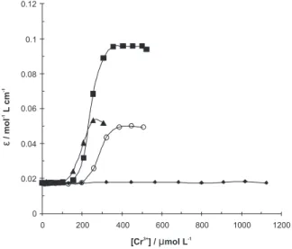
The aminoacid residue responsible for the major absorbance of proteins in the near-UV region is Trp, with a maximum at ca. 280 nm.22 Figure 1 shows the
absorbance change at 280 nm of BLG-A upon titration by aliquots of a fixed stock concentration of Cr3+
ion (10 mmol L−1) at different intentionally chosen
temperatures of 27 (room temperature), 37 (physiologic temperature), 42 (fever temperature) and 47 °C (upper
the fever temperature). At 27 °C, as the aliquot solutions of Cr3+ion are increased in the titration mixture, the
absorbance of the protein did not change. On the other hand, Cr3+ ion induces signiicant change in ε
280 at higher
temperatures. The values of [Cr3+]
1/2, the midpoint of
transition, were estimated, which are given in Table 1. It seems in this table that [Cr3+]
1/2 deceases with an increase
in temperature. In order to evaluate the effect of Cr ion on the protein stability, it was estimated the free energy of chemical unfolding using Pace’s analysis.23 In brief,
it is assumed that the Cr ion-induced denaturation is a two-state process (equation 1),
Native (N) ⇔ Denatured (D) (1)
This process was described as a single denaturant-dependent step according to the two-step theory.24-26 Each
transition curve shown in Figure 1 was analyzed for Fd, the denatured fraction, and KD, the equilibrium constant of denaturation using the relations, showed in equations 2 and 3, respectively:
Table 1. Thermodynamic parameters of BLG-A under different solvent conditions
Temperature / °C a[Cr3+]
1/2 / µmol L−1 b ∆G˚
H20/
kJ mol−1 cTm / °C
27 224.2 - 83
37 130.3 23.0 85
42 72.7 24.0 84
47 36.8 20.8 88
Native BLG at 27 - - 86
a The midpoint concentrations of Cr3+, b the stability value of the protein in the absence of denaturant (Cr3+) and c the value of melting temperature.
(YN − Yobs)
Fd = –––––––––– (2)
(YN − YD)
Fd (YN − Yobs)
KD = –––––– = –––––––––– (3)
(1−Fd) (Yobs − YD)
where Yobs is the observed variable parameter (e.g. absor-bance) and YN and YD are the values of Y characteristics of the fully native and denatured conformation, respectively. The ∆G° change is given by the following equation 4:
∆G°= −RT lnKD (4)
where R is the universal gas constant, and T is the absolute temperature. ∆G0H
2Ovaries linearly with denaturant
concentration over a limited region. The simplest method of estimating the conformational stability of the protein in the absence of denaturant, is to assume that ∆G0
H2Oversus
denaturant concentration in the transition region is a linear plot.27 The ∆G0
H2Ovalues obtained from the linear extrapolation
(Figure 2) are given in Table 1. This table shows that the ∆G0
H2Ovalues of the native BLG-A at 42 °C are greater than
those of the protein at 37 °C and 47 °C (approximately 1.0 and 3.2 kJ mol−1, respectively). Previous reports represented
that BLG denatured via three-state dissociation coupled unfolding (DCU) process involving the formation of stable monomer as intermediate; dimer ⇔ monomer ⇔ unfolding state. The Gibbs free energy change for denaturing BLG (∆G° DCU) obtained by Apenten28 calculated 60.0 kJ mol−1
at pH 7.0 and 72.0 kJ mol−1 at pH 2.6. By comparison, the
dissociation free energies of dimeric BLG are 23.0, 24.0 and 20.8 kJ mol−1 at 37, 42 and 47 ºC, respectively at pH 7.0.
Since BLG at neutral pH (our experimental conditions) is a dimer and we have calculated the Gibbs free energy of BLG in the presence of different concentrations of Cr3+ at different
temperatures (Table 1), then we can conclude that Cr3+ ions
can induce the dissociation of the subunits in dimeric form of BLG at different temperatures.Regarding to these data, we used other techniques (far- and near-UV CD and luorescence spectroscopy) that are capable of sensing different structural properties of the BLG-A at different temperatures in the presence and absence of Cr3+ ion.
Fluorescence measurements
Fluorescence spectroscopy is an useful technique to study the structure, dynamics and binding properties of protein molecules in solution. The intrinsic luorescence of tryptophanyl residues is a particularly sensitive method to perform this kind of studies. BLG has two tryptophanyl residues. From the Papiez et al.29 crystal structure, Trp19 is
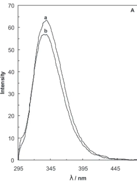
at the base of the central hydrophobic calyx of the protein, while Trp 61 is part of an external loop. The intrinsic luorescence of BLG is then almost exclusively attributed to Trp19, positioned in a more apolar environment than Trp 61.29 Figure 3 (A-D) shows the effect of the midpoint
concentration of Cr3+ on the intrinsic Trp luorescence of
BLG-A at different temperatures (27, 37, 42 and 47 °C). It is seen in this igure that the addition of Cr3+ induces a
decrease (approximately 6%) at 27 °C (Figure 3A) and an increase at other temperatures of 37, 42 and 47 °C (Figure B, C and D) in the emission of thenative BLG. Figure 3E shows the effects of the temperature (27°C to 47°C) on intrinsic luorescence of the native BLG-A. It
Figure 2. Plot of ∆G°versus[Cr3+] at different temperatures, 37 (), 42 () and 47 () in 50 mmol L−1 sodium chloride solution.
is seen in this igure that an increase in temperature can signiicantly reduce the intrinsic luorescence of the native BLG-A (approximately 51%). Hence; it can be concluded that the tertiary structure of BLG is altered on heating. It has been reported that Trp 61 has a minor contribution to the overall luorescence emission of BLG.30 This can be
due to the location of Trp 61 near a disulide bond (Cys 66 and 160) or near the guanidine group of Arg 124 (3-4 Å distance from Trp 19) indole ring,22 which can quench its
emission and/or to the self-quenching of Trp 61 of the other monomer in the BLG dimer form. Also, the position of the emission maximum of the native BLG at 335 nm is not a characteristic of a completely nonpolar medium, suggesting some contribution to the spectrum of the more
surface exposed Trp-61. The observation that luorescence intensity decreases on increasing the temperature (27 up to 47°C) might be due to the changing environment of Trp 19 (Figure 3E).
Is the calyx structure of the native BLG in the presence of Cr3+ ion slightly loosed on heating? In order
to answer this question, we use ANS luorescence intensity measurements in our studies. The ANS is a molecule that shares both hydrophilic and hydrophobic characters and whose luorescence response appears as a convenient and sensitive tool apt to investigate the nature of proteins binding regions. Small, albeit signiicant, changes in ANS luorescence, when changing the protein environment, appear to relect a modiied ligand-protein interaction, Figure 3B. The effect of midpoint concentration of Cr3+ ion on the intrinsic
luorescence of BLG-A (5 µmol L−1) in 50 mmol L−1 sodium chloride solution (pH 7.0) at 37 °C. [Cr3+] = 0 (a) and [Cr3+] = 130.3 µmol L−1 (b) .
Figure 3C. The effect of midpoint concentration of Cr3+ ion on the intrinsic luorescence of BLG-A (5 µmol L−1) in 50 mmol L−1 sodium chloride solution (pH 7.0) at 42 °C. [Cr3+] = 0 (a) and [Cr3+] = 72.7 µmol L−1 (b).
Figure 3D. The effect of midpoint concentration of Cr3+ ion on the intrinsic luorescence of BLG-A (5 µmol L−1) in 50 mmol L−1 sodium chloride solution (pH 7.0) at 47 °C. [Cr3+] = 0 (a) and [Cr3+] = 36.8 µmol L−1 (b).
which can be due to changes in the probe response.31 ANS
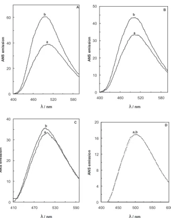
is frequently used to demonstrate the presence of partially unfolded conformations of globular proteins. This is because ANS binds to solvent-exposed hydrophobic clusters, which results in a considerable increase in ANS luorescence intensity and in a blue shift of the luorescence emission maximum.32 Figure 4 (A-D) shows
the ANS spectra in the presence of midpoint concentration of Cr3+ and the absence of it on BLG-A at different
temperatures. It can be seen that Cr3+ considerably
enhances the ANS luorescence intensity of the protein at all temperatures except at 47 °C (the emission of ANS in the absence and presence of Cr3+ ions does not show any
change). Cr3+ ions induce a pronounced blue shift (ca. 3
in 27°C to ca. 7 nm in 37 and 42°C) in the λmax of ANS. Hence, binding of chromium ions leads to a partially unfolding in the protein resulted from solvent-exposed hydrophobic patches.
Far-UV CD measurements
CD has proved to be an ideal technique for monitoring conformational changes in proteins, which can occur as a result of changes in experimental parameters such as
pH, temperature and binding of ligands.22 The far-UV CD
spectra characterize the secondary structure of proteins due to the peptide bond absorption. The CD spectrum of BLG is typical of a protein that is composed of antiparallel β-structure and shows a minimum at 217 nm.33 The
far-UV CD spectra of BLG-A at different temperatures in the absence and in the presence of midpoint concentrations of Cr3+ ions (605 µmol L−1 for 27°C, 352 µmol L−1 for 37°C,
196 µmol L−1 for 42°C and 99.3 µmol L−1 for 47°C) are
shown in Figures 5A and B, respectively. Increasing of the temperature from 27°C to 47°C (in the absence and presence of Cr3+ ions) does not show any signiicant changes
in the content of regular secondary structure of the BLG-A.
Near-UV CD measurements
The near-UV CD of proteins arises from the environments of each aromatic amino acid side chains as well as possible contributions from disulphide bonds, or non-protein cofactors, which might absorb in this spectral region and Figure 4. ANS luorescence spectra measured of 5 µmol L−1 BLG-A (a)
and BLG-A (5 µmol L−1) incubated with midpoint concentration of Cr3+ (b) at 27 °C (A), 37 °C (B), 42 °C (C) and 47 °C (D) in 50 mmol L−1 sodium chloride solution.
thus gives information about the tertiary structure of the protein.33 The near-UV CD spectrum of BLG alone shows
a pattern of negative peaks between 260 and 320 nm (Figure 6A) due to the presence of aromatic residue: two tryptophans (Trp19 and 61), four tyrosines (Tyr 20, 42, 99 and 102) and four phenylalanines (Phe 82, 105, 136 and 151). Although all of them can contribute to the CD spectrum, Trp residues are often the major determinants of the near-UV CD curve, Trp signals are generally more intense than those of Tyr and Phe and they occur between 250 and 300 nm, whereas Phe and Tyr usually do not absorb above 270 nm and 290 nm, respectively.22,33-35 According
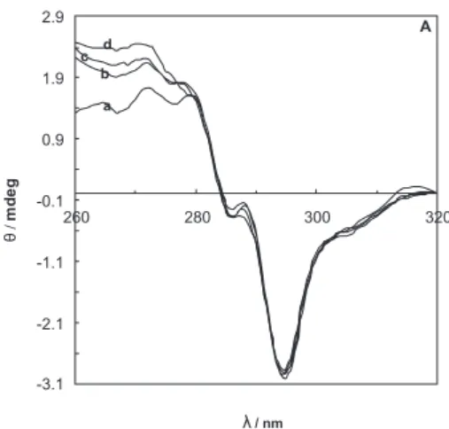
to this, the negative bands at 293 nm and 285 nm can be assigned to asymmetrically perturbed tryptophans while peaks below 280 nm are likely the result of the chiral environment of Phe and Tyr residues.35 The near-UV CD
spectrum of BLG-A at different temperatures in the absence and in the presence of midpoint concentrations of Cr3+
ions (2.55 mmol L−1 for 27°C, 1.48 mmol L−1 for 37°C,
0.42 mmol L−1 for 42°C and 0.063 mmol L−1 for 47°C)
are shown in Figure 6A and 6B. Figure 6A shows that an increase in temperature does not cause any considerably change in the [θ]293 and [θ]285 values, but causes signiicance decrease in [θ]275. The signiicance decrease in [θ]275 means that the position of Tyr residues of protein can change and move into less hydrophobic environment on increasing the temperature,35 and BLG-A undergoes a conformational
transition affecting its tertiary structure. Since the decrease in luorescence emission on increasing the temperature might be due to a change in the environment of Trp19 which is located near the Tyr 20, it may be concluded that a change in the structure of calyx (Trp 19 environment) may affect the adjacent residue (Tyr 20) and move it into a less hydrophobic environment (Figure 7). From these data, it seems that the signiicance decrease in [θ]275 of the BLG-A probably resulted from the changing in the location of Tyr 20. The near-UV CD spectrum of BLG-A at different temperatures in the presence of midpoint concentrations of Cr3+ ions shows considerable changes in [θ]
293 , [θ]285
and [θ]275 (Figure 6B), suggesting a change in the tertiary structure of the protein.
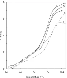
The change in the mean residue ellipticity of BLG in the absence and presence of different concentrations of Cr(III) (the concentrations equivalent to midpoint concentrations of Cr3+ ions at 27, 37, 42 and 47°C) at 293 nm as a function
of temperature in the range, 25-100 °C is shown in Figure 8. The melting points (Tm) are listed in Table 1. This table shows that high concentrations (equivalent to midpoint concentrations of 27, 37 and 42°C) of Cr3+ ions can reduce
the thermal stability of the BLG-A, whereas in the lowest concentration (equivalent to midpoint concentration of 47°C) can increase the thermal stability of the protein. Figure 6A. Near-UV circular dichroism spectra of 57 µmol L−1 BLG-A
measured at different temperatures of 27 (a), 37 (b), 42 (c) and 47 °C (d) in 50 mmol L−1 sodium chloride solution.
Figure 6B. Near-UV circular dichroism spectra of 57 µmol L−1 BLG-A incubated with different midpoint concentrations of Cr3+ at 27 (a), 37 (b), 42 (c) and 47 °C (d) in 50 mmol L−1 sodium chloride solution.
Conclusions
The above results on BLG in the absence and in the presence of Cr3+ ions at different temperatures allow to
conclude that an increase in temperature does not perturb the secondary structures of the protein, but signiicantly alters its tertiary structure.
Acknowledgements
The inancial supports of the Research Council of University of Tehran and the Iran National Science Foundation (INSF) are highly appreciated.
References
1. Hong, Y. H.; Creamer, L. K; Int. Dairy J.2002,12,345.
2. Medrano, A.; Abirached, C.; Panizzolo, L.; Moyna, P.; Añón, M.C; Food. Chem.2009, 113, 127.
3. Townend, R; Acta. Biochem. Biophys. 1965, 109, 1. 4. Wit, J.N; Trends Food. Sci. Technol. 2009, 20, 27.
5. Divsalar, A.;Saboury, A.A.; Moosavi- Movahedi, A.A.; Mansoori-Torshizi, H; Int. J. Biol. Macromol.2006, 38, 9.
6. Brown, E. D; J. Dairy Sci.1984, 67, 713.
7. Dodin, G.; Andrieux, M.; Al Kabbani, H; Eur. J. Biochem.1990, 193, 697.
Figure 8. Effect of temperature on the mean residue ellipticity at 293 (mdeg) for the native BLG-A (in the absence of Cr3+ ion), BLG-A after 3 min incubation with midpoint concentrations of Cr3+ at 27 (b), 37 (c), 42 (d) and 47 °C (e) in 50 mmol L−1 sodium chloride solution. The protein concentration was 57 µmol L−1.
8. Dufour, E.; Haertle, T; J. Agric. Food. Chem.1990, 38, 1691. 9. Farrel, H. M.; Behe, M. J.; Enyart, J. A; J. Dairy Sci. 1987, 70,
252.
10. Ono, J.; Doi, K.; Nagasawa, T; Agric. Biol. Chem.1975, 39, 2149.
11. Powell Baker, H.; Saroff, H. A; Biochemistry1965, 4, 1670. 12. Lu, Y.; Valentine, J. S; Curr. Opin. Struct. Biol.1997, 7, 495.
13. Myers, J. M.; Antholine, W. E.; Myers, C. R; Toxicology2008,
246, 222.
14. Shirvastava, R.; Upretic, R. K.; Chaturvedi, U. C; FEMS Immunol. Med. Microbiol.2002,34, 1.
15. Tamblyn, L.; Li, E.; Sarras, H.; Srikanth, P.; Hande, M. P.; McPherson, J. P; Mutat. Res.2009, 660, 57.
16. Gayatari, R.; Sharma, A. K.; Rajarm, R.; Ramasami, T; Biochem. Biophys. Res. Com.2001, 283, 229.
17. Anderson, R.A; Diabetes Metab. 2000, 26, 22.
18. Divsalar, A.; Saboury, A.A;. Moosavi-Movahed, A.A; Protein J. 2006, 25, 157.
19. Brownlow, S.; Cabral, J. H. M.; Cooper, R.; Flower, D. R.; Yewdall, S. J.; Polikarpov, I.; North, A. C. T.; Sawyer, L; Structure 1997,5, 481.
20. Manavalan, P.; Johnson, C .J. R; Anal. Biochem.1987, 167, 76. 21. Yang, J. T.; Wu, C. S. C.; Martinez, H. M; Methods Enzymol.
1986, 130, 208.
22. Viseu, M. I.; Carvalho, T. I.; Costa, S. M. B; Biophys. J.2004, 86, 2392.
23. Pace, C. N; TibTech. 1990, 8, 93.
24. Saboury, A. A.; Moosavi-Movahedi, A. A; Biochem. Educ.1995,
23, 164.
25. Moosavi-Movahedi, A. A.; Nazari, K.;, Saboury, A. A; Colloids Surf., B1997, 9, 123.
26. Ahmad. F; J. Iran. Chem. Soc.2004, 1, 99.
27. Pace, C. N.; Shiley, B. A.; Thomson, J. A; In Protein Structure - a Practical Approach, Creighton T. E ed.; IRL Press: Oxford, 1990. 28. Apenten, P. K. O.; Khokhar, S.; Galani, D; Food Hydrocolloids
2002, 16, 95.
29. Oliveira, K. M. G.; Valente-Mesquita, V. L.; Botelho, M. M.; Sawyer, L.; Ferreira, S. T.; Polikarpov, I; Eur. J. Biochem.2001,
268, 477.
30. Zsila, F.; Imre, T.; Szabo, P. T.; Bikadi, Z.; Simonia, M; FEBS Lett. 2002, 520, 81.
31. Creamer, L.K; Biochemistry1995,34, 7170.
32. Matulis, D.; Baumann, C. G.; Bloomield, V. A.; Lovrien, R. E;
Biopolymers 1999, 49, 451.
33. Uversky, V. N.; Li, J.; Fink, A. L; J. Biol. Chem.2001, 47,44284.
34. Kelly, S. M.; Price, N. C; Biochem. Biophys. Acta1997, 1338, 161.
35. Kelly, S. M.; Price, N. C; Curr. Protein Pept. Sci.2000, 1, 349.
Received: November 25, 2008