Article
Printed in Brazil - ©2014 Sociedade Brasileira de Química0103 - 5053 $6.00+0.00A
*e-mail: [email protected], [email protected]
Experimental and NMR Theoretical Methodology Applied to Geometric Analysis of
the Bioactive Clerodane
trans
-Dehydrocrotonin
Breno Almeida Soares,a Caio Lima Firme,*,a Maria Aparecida Medeiros Maciel,a,b
Carlos R. Kaiser,c Eduardo Schillingd and Adailton J. Bortoluzzid
aUniversidade Federal do Rio Grande do Norte, Instituto de Química,
Campus Lagoa Nova, 59072-970 Natal-RN, Brazil
bPrograma de Pós-graduação em Biotecnologia, Universidade Potiguar Laureate International
Universities, Campus Salgado Filho, 59075-000 Natal-RN, Brazil
cUniversidade Federal do Rio de Janeiro, Instituto de Química,
Ilha do Fundão, 21941-909 Rio de Janeiro-RJ, Brazil
dUniversidade Federal de Santa Catarina, Departamento de Química,
Campus Universitário Trindade, 88040-900 Florianópolis-SC, Brazil
trans-Desidrocrotonina (t-DCTN), um bioditerpeno do tipo 19-nor-clerodano isolado de Croton cajucara Benth, representa um dos clerodanos da atualidade com maiores índices de investigações
científicas. Neste trabalho, uma nova abordagem unindo dados de difração de raios X, dados de ressonância magnética nuclear (NMR) e dados teóricos foi aplicada para a completa caracterização
da t-DCTN. Para tanto, a geometria de t-DCTN foi reavaliada por difração de raios X e NMR de
1H e 13C e foi comparada com os dados teóricos obtidos por B3LYP/6-311G++(d,p). A subsequente
avaliação dos valores teóricos e experimentais de deslocamentos químicos de NMR e constantes de acoplamento spin-spin apresentou correlações muito boas entre propriedades magnéticas teóricas e
experimentais. Adicionalmente, os índices de deslocalização entre átomos de hidrogênio, δ(Η,Η’),
correlacionaram bem com os dados experimentais e calculados de constante de acoplamento. Uma análise topológica complementar utilizando a teoria quântica de átomos em moléculas (QTAIM)
mostrou interações intramoleculares para t-DCTN.
trans-Dehydrocrotonin (t-DCTN) a bioactive 19-nor-diterpenoid clerodane type isolated from Croton cajucara Benth, is one of the most investigated clerodane in the current literature. In this
work, a new approach joining X-ray diffraction data, nuclear magnetic resonance (NMR) data
and theoretical calculations was applied to the thorough characterization of t-DCTN. For that,
the geometry of t-DCTN was reevaluated by X-ray diffraction as well as 1H and 13C NMR data,
whose geometrical parameters where compared to those obtained from B3LYP/6-311G++(d,p)
level of theory. From the evaluation of both calculated and experimental values of 1H and 13C NMR
chemical shifts and spin-spin coupling constants, it was found very good correlations between
theoretical and experimental magnetic properties of t-DCTN. Additionally, the delocalization
indexes between hydrogen atoms correlated accurately with theoretical and experimental spin-spin coupling constants. An additional topological analysis from quantum theory of atoms in molecules
(QTAIM) showed intramolecular interactions for t-DCTN.
Keywords: Croton cajucara, trans-dehydrocrotonin, NMR, DFT calculations, QTAIM, X-ray
Introduction
The clerodane group of diterpenes includes more than 800 isolated compounds and a significant number showed to have biological activity such as antimicrobial,1 psychotropic,2 antiulcer3 and antitumor.4 Actually, the 19-nor-clerodane trans-dehydrocrotonin (t-DCTN), a
furan clerodane skeleton type diterpene (Figure 1), is one of the most important bioactive clerodane reported in the current literature. This natural compound isolated from
Croton cajucara Benth (Euphorbiaceae), a widely grown
tree in the Amazonian region of Northern Brazil, became an important target for pre-clinical researches. In fact,
pharmacological studies examining t-DCTN confirmed
its anti-inflammatory, analgesic, antitumor, antiulcer, hypolipidemic and cardioprotective effect.5,6t-DCTN has also been shown to have antimutagenic activity5-7 and did not induce clastogenic, anticlastogenic, apoptotic and cytotoxic activities.8
In general, a complete characterization of natural terpenoids compounds requires a large amount of work, in which 1D and 2D-nuclear magnetic resonance (NMR) techniques can be combined with X-ray diffraction analysis. Since 1976, when Ferguson and Marsh reported the first crystallographic analysis of a diterpenoid type clerodane molecule,9 a plenty of reports arises describing clerodane molecules by crystallographic analysis in combination with NMR techniques. Owing to the complexity of clerodane structural molecule, the X-ray diffraction analysis is an important tool for a reliable and complete structural analysis of a new natural or semi-synthetic organic compound. In this context, the use of theoretical quantum chemistry
methods becomes a complementary tool for structural analysis of organic compounds.3,10-26
The NMR spectral data of a new compound provide an useful information for its molecular identity and geometry. In fact, NMR parameters such as chemical shifts and spin-spin coupling constants (SSCC), as well as theoretical calculations and 1H spectrum simulations give support for the total characterization of complex molecules. Regarding the SSCC parameter, it plays an important role on the conformational analysis and elucidation of several types of molecules,27-31 mainly when it is correlated with theoretical SSCC.32-43
Most of the theoretical descriptions of spin spin coupling constants follows the Ramsey and Purcell interpretation44 and Ramsey formulation.45 In this method, the coupling constants are calculated by adding four different terms: (1) diamagnetic spin orbit (DSO) as well as (2) the paramagnetic spin orbit (PSO), which represent the interactions of the magnetic field of the nuclei mediated by the electron orbital motion, (3) the Fermi contact (FC), which is also a response property reflecting the interaction between the electron spin magnetic moment close to the nucleus and the magnetic field at the nucleus, and (4) the spin dipole (SD), which describes the interactions between the nuclear magnetic moments as mediated by the electronic spin angular moment. For an accurate quantum chemical description of SSCCs all the terms cited above must be considered.46-49 In addition, the delocalization index between two hydrogen atoms, δ(H,H′), can also be used in the theoretical analysis of the SSCCs.50
The quantum theory of atoms in molecules (QTAIM)51 is based on an observable, the electron density, and it has been
used for several experimental applications, for example, the estimate of the intensity of infrared (IR) spectrum52,53 and magnetic susceptibility,54 as well as the calculation of topological information from X-ray diffraction data55 and the linear correlation between delocalization index
δ(H,H′) and proton-proton coupling constants.56-58 The linear correlation between delocalization index and fluorine fluorine coupling constants was also described.59
Unambiguous NMR assignments for t-DCTN and its
stereochemistry determination were previously reported
using a powerful NMR equipment (600 MHz for 1H),
2D-NMR experiments, AM1 calculations and 1H NMR
spectrum simulations.60 In the present work, we have
performed X-ray diffraction of t-DCTN and many
theoretical calculations using B3LYP. The geometric skeleton of t-DCTN was re-examined by means of several
correlations involving experimental and theoretical data in order to provide additional structural information such as bond length and dihedral angles measurements which are extensively used in the conformational analysis of biomolecules applied to its pharmacological responses.3,12,24 Correlations between theoretical results and corresponding experimental data plus topological analysis from QTAIM are herein discussed.
Experimental
Material and methods
Plant material was collected in Jacundá City located at Pará State (Amazonian region, Brazil), and identified by Nelson A. Rosa.5 A voucher specimen (no. 247) has been stored at the Herbarium of the Museu Paraense Emílio Goeldi (Belém, PA, Brazil). The isolation of
t-DCTN was performed according to the literature5 and
its recrystallization (from a mixture of hexane:ether 7:3) yielded suitable crystal for X-ray diffraction. The assignments for structure elucidation were previously determined from 1H (600 MHz) and 13C (150 MHz) NMR spectroscopy.60
Single crystal X-ray diffraction
The crystal data were measured on an Enraf-Nonius CAD4 diffractometer using graphite monochromated Mo-Kα radiation (λ = 0.71069 Å) at room temperature. A
colorless crystal with dimensions of 0.40 × 0.33 × 0.30 mm was isolated from a homogeneous crystalline sample of
t-DCTN. Unit-cell parameters were determined from
centering of 25 reflections in the θ angle ranging from 4.92 to 12.03o and refined by the least-squares method
according to standard procedure.61 Intensities were collected using the ω-2θ scan technique. All diffracted intensities were corrected by Lorentz and polarization effects. The structure was solved by direct methods and was refined by the full-matrix least-squares method using
SIR97 and SHELXL97.62,63 All non-hydrogen atoms
were refined with anisotropic displacement parameters. Hydrogen atoms were placed at idealized positions using standard geometric criteria. The PLATON program64 was used to generate the picture of the molecular structure. Further relevant crystallographic data are summarized in Table 1.
Crystallographic data (atomic coordinates and equivalent isotropic displacement parameters, calculated hydrogen atom parameters, anisotropic thermal parameters and bond lengths and angles) have been deposited at the Cambridge Crystallographic Data Center (deposition number CCDC 276648). Copies of this information may be obtained free of charge from: CCDC, 12 Union Road, Cambridge, CB2 1EZ, UK (Fax: +44-1223-336-033; e-mail: [email protected] or http://www.ccdc.cam.ac.uk).
Table 1. Crystal data and structure refinement for t-DCTN
Empirical formula C19H22O4
Formula weight 314.37
Temperature / K 293(2) Wavelength / Å 0.71073 Crystal system Orthorhombic Space group P212121
a / Å 7.3869(8)
b / Å 13.8966(13)
c / Å 16.018(3)
Volume / Å3 1644.3(4)
Z 4
ρ / (g cm3) 1.270
µ / mm-1 0.088
F(000) 672
Crystal size 0.40 × 0.33 × 0.30 mm3 Theta range for data collection 1.94 to 27.97o
Index ranges -9 ≤ h ≤ 0. -18 ≤ k ≤ 0. -21 ≤ l ≤ 0 Reflections collected / unique 2184 / 2184 [R(int) = 0.0000] Completeness to theta = 27.97° 96.2 %
Absorption correction None
Refinement method Full-matrix least-squares on F2
Data / restraints / parameters 2184 / 0 / 208 Goodness-of-fit on F2 0.982
Computational details
The geometry of the studied species was optimized using standard techniques.65 Vibrational analysis of t-DCTN optimized geometry was carried out to determine whether it was a true minimum or a transition state. No imaginary frequency was obtained confirming that a minimum was found in the potential energy surface. The calculations
were performed at B3LYP/6-311G++(d,p)66-71 using
GAUSSIAN 09 package.72 From the Kohn-Sham orbitals
of t-DCTN optimized structure, the electron density was
obtained and then used for the calculation of t-DCTN
topological data by means of the AIM2000 software.73
Results and Discussion
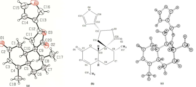
The DFT procedures for computing proton-proton coupling constants and/or chemical shifts of unknown structures or targets from synthetic studies, have been extensively reported.32-38 In this work, aiming to describe the most stable conformational status of t-DCTN (Figure 1)
more thoroughly, we have examined the combined set of X-ray data, NMR spectra and computational calculations using B3LYP/6-311G++(d,p) level of theory. A single crystal X-ray diffraction was undertaken in order to re-examine the asymmetric centers (C5, C8, C9,
C10 and C12) of t-DCTN. The PLATON perspective
drawing (Figure 1a) is in agreement with the geometric configuration supported by NMR spectra of t-DCTN. The
theoretical analysis of geometric and magnetic properties of t-DCTN gives additional support for its experimental
characterization since it confirms the structure of the most stable conformer of t-DCTN.
Figure 1 shows the crystallographic structure of
t-DCTN (Figure 1a), the corresponding skeleton formula
(Figure 1b) and its optimized structure obtained from B3LYP/6-311G++(d,p) (Figure 1c). In the perspective drawing (Figure 1a and 1c), the conformation of the decalin system chair is distorted due to ∆3,4 double bond (located at C3 and C4 in the so-called ring A) and also the spiro-lactone at C9 (in the so-called ring B).
Geometrical data available in the Supplementary Information indicate that the hydrogen atoms at C10 and C5 positions are on the opposite side of A/B decalin system, assuming trans configuration (Table S1 from
Supplementary Information). In addition, the bond lengths and angles are as expected for a trans geometry for t-DCTN.
The existence of the spiro arrangement in lactone moiety can be noticed by the fact that C9 is shared by the lactone group itself and also by ring B of decalin system. The α,β-unsaturated carbonyl group located in C2, C3, C4
and O1 atoms of decalin ring A could be observed revealing similar bond lengths of 3-(4-chloro-benzene-sulfonamido)-5-methyl-cyclo-hex-2-en-1-one, whose geometric and topological parameters were described by Jackson and
co-workers.74 The C=O, C=C and C–C bond lengths of
3-(4-chloro-benzene-sulfonamido)-5-methyl-cyclo-hex-2-en-1-one (1.251, 1.356 and 1.437 Å, respectively) are comparable to those corresponding bonds in ring A of
t-DCTN (1.215, 1.325 and 1.449 Å, respectively).
The minimum in the potential energy surface of t-DCTN
was obtained by B3LYP/6-311G++(d,p) level of theory whose geometrical parameters were compared with those from X-ray diffraction (Table S1 from Supplementary Information). The theoretical and experimental parameters for t-DCTN are very similar, except for some angle values
from furan and lactone moieties. The O3–C12–C11, C15–C14–C13 and C13–C16–C14 angles have nearly two degrees of discrepancy when comparing theoretical and experimental results (See Supplementary Information). The root mean square deviation (RMSD) of t-DCTN bond
lengths and bond angles was calculated in order to measure the accuracy of theoretical results in comparison with X-ray data. The RMSD values are 0.0153 and 0.9333 for bond lengths and bond angles, respectively. These values show the precision of the chosen level of theory for geometric calculations of t-DCTN and also indicate the relatively
smaller precision of calculated bond angles with respect to experimental values.
As a consequence of agreement between experimental and theoretical results, Figure 1a (PLATON thermal ellipsoids plots) and Figure 1c (an optimized structure) indicate similar nuclear configuration for t-DCTN. Both
stereo views showed a visual similarity in arrangements of the decalin and lactone moieties, as well as for furan ring, which is out of plane for both representations.
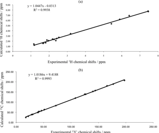
Theoretical magnetic properties (chemical
shifts and SSCC) of t-DCTN were obtained from
B3LYP/6-311G++(d,p) level of theory and compared with corresponding experimental values (See Supplementary Information).
Aiming to reinforce the discussion on the absolute
configuration of t-DCTN, correlations between the
that B3LYP/6-311G++(d,p) level of theory suffices for the analysis of magnetic and geometric properties of the evaluated clerodane t-DCTN.
The vicinal or long range coupling constants of t-DCTN
were calculated from the gauge-independent atomic orbital (GIAO) method including the four contributing terms (FC, PSO, DSO, SD)46 at the B3LYP/6-311G++(d,p) level of theory. The B3LYP/GIAO method provides accurate coupling constant values in many systems, being most of them related to rigid structure and/or aromatic molecular systems.37-40
A reasonably good correlation between calculated and experimental coupling constants was obtained (Figure 3), except for those values involving hydrogen atoms 8 and 17, whose differences between experimental and theoretical values were larger than three ppm. Therefore, the B3LYP/6-311G++(d,p)/GIAO method, including the four contributing terms (FC, PSO, DSO, SD), suffices to describe magnetic properties of low symmetry and flexible structures similar to t-DCTN.
The delocalization index from QTAIM is the quantity of shared electrons in an atomic pair, bonded or non-bonded,
Figure 2. (a) Plot of experimental vs. calculated (B3LYP/6-311G++(d,p)) 1H NMR chemical shifts and (b) plot of experimental vs. theoretical 13C NMR
chemical shifts for t-DCTN.
and it has been used to establish a relation with coupling
constants of many molecular systems.56 There are
excellent correlations observed in literature37,56-58 between delocalization index of vicinal or long range hydrogen atoms δ(H,H’) and corresponding experimental coupling constants. Additionaly, fluorine fluorine coupling constants also showed satisfactory correlation with delocalization index data for aromatic systems.59
Figures 4a and 4b shows the plots of experimental and theoretical coupling constants versus corresponding delocalization index of vicinal or long range hydrogen
atoms of t-DCTN, respectively. Both of them give a
reasonably good coefficient of determination, meaning that the amount of shared charge density between long or vicinal hydrogen atoms is related with the magnitude of the corresponding coupling constant, i.e., a relation between electronic and magnetic properties of t-DCTN is established
in Figure 4. According to Gutowsky and collaborators,75 the spin-spin coupling arises from second-order interaction between the nuclear magnetic moments and some magnetic field internal to the molecule through electrons along chemical bonding. The results from Figure 4 suggest that the influence of weak electron interaction, through field
effect, between the interacting nuclei plays some role in long range or vicinal spin-spin coupling as well, which concords with J-couplings in hydrogen bonds.76,77
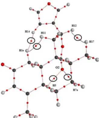
Aiming at the investigation of possible intramolecular interactions in t-DCTN, we also carried out its topological
analysis based on the gradient of the charge density distribution. Figure 5 shows the molecular graph of
t-DCTN which contains critical points of the charge
density and bond paths. Critical point is a mathematical point of a determined function, whose gradients, with respect to their coordinates, are zero. The topology of charge density may have four types of critical points: the nuclear attractor critical point (where it is located an atomic nucleus), the bond critical point (a critical point between two linking atoms), the ring critical point (a critical point within a ring) and the cage critical point (a critical point inside a molecular cage). The bond path is an atomic interaction line linking two nuclear critical points (or atomic basins) and one bond critical point between them. It corresponds to the maximum charge density compared to vicinal transversal region.51
Figure 5 shows the molecular graph of the t-DCTN
which has five bond critical points associated with
Figure 4. (a) Plot of experimental spin-spin coupling constants of vicinal or long range protons [Jn (H,H′)] vs. corresponding delocalization index values
intramolecular closed-shell interactions, where three out of them are hydrogen-hydrogen bonds (Table 2).
An important applicability of QTAIM is to quantify and qualify bonded interactions based on five topological parameters obtained at the analyzed bond critical point:78-81 (1) the value of the charge density of the critical point (ρb); (2) the value and the sign of the Laplacian (∇2ρ) of the charge density; (3) the ratio |λ1|/λ3, where λ1 and λ3 are eigenvalues of Hessian matrix of the charge density; (4) the ratio Gb / ρb, where Gb is the kinetic energy density; and (5) the total energy density (Hb) at the bond critical point. In the case where ∇2ρ > 0, ρ
b is relatively low (ρb < 6 × 10⁻² a.u.), the ratio |λ1|/λ3 < 1, the ratio Gb / ρb > 1 or close to 1, and Hb has a positive value, close to zero, the chemical interaction is defined as closed shell interaction and it corresponds, for example, to hydrogen bond, ionic bond and van der Waals interactions.78-81 Table 2 shows these five topological data for all intramolecular bonded interaction in t-DCTN
(Figure 5) which are certainly related to the highest stability of t-DCTN found in its NMR and X-ray data.
All values in Table 2 are in agreement with the topological characterization of closed shell interaction for the five interatomic interactions.78-81 The values of G
b/
ρb in Table 2 are in the same range to that found in closed shell interactions in metallocenes, involving metal atom and π ligand carbon atom.82 The most stable conformer of t-DCTN presents five intramolecular interactions:
three H-H bonds, linking hydrogen atoms from decalin system to hydrogen atoms from spiro-lactone and furan rings, and two (C)O--H(C) bonded interactions linking the lactone carbonyl oxygen to two axial hydrogen atoms from decalin ring. It is demonstrated in literature that H–H bonding contributes as a stabilizing factor to the energy of a chemical system.81,83 Matta and co-workers have investigated intramolecular H–H bonding in several compounds showing its stabilizing effect whose values of the five topological parameters listed in Table 2 were similar to the H–H bonds in t-DCTN.
Regarding the (C)O--H(C) bonded interactions we compared the charge density of bond critical points and Laplacian of the charge density between (C)O--H(C) bonded interactions and the corresponding values from hydrogen bonds (O--HO) of substituted malonaldehyde enols.84 The charge density of (C)O--H(C) bonded interactions are nearly one-fifth of the averaged charge density value of hydrogen bonds of substituted malonaldehyde enols. On the other hand, according to less positive values of ∇2ρ of the bond critical point related to (C)O--H(C) bonded interactions in t-DCTN, the corresponding distribution of
the charge density is less dispersed in (C)O--H(C) bonded interactions in t-DCTN than that in hydrogen bonds of
substituted malonaldehyde enols.84
Therefore, the sum of the charge density in the five intramolecular interactions in t-DCTN is nearly
equivalent to the charge density of one hydrogen bond of malonaldehyde enol,84 which shows the cooperative effect of the five weak intramolecular interactions in determining the most stable conformer of t-DCTN. Three intramolecular
interactions (H–H bonds) are not influenced by polarity
Figure 5. Molecular graph for t-DCTN indicating the bond paths analyzed in Table 2.
Table 2. Values of the charge density of bond critical points (ρb), the corresponding Laplacian of the charge density (∇2ρ), the ratio |λ1| / λ3, the ratio
Gb / ρb, and the total energy density (Hb) of all intramolecular bonded interactions in t-DCTN
Interactions ρb× 102 / a.u. ∇2ρ / a.u. |λ
1| / λ3 Gb / ρb Hb× 102 / a.u.
H1e–H11 1.285 0.045 0.194 0.721 1.653
H1e–H14 0.384 0.012 0.175 0.632 0.487
H17–H12 0.771 0.026 0.162 0.705 0.971
O2–H7a 0.882 0.029 0.167 0.722 1.186
while it influences the other two interactions involving oxygen atom. Indeed, the most stable conformer of t-DCTN
is the one obtained by means of NMR and X-ray whose experimental values are in very good agreement with the corresponding theoretical data. Then, our theoretical calculations reinforced the characterization of the most stable conformer and also enabled a clear understanding of the electronic structure of t-DCTN.
Currently, there is a number of Croton cajucara
Benth researches devoted to its chemical, biochemical, pharmacological and more recently potential advantages of molecular incorporation into drug delivery systems, specifically DCTN-load studies.85-88 In fact, concerning to the biotechnological advanced context, our recent work reported to trans-dehydrocrotonin cover its encapsulation
in liposomes with a significant enhancement of the antitumor activity of this 19-nor-clerodane-type diterpene.85
Since the stability of t-DCTN loaded in different
biological formulations had been investigated aiming its pharmacologic improvement, this present work attracts significant importance on physicochemical characteristics of this compound to be applied in the advancement of this drug uses in therapy. For that, in this present work, a new approach joining two different methodologies, previously applied to organic compounds, has been developed and validated for the thorough characterization of some complex organic compounds such as the natural bioactive
t-DCTN.
Conclusions
The crystallographic and theoretical data are in agreement with the previously detailed NMR analysis
and characterization of the structure of t-DCTN.
Experimental and theoretical geometric parameters present nearly negligible discrepancies, indicating that B3LYP/6-311G++(d,p) is a satisfactory level of theory for the calculation of the optimized geometry of complex molecules such as t-DCTN. Those results confirmed the
stereochemistry of the decaline and lactone units and a hindered rotation around the C12–C13 bond in t-DCTN.
There are very good correlations involving experimental and theoretical magnetic properties (NMR nuclear shielding and spin-spin coupling constants), indicating that: (1) the B3LYP/6-311G++(d,p)/GIAO method is satisfactory for the calculation of NMR nuclear shielding and; (2) the B3LYP/311G++(d,p)/GIAO method is satisfactory for the calculation of spin-spin coupling constants.
The values of QTAIM delocalization indexes involving vicinal or long range hydrogen atoms correlate well with both corresponding theoretical and experimental spin-spin
coupling constants of t-DCTN, indicating that the amount
of charge density between each proton pair (being vicinal or not) is directly proportional to the spin-spin coupling constant.
From the topological analysis of t-DCTN there are five
intramolecular bonded interactions in t-DCTN (three out
of them being hydrogen-hydrogen bonds) which possibly influence in the optimal nuclear configuration and the geometric structure of t-DCTN.
A new approach joining two different methodologies, previously applied to organic compounds, has been developed and validated in this work for the thorough characterization of complex organic compounds.
Supplementary Information
Selected bond length and bond angles from X-ray diffraction and B3LYP method, experimental and theoretical 1H and 13C NMR chemical shifts, experimental and theoretical NMR spin-spin coupled constants, Z-matrix and crystallographic data for t-DCTN. This material is
available free of charge athttp://jbcs.sbq.org.br as PDF file.
Acknowledgments
The authors are grateful to FAPERN (Fundação de Amparo à Pesquisa do Estado do Rio Grande do Norte), CAPES (Coordenação de Aperfeiçoamento de Pessoal de Nível Superior) and CNPq (Conselho Nacional de Desenvolvimento Científico e Tecnológico) for financial support.
References
1. Murthy, M. M.; Subramanyam, M.; Bindu, M. H.; Annapurna, J.; Fitoterapia 2005, 76, 336.
2. Shirota, O.; Nagamatsu, K.; Sekita, S.; J. Nat. Prod. 2006, 69, 1782.
3. Brasil, D. S. B.; Alves, C. N.; Guilhon, G. M. S. P.; Muller, A. H.; Secco, R. S.; Peris, G.; Llusar, R.; Int. J. Quan. Chem. 2008, 108, 2564.
4. Ravikumar, Y. S.; Mahadevan, K. M.; Manjunatha, H.; Satyanarayana, N. D.; Phytomedicine 2010, 17, 513.
5. Maciel, M. A. M.; Pinto, A. C.; Arruda, A. C.; Pamplona, S. G.; Vanderlinde, F. A.; Lapa, A. J.; Echevarria, A.; Grynberg, N. F.; Cólus, I. M. S.; Farias, R. A. F.; Costa, A. M. L.; Rao, V. S. N.; J. Ethnopharmacol. 2000, 70, 41.
6. Costa, M. P.; Magalhães, N. S. S.; Gomes, F. E. S.; Maciel, M. A. M.; Braz. J. Pharmacog. 2007, 17, 275.
8. Poersch, A.; Santos, F. V.; Maciel, M. A. M.; Câmara, J. K. P.; Dantas, T. N. C.; Cólus, I. M. S.; Mutat. Res. 2007, 629, 14. 9. Ferguson, G.; Marsh, W. C.; Cryst. Struct. Commun. 1976, 5,
35.
10. Herz, W.; Pilotti, A. M.; Soderholm, A. C.; Shuhama, I. K.; Vichnewski, W.; J. Org. Chem. 1977, 42, 3913.
11. Busygin, I.; Nieminen, V.; Taskinen, A.; Sinkkonen, J.; Toukoniitty, E.; Sillanpaa, R.; Murzin, D. Y.; Leino, R.; J. Org. Chem. 2008, 73, 6559.
12. Roslund, M. U.; Klika, K. D.; Lehtila, R. L.; Tahtinen, P.; Sillanpaa, R.; Leino, R.; J. Org. Chem. 2004, 69, 18.
13. Nishino, C.; Manabe, S.; Kazui, M.; Matsuzaki, T.; Tetrahedron Lett. 1984, 25, 2809.
14. Soriano-Garcia, M.; Toscano, R. A.; Esquivel, B.; Hernandez, M.; Rodriguez-Hahn, L.; Acta Crystallogr., Sect. C: Cryst. Struct. Commun. 1987, 43, 272.
15. Manriquez, V.; San-Martin, A.; Rovirosa, R.; von Schnering, H. G.; Peters, K. Acta Crystallogr., Sect. C: Cryst. Struct. Commun. 1990, 46, 1170.
16. Ning, X.; Zhi-Da, M.; Shou-Xun, Z.; Bing, W.; Qi-Tai, Z.; Pei, Z.; Phytochemistry 1991, 30, 1963.
17. Toscano, R. A.; Sanchez, A. A.; Esquivel, B.; Esquivel, O.; Rodriguez-Hahn, L.; Acta Crystallogr., Sect. C: Cryst. Struct. Commun. 1994, 50, 1794.
18. Huan-Ming, C.; Zhi-Da, M.; Iinuma, M.; Tanaka, T.; Heterocycles 1996, 43, 611.
19. Ye, D.; Shu-Lin, P.; Qiang, Z.; Xun, L.; Li-Sheng, D.; Helv. Chim. Acta. 2002, 85, 2547.
20. Januario, A. H.; Santos, S. L.; Marcussi, S.; Mazzi, M. V.; Pietro, R. C. R. L.; Sato, D. N.; Ellena, J.; Sampaio, S. V.; Franca, S. C.; Soares, A. M.; Chem. Biol. Interact. 2004, 150, 243.
21. Sathe, M.; Kaushik, M. P.; Bioorg. Med. Chem. Lett. 2010, 20, 1312.
22. Cantrell, C. L.; Klun, J. A.; Pridgeon, J.; Becnel, J.; Green III, S.; Fronczek, F. R.; Chem. Biodivers. 2009, 6, 447.
23. Liu, H.; Chou, G.; Guo, Y.; Ji, L.; Wang, J.; Wang, Z.; Phytochemistry 2010, 71, 1174.
24. Brasil, D. S. B.; Müller, A. H.; Guilhon, G. M. S. P.; Alves, C. N.; Peris, G.; Llusard, R.; Moline, V.; J. Braz. Chem. Soc. 2010, 21, 731.
25. Brasil, D. S. B.; Moreira, R. Y. O.; Müller, A. H.; Alves, C. N.; Int. J. Quan. Chem. 2006, 106, 2706.
26. Guo, P.; Li, Y.; Xu, J.; Liu, C.; Ma, Y.; Guo, Y.; J. Nat. Prod. 2011, 74, 1575.
27. Günther, H.; NMR Spectroscopy: Basic Principles. Concepts and Applications in Chemistry, 2nd ed.; John Wiley & Sons:
New York, 1995.
28. Muller, N.; Pritchard, D. E.; J. Chem. Phys. 1959,31, 768. 29. Karplus, M.; Anderson, D. H.; J. Chem. Phys. 1959, 30, 6. 30. Karplus, M.; J. Chem. Phys. 1959, 30, 11.
31. Karplus, M.; J. Am. Chem. Soc. 1963, 85, 2870.
32. Barone, V.; Peralta, J. E.; Contreras, R. H.; Snyder, J. P.; J. Phys. Chem. A. 2002, 106, 5607.
33. Pihlaja, K.; Tahtinen, P.; Klika, K. D.; Jokela, T.; Salakka, A.; Wahala, K.; J. Org. Chem. 2003, 68, 6864.
34. Bally, T.; Rablen, P. R.; J. Org. Chem. 2011, 76, 4818. 35. López-Vallejo, F.; Fragoso-Serrano, M.; Suárez-Ortiz, G. A.;
Hernández-Rojas, A. C.; Cerda-García-Rojas, C. M.; Pereda-Miranda, R.; J. Org. Chem. 2011, 76, 6057.
36. Pu, J.; Huang, S.; Ren, J.; Xiao, W.; Li, L. M.; Li, R.; Li, L. B.; Liao, T.; Lou, L.; Zhu, H.; Sun, H.; J. Nat. Prod. 2007, 70, 1706. 37. S á n c h e z - M e n d o z a , E . ; H e r n á n d e z - Tr u j i l l o , J . ;
del Río-Portilla, F.; J. Phys. Chem. A. 2007, 111, 8264. 38. Del Bene, J. E.; Alkorta, I.; Elguero, J.; J. Phys. Chem. A. 2010,
114, 3713.
39. Alkorta, I.; Elguero, J.; Limbach, H.; Shenderovich, I. G.; Winkler, T.; Magn. Reson. Chem. 2009, 47, 585.
40. Czernek, J.; Lang, J.; Sklenář, V.; J. Phys. Chem. A. 2000, 104, 2788.
41. Gräfenstein, J.; Tuttle, T.; Cremer, D.; Phys. Chem. Chem. Phys. 2005, 7, 452.
42. Gräfenstein, J.; Kraka, E.; Cremer, D.; J. Phys. Chem. A. 2004, 108, 4520.
43. Bouř, P.; Raich, I.; Kaminský, J.; Hrabal, R.; Čejka, J.; Sychrovský, V.; J. Phys. Chem. A. 2004, 108, 6365.
44. Ramsey, N. F.; Purcell, E. M.; Phys. Rev. 1952, 85, 143. 45. Ramsey, N. F.; Phys. Rev. 1953, 91, 303.
46. Autschbach, J.; Le Guennic, B.; J. Chem. Ed. 2007, 84, 156. 47. Cremer, D.; Gräfenstein, J.; Phys. Chem. Chem. Phys. 2007, 9,
2791.
48. Helgaker, T.; Watson, M.; Handy, N. C.; J. Chem. Phys. 2000, 113, 9402.
49. Sychrovský, V.; Gräfenstein, J.; Cremer, D.; J. Chem. Phys. 2000, 113, 3530.
50. Bader, R. F. W.; Streitwieser, A.; Neuhaus, A.; Laidig, K. E.; Speers, P.; J. Am. Chem. Soc. 1996, 118, 4959.
51. Bader, R. F. W.; Atoms in Molecules: A Quantum Theory; Oxford University Press: Oxford, 1994.
52. Haiduke, R. L. A.; de Oliveira, A. E.; Bruns, R. E.; J. Phys. Chem. A. 2004, 108, 6788.
53. Haiduke, R. L. A.; Bruns, R. E.; J. Phys. Chem. A. 2005, 109, 2680.
54. Bader, R. F. W.; Keith, T. A.; J. Chem. Phys. 1993, 99, 3683. 55. Koritsanszky, T. S.; Coppens, P.; Chem. Rev. 2001, 101, 1583. 56. Matta, C. F.; Hernández-Trujillo, J.; Bader, R. F. W.; J. Phys.
Chem. A. 2002, 106, 7369.
57. Mandado, M.; Blockhuys, F.; Alsenoy, C. V.; Chem. Phys. Lett. 2006, 430, 454.
58. Sánchez-Mendoza, E.; Hernández-Trujillo, J.; Magn. Reson. Chem. 2010, 48, 866.
60. Maciel, M. A. M.; Pinto, A. C.; Kaiser, C. R.; Magn. Reson. Chem. 2003, 41, 278.
61. Enraf-Nonius; CAD-4 Software; Enraf-Nonius, Delft, The Netherlands, 1989.
62. Altomare, A.; Burla, M. C.; Camalli, M.; Cascarano, G.; Giacovazzo, C.; Guagliard, A.; Moliterni, A. G. G.; Spagna, R.; J. Appl. Crystallogr. 1999, 32, 115.
63. Sheldrick, G.; SHELXL-97 Program for Crystal Structure Refinement; University of Göttingen, Germany, 1997. 64. Spek, A. L.; Acta Crystallogr., Sect. D: Biol. Crystallogr. 2009,
65, 148.
65. Fletcher, R.; Practical Methods of Optimization; Wiley: New York, 1980.
66. Becke, A. D.; J. Chem. Phys. 1993, 98, 5648. 67. Becke, A. D.; J. Chem. Phys. 1993, 98, 1372. 68. Dunning, J. T. H.; J. Chem. Phys. 1989, 90, 1007.
69. Head-Gordon, M.; Pople, J. A.; Frisch, M. J.; Chem. Phys. Lett. 1988, 153, 503.
70. Frisch, M. J.; Head-Gordon, M.; Pople, J. A.; Chem. Phys. Lett. 1990, 166, 281.
71. Frisch, M. J.; Head-Gordon, M.; Pople, J. A.; Chem. Phys. Lett. 1990, 166, 275.
72. Frisch, M. J.; Trucks, G. W.; Schlegel, H. B.; Scuseria, G. E.; Robb, M. A.; Cheeseman, J. R.; Scalmani, G.; Barone, V.; Mennucci, B.; Petersson, G. A.; Nakatsuji, H.; Caricato, M.; Li, X.; Hratchian, H. P.; Izmaylov, A. F.; Bloino, J.; Zheng, G.; Sonnenberg, J. L.; Hada, M.; Ehara, M.; Toyota, K.; Fukuda, R.; Hasegawa, J.; Ishida, M.; Nakajima, T.; Honda, Y.; Kitao, O.; Nakai, H.; Vreven, T.; Montgomery, J. A., Jr.; Peralta, J. E.; Ogliaro, F.; Bearpark, M.; Heyd, J. J.; Brothers, E.; Kudin, K. N.; Staroverov, V. N.; Kobayashi, R.; Normand, J.; Raghavachari, K.; Rendell, A.; Burant, J. C.; Iyengar, S. S.; Tomasi, J.; Cossi, M.; Rega, N.; Millam, N. J.; Klene, M.; Knox, J. E.; Cross, J. B.; Bakken, V.; Adamo, C.; Jaramillo, J.; Gomperts, R.; Stratmann, R. E.; Yazyev, O.; Austin, A. J.; Cammi, R.; Pomelli, C.; Ochterski, J. W.; Martin, R. L.; Morokuma, K.; Zakrzewski, V. G.; Voth, G. A.; Salvador, P.; Dannenberg, J. J.; Dapprich, S.; Daniels, A. D.; Farkas, Ö.; Foresman, J. B.; Ortiz, J. V.; Cioslowski, J.; Fox, D. J.; Gaussian 09, Rev A.02; Gaussian, Inc.: Wallingford, CT, 2009.
73. Biegler-König, F.; Schönbohm, J.; Bayles, D.; J. Comput. Chem. 2001, 22, 545.
74. Jackson, P. L.; North, H.; Alexander, M. S.; Assey, G. E.; Scott, K. R.; Butcher, R. J.; Acta Crystallogr., Sect. E: Struct. Rep. Online. 2011, 67, 2272.
75. Gutowsky, H. S.; McCall, D. W.; Slichter, C. P.; Phys. Rev. 1952, 84, 589.
76. Blake, P. R.; Lee, B.; Summers, M. F.; Adams, M. W. W.; Park, J. B.; Zhou, Z. H.; Bax, A.; J. Biomol. NMR. 1992, 2, 527. 77. Blake, P. R.; Park, J. B.; Adams, M. W. W.; Summers, M. F.;
J. Am. Chem. Soc. 1992, 114, 4931.
78. Bader, R. F. W.; Essén, H.; J. Chem. Phys. 1984, 80, 1943. 79. Espinosa, E.; Alkorta, I.; Elguero, J.; Molins, E.; J. Chem. Phys.
2002, 117, 5529.
80. Gibbs, G. V; Spackman, M. A.; Jayatilaka, D.; Rosso, K. M.; Cox, D. F.; Tech, V.; J. Phys. Chem. A. 2006, 110, 12259. 81. Matta, C. F.; Hernández-Trujillo, J.; Tang. T.; Bader, R. F. W.;
Chem. Eur. J. 2003, 9, 1940.
82. Firme, C. L.; Pontes, D. D. L.; Antunes, O. A. C.; Chem. Phys. Lett. 2010, 499, 193.
83. Hernández-Trujillo, J.; Matta, C. F.; Struct Chem. 2007, 18, 849.
84. Mariam, Y. H.; Musin, R. N.; J. Phys. Chem. A. 2008, 112, 134. 85. Lapenda, T. L. S.; Morais, W. A.; Almeida, F. J. F.; Ferraz,
M. S.; Lira, M. C. B.; Santos, N. P. S.; Maciel, M. A. M.; Santos-Magalhães, N. S.; J. Biomed. Nanotechnol. 2013, 9, 499. 86. Nascimento-Filho, J. M.; de Melo, C. P.; Santos-Magalhães, V. R.; Maciel, M. A. M.; Andrade, C. A. S.; Colloids Surf., A. 2010, 358, 42.
87. Canegim, B. H.; Serpeloni, J. M.; Maciel, M. A. M.; Cólus, I. M. S.; Mesquita, S. F. P.; J. Med. Plants Res. 2011, 5, 3277. 88. Lapenda, T. L. S.; Morais, W. A.; Lira, M. C. B.; Maciel,
M. A. M.; Santos-Magalhães, N. S.; Lat. Am. J. Pharm. 2012, 31, 97.
Submitted: August 4, 2013
Supplementary Information
0103 - 5053 $6.00+0.00S
I
*e-mail: [email protected], [email protected]
Experimental and NMR Theoretical Methodology Applied to Geometric Analysis of
the Bioactive Clerodane
trans
-Dehydrocrotonin
Breno Almeida Soares,a Caio Lima Firme,*,a Maria Aparecida Medeiros Maciel,a,b
Carlos R. Kaiser,c Eduardo Schillingd and Adailton J. Bortoluzzid
aUniversidade Federal do Rio Grande do Norte, Instituto de Química, Campus Lagoa Nova, 59072-970 Natal-RN, Brazil
bPrograma de Pós-graduação em Biotecnologia, Universidade Potiguar Laureate International Universities,
Campus Salgado Filho, 59075-000 Natal-RN, Brazil
cUniversidade Federal do Rio de Janeiro, Instituto de Química, Ilha do Fundão, 21941-909 Rio de Janeiro-RJ, Brazil
dUniversidade Federal de Santa Catarina, Departamento de Química, Campus Universitário Trindade,
88040-900 Florianópolis-SC, Brazil
Bond length and bond angles data for t-DCTN
Table S1. Selected bond length and bond angles of t- DCTN from X-ray diffraction and B3LYP method
Atoms Bond length /Å Atoms Bond angles / o
X-ray B3LYP X-ray B3LYP
1H and 13C NMR chemical shift data, in ppm,
for t-DCTN
Following the IUPAC diterpene numeration rule, both carbon (C) and hydrogen (H) atoms have the same numeration. The numbered skeleton structure of t-DCTN
is indicated below Table S2.
Table S2. Experimental59 and theoretical 1H and 13C NMR chemical shifts
(ppm) for t-DCTN
Atoms Experimental Theoretical
δ C δ H δ C δ H
1(p)a
39.76 2.19 48.71 2.38
1(p)e 2.54 2.70
2 197.50 – 206.96 –
3 126.73 5.89 138.58 6.19
4 165.70 – 180.28 –
5 39.59 3.18 49.84 3.42
6a
28.23 1.18 38.44 1.21
6e 2.27 2.32
7a
30.13 1.88 39.43 2.20
7e 1.66 1.56
8 41.74 1.69 54.05 1.59
9 51.40 – 63.10 –
10 46.11 1.81 57.73 1.67
11
40.53 2.42 51.67 2.49
11’ 2.37 2.09
12 72.31 5.43 82.50 5.33
13 125.06 – 138.87 –
14 107.98 6.41 119.78 6.91
15 144.26 7.45 156.99 7.78
16 139.93 7.46 152.59 7.69
17 17.57 1.16 24.24 1.43
18 21.90 1.97 29.96 2.07
20 176.90 – 188.61 –
a: axial; e: equatorial; (p): pseudo (related to some hydrogens of decalin).
NMR spin-spin coupling constant data for
t-DCTN
Table S3. Experimental59 and theoretical (B3LYP/6-311++(d,p)) NMR
spin-spin coupling constants and delocalization indexes (DI) for hydrogen pairs in t-DCTN.
H pairs Experimental(J2-J4) / HzB3LYP DI × 103 / a.u.
H1(p)a–H10 13.8 12.13 12.096
H1(p)e–H10 2.8 2.71 2.662
H3–H18 1.27 1.96 1.245
H3–H5 1.19 2.99 1.253
H5–H6a 12.5 10.51 11.098
H5–H10 10.7 8.42 10.546
H5–H6e 3.35 3.58 3.328
H5–H18 1.2 2.07 2.303
H6a–H7a 12.7 11.41 11.808
H6a–H7e 3.61 3.51 4.134
H6e–H7a 3.6 3.601 3.882
H6e–H7e 3.3 3.13 3.765
H7a–H8 12.31 10.5 11.877
H7e–H8 3.58 3.38 3.991
H8–H17 6.8 3.61 3.531
H11- H12 8.62 7.82 6.998
H11’- H12 8.65 8.1 7.309
H14–H15 1.83 1.72 2.101
H14–H16 0.89 0.34 1.651
H15–H16 1.66 0.87 1.919
a: axial; e: equatorial; (p): pseudo (related to some hydrogens of decalin).
Z-Matrix structure for t-DCTN
0 1 C
C 1 B1
C 2 B2 1 A1
C 3 B3 2 A2 1 D1
C 4 B4 3 A3 2 D2
C 1 B5 2 A4 3 D3
H 4 B6 3 A5 2 D4
H 4 B7 3 A6 2 D5
H 6 B8 1 A7 2 D6
C 3 B9 2 A8 1 D7
C 2 B10 1 A9 6 D8
H 11 B11 2 A10 1 D9
H 11 B12 2 A11 1 D10
C 11 B13 2 A12 1 D11
H 14 B14 11 A13 2 D12
H 14 B15 11 A14 2 D13
C 14 B16 11 A15 2 D14
C 1 B18 6 A17 5 D16
H 19 B19 1 A18 6 D17
H 19 B20 1 A19 6 D18
H 19 B21 1 A20 6 D19
C 17 B22 14 A21 11 D20
H 23 B23 17 A22 14 D21
H 23 B24 17 A23 14 D22
H 23 B25 17 A24 14 D23
C 10 B26 3 A25 2 D24
H 27 B27 10 A26 3 D25
H 27 B28 10 A27 3 D26
C 10 B29 3 A28 2 D27
C 27 B30 10 A29 3 D28
O 30 B31 10 A30 3 D29
O 30 B32 10 A31 3 D30
H 31 B33 27 A32 10 D31
C 31 B34 27 A33 10 D32
C 35 B35 31 A34 27 D33
C 36 B36 35 A35 31 D34
H 36 B37 35 A36 31 D35
H 37 B38 36 A37 35 D36
O 5 B39 4 A38 3 D37
C 37 B40 36 A39 35 D38
H 41 B41 37 A40 36 D39
O 36 B42 35 A41 31 D40
H 3 B43 2 A42 1 D41
H 2 B44 1 A43 6 D42
B1 1.51803783 B2 1.54061592 B3 1.53193224 B4 1.52007448 B5 1.34608977 B6 1.09728912 B7 1.09256316 B8 1.08574024 B9 1.56993713 B10 1.55661404 B11 1.09383555 B12 1.09449277 B13 1.54115916 B14 1.09400100 B15 1.09367837 B16 1.53322782 B17 1.09778594 B18 1.50502647 B19 1.09394349 B20 1.09618721 B21 1.09068923 B22 1.53524731 B23 1.09273484
A31 126.99016135 A32 110.89217926 A33 115.66090559 A34 125.89934788 A35 74.06765611 A36 133.09204790 A37 152.39385804 A38 121.97161267 A39 74.03234761 A40 126.16683329 A41 110.73877362 A42 75.96629185 A43 72.35721771 D1 37.54160444 D2 -52.92819769 D3 -8.25901089 D4 64.67609839 D5 -174.70620113 D6 176.89299873 D7 167.91964740 D8 -131.29385604 D9 -80.26247743 D10 36.20283290 D11 157.31493184 D12 153.26980256 D13 -89.85979495 D14 31.91852487 D15 49.47923124 D16 173.36010050 D17 116.48343566 D18 -125.04497806 D19 -4.71117747 D20 166.33689976 D21 -176.04242096 D22 -57.03521595 D23 62.01254246 D24 -92.06387423 D25 103.77711042 D26 -17.24782738 D27 154.05896017 D28 -135.44106634 D29 134.43272039 D30 -45.88150421 D31 -94.57220085 D32 141.18187816 D33 120.13365900 D34 -179.00647259 D35 1.28643125 D36 -178.85908031 D37 -145.26477332 D38 -0.07136514
D39 -178.54842871 D40 -178.68946042 D41 101.87341666 D42 -69.70769645
CIF data for t-DCTN
data_publica
_audit_creation_method SHELXL-97 _chemical_name_systematic
; ? ;
_chemical_name_common ? _chemical_melting_point ?
_chemical_formula_moiety ‘C19 H22 O4’ _chemical_formula_sum
‘C19 H22 O4’
_chemical_formula_weight 314.37
loop_
_atom_type_symbol _atom_type_description
_atom_type_scat_dispersion_real _atom_type_scat_dispersion_imag _atom_type_scat_source
‘C’ ‘C’ 0.0033 0.0000
‘International Tables Vol C Tables 4.2.6.8 and 6.1.1.4’ ‘H’ ‘H’ 0.0000 0.0000
‘International Tables Vol C Tables 4.2.6.8 and 6.1.1.4’ ‘O’ ‘O’ 0.0106 0.0000
‘International Tables Vol C Tables 4.2.6.8 and 6.1.1.4’
_symmetry_cell_setting orthorhombic _symmetry_space_group_name_H-M ‘P 21 21 21’ _symmetry_space_group_name_Hall ‘P 2ac 2ab’
loop_
_symmetry_equiv_pos_as_xyz ‘x, y, z’
‘-x+1/2, -y, z+1/2’ ‘x+1/2, -y+1/2, -z’ ‘-x, y+1/2, -z+1/2’
_cell_angle_gamma 90.00 _cell_volume 1644.3(4) _cell_formula_units_Z 4
_cell_measurement_temperature 293(2) _cell_measurement_reflns_used 25 _cell_measurement_theta_min 4.92 _cell_measurement_theta_max 12.03
_exptl_crystal_description ‘irregular’ _exptl_crystal_colour colorless _exptl_crystal_size_max 0.40 _exptl_crystal_size_mid 0.33 _exptl_crystal_size_min 0.30 _exptl_crystal_density_meas ? _exptl_crystal_density_diffrn 1.270
_exptl_crystal_density_method ‘not measured’ _exptl_crystal_F_000 672
_exptl_absorpt_coefficient_mu 0.088 _exptl_absorpt_correction_type none _exptl_absorpt_correction_T_min ? _exptl_absorpt_correction_T_max ? _exptl_absorpt_process_details ?
_exptl_special_details ;
? ;
_diffrn_ambient_temperature 293(2) _diffrn_radiation_wavelength 0.71073 _diffrn_radiation_type MoK\a
_diffrn_radiation_source ‘fine-focus sealed tube’ _diffrn_radiation_monochromator graphite
_diffrn_measurement_device_type ? _diffrn_measurement_method \w--2\q _diffrn_detector_area_resol_mean ? _diffrn_standards_number 3 _diffrn_standards_interval_count 200 _diffrn_standards_interval_time ? _diffrn_standards_decay_% 1 _diffrn_reflns_number 2184 _diffrn_reflns_av_R_equivalents 0.0000 _diffrn_reflns_av_sigmaI/netI 0.0756 _diffrn_reflns_limit_h_min -9 _diffrn_reflns_limit_h_max 0 _diffrn_reflns_limit_k_min -18 _diffrn_reflns_limit_k_max 0 _diffrn_reflns_limit_l_min -21 _diffrn_reflns_limit_l_max 0 _diffrn_reflns_theta_min 1.94 _diffrn_reflns_theta_max 27.97
_reflns_number_total 2184 _reflns_number_gt 940 _reflns_threshold_expression >2\s(I)
_computing_data_collection ? _computing_cell_refinement ? _computing_data_reduction ? _computing_structure_solution ?
_computing_structure_refinement ‘SHELXL-97 (Sheldrick, 2008)’
_computing_molecular_graphics ? _computing_publication_material ?
_refine_ls_structure_factor_coef Fsqd _refine_ls_matrix_type full _refine_ls_weighting_scheme calc _refine_ls_weighting_details
‘calc w=1/[\s^2^(Fo^2^)+(0.0573P)^2^+0.0000P] where P=(Fo^2^+2Fc^2^)/3’
_atom_sites_solution_primary direct _atom_sites_solution_secondary difmap _atom_sites_solution_hydrogens geom _refine_ls_hydrogen_treatment constr _refine_ls_extinction_method none _refine_ls_extinction_coef ? _chemical_absolute_configuration unk _refine_ls_number_reflns 2184 _refine_ls_number_parameters 208 _refine_ls_number_restraints 0 _refine_ls_R_factor_all 0.1753 _refine_ls_R_factor_gt 0.0502 _refine_ls_wR_factor_ref 0.1386 _refine_ls_wR_factor_gt 0.1076 _refine_ls_goodness_of_fit_ref 0.986 _refine_ls_restrained_S_all 0.986 _refine_ls_shift/su_max 0.000 _refine_ls_shift/su_mean 0.000
loop_
_atom_site_label _atom_site_type_symbol _atom_site_fract_x _atom_site_fract_y _atom_site_fract_z
_atom_site_U_iso_or_equiv _atom_site_adp_type _atom_site_occupancy
_atom_site_symmetry_multiplicity _atom_site_calc_flag
_atom_site_disorder_group
C1 C 0.2078(6) 0.6281(3) 0.0303(2) 0.0632(11) Uani 1 1 d . . .
H1A H 0.2704 0.5694 0.0147 0.076 Uiso 1 1 calc R . . H1B H 0.0808 0.6125 0.0379 0.076 Uiso 1 1 calc R . . C2 C 0.2253(6) 0.7000(3) -0.0396(3) 0.0655(12) Uani 1 1 d . . .
C3 C 0.2256(6) 0.8004(3) -0.0152(3) 0.0714(13) Uani 1 1 d . . .
H3 H 0.2399 0.8465 -0.0569 0.086 Uiso 1 1 calc R . . C4 C 0.2067(6) 0.8308(3) 0.0627(3) 0.0693(12) Uani 1 1 d . . .
C5 C 0.1948(6) 0.7613(3) 0.1346(2) 0.0631(11) Uani 1 1 d . . .
H5 H 0.0662 0.7487 0.1450 0.076 Uiso 1 1 calc R . . C6 C 0.2756(7) 0.8012(3) 0.2151(3) 0.0840(14) Uani 1 1 d . . .
H6A H 0.4029 0.8154 0.2067 0.101 Uiso 1 1 calc R . . H6B H 0.2147 0.8607 0.2299 0.101 Uiso 1 1 calc R . . C7 C 0.2551(7) 0.7288(4) 0.2857(3) 0.0896(15) Uani 1 1 d . . .
H7A H 0.3100 0.7551 0.3358 0.108 Uiso 1 1 calc R . . H7B H 0.1272 0.7194 0.2970 0.108 Uiso 1 1 calc R . . C8 C 0.3404(6) 0.6327(4) 0.2667(3) 0.0796(14) Uani 1 1 d . . .
H8 H 0.4706 0.6445 0.2606 0.095 Uiso 1 1 calc R . . C9 C 0.2761(5) 0.5887(3) 0.1832(2) 0.0618(11) Uani 1 1 d . . .
C10 C 0.2845(6) 0.6646(3) 0.1127(2) 0.0562(10) Uani 1 1 d . . .
H10 H 0.4130 0.6781 0.1030 0.067 Uiso 1 1 calc R . . C11 C 0.3874(6) 0.4977(3) 0.1627(3) 0.0728(12) Uani 1 1 d . . .
H11A H 0.4178 0.4962 0.1038 0.087 Uiso 1 1 calc R . . H11B H 0.4987 0.4969 0.1947 0.087 Uiso 1 1 calc R . . C12 C 0.2700(6) 0.4115(3) 0.1853(3) 0.0732(12) Uani 1 1 d . . .
H12 H 0.2979 0.3917 0.2426 0.088 Uiso 1 1 calc R . . C13 C 0.2841(7) 0.3274(4) 0.1304(3) 0.0764(13) Uani 1 1 d . . .
C14 C 0.3384(8) 0.3191(4) 0.0454(3) 0.1034(18) Uani 1 1 d . . .
H14 H 0.3731 0.3697 0.0109 0.124 Uiso 1 1 calc R . . C15 C 0.3313(9) 0.2267(5) 0.0238(4) 0.121(2) Uani 1 1 d . . .
H15 H 0.3619 0.2024 -0.0284 0.145 Uiso 1 1 calc R . . C16 C 0.2486(7) 0.2361(4) 0.1525(4) 0.0949(16) Uani 1 1 d . . .
H16 H 0.2114 0.2181 0.2057 0.114 Uiso 1 1 calc R . . C17 C 0.3189(8) 0.5654(4) 0.3422(3) 0.115(2) Uani 1 1 d . . .
H17A H 0.3733 0.5043 0.3299 0.172 Uiso 1 1 calc R . . H17B H 0.1926 0.5565 0.3540 0.172 Uiso 1 1 calc R . . H17C H 0.3773 0.5933 0.3900 0.172 Uiso 1 1 calc R . . C18 C 0.1881(8) 0.9364(3) 0.0805(3) 0.1055(18) Uani 1 1 d . . .
H18A H 0.1756 0.9461 0.1395 0.158 Uiso 0.50 1 calc PR . . H18B H 0.0829 0.9609 0.0524 0.158 Uiso 0.50 1 calc PR . . H18C H 0.2937 0.9696 0.0608 0.158 Uiso 0.50 1 calc PR . . H18D H 0.1925 0.9717 0.0290 0.158 Uiso 0.50 1 calc PR . . H18E H 0.2853 0.9568 0.1161 0.158 Uiso 0.50 1 calc PR . . H18F H 0.0744 0.9482 0.1077 0.158 Uiso 0.50 1 calc PR . . C20 C 0.0852(6) 0.5476(4) 0.1903(3) 0.0646(11) Uani 1 1 d . . .
O1 O 0.2325(5) 0.6745(2) -0.11208(19) 0.0905(10) Uani 1 1 d . . .
O2 O -0.0543(4) 0.5905(2) 0.19977(19) 0.0798(9) Uani 1 1 d . . .
O3 O 0.0855(4) 0.4511(2) 0.1829(2) 0.0809(10) Uani 1 1 d . . .
O4 O 0.2726(6) 0.1733(3) 0.0894(3) 0.1154(13) Uani 1 1 d . . .
loop_
_atom_site_aniso_label _atom_site_aniso_U_11 _atom_site_aniso_U_22 _atom_site_aniso_U_33 _atom_site_aniso_U_23 _atom_site_aniso_U_13 _atom_site_aniso_U_12
C1 0.059(3) 0.061(2) 0.070(2) -0.003(2) 0.001(2) 0.005(2) C2 0.055(3) 0.077(3) 0.065(3) 0.000(2) -0.001(3) -0.002(2) C3 0.059(3) 0.066(3) 0.089(3) 0.020(3) -0.009(3) -0.010(2) C4 0.049(3) 0.059(3) 0.100(3) -0.006(3) -0.004(3) -0.009(2)
C5 0.047(2) 0.062(3) 0.080(3) -0.012(2) 0.005(2) -0.004(2) C6 0.073(3) 0.086(3) 0.093(3) -0.027(3) -0.006(3) -0.006(3)
C7 0.079(3) 0.121(4) 0.069(3) -0.023(3) 0.000(3) -0.018(4) C8 0.065(3) 0.108(4) 0.066(3) -0.001(3) -0.005(2) -0.010(3)
C9 0.045(2) 0.079(3) 0.061(2) 0.003(2) -0.001(2) 0.000(2) C10 0.045(2) 0.064(2) 0.060(2) -0.003(2) 0.006(2) -0.006(2)
C11 0.053(2) 0.085(3) 0.080(3) 0.012(3) -0.007(2) 0.002(3) C12 0.060(3) 0.078(3) 0.082(3) 0.022(3) -0.011(3) 0.007(3) C13 0.068(3) 0.069(3) 0.092(4) 0.019(3) -0.012(3) -0.002(3)
C17 0.119(5) 0.158(5) 0.067(3) 0.013(4) -0.014(3) -0.038(5)
C18 0.112(4) 0.059(3) 0.145(5) -0.013(3) -0.017(4) 0.003(3)
C20 0.054(3) 0.077(3) 0.063(3) 0.006(3) 0.005(3) -0.006(3) O1 0.109(3) 0.099(2) 0.0640(18) -0.0010(18) -0.006(2) -0.004(2)
O2 0.057(2) 0.090(2) 0.093(2) -0.0023(19) 0.0137(17) 0.0001(18)
O3 0.063(2) 0.077(2) 0.103(3) 0.018(2) 0.009(2) -0.0064(19)
O4 0.102(3) 0.079(2) 0.165(4) 0.009(3) -0.025(3) 0.005(3)
loop_
_geom_bond_atom_site_label_1 _geom_bond_atom_site_label_2 _geom_bond_distance
_geom_bond_site_symmetry_2 _geom_bond_publ_flag C1 C2 1.506(5) . ? C1 C10 1.524(5) . ? C2 O1 1.215(5) . ? C2 C3 1.450(5) . ? C3 C4 1.324(6) . ? C4 C18 1.501(6) . ? C4 C5 1.506(6) . ? C5 C6 1.526(5) . ? C5 C10 1.538(5) . ? C6 C7 1.521(6) . ? C7 C8 1.508(6) . ? C8 C17 1.538(6) . ? C8 C9 1.545(5) . ? C9 C19 1.526(6) . ? C9 C11 1.544(5) . ? C9 C10 1.547(5) . ? C11 C12 1.522(6) . ? C12 C13 1.467(6) . ? C12 O3 1.470(5) . ? C13 C16 1.343(6) . ? C13 C14 1.423(7) . ? C14 C15 1.331(7) . ? C15 O4 1.357(7) . ? C16 O4 1.346(6) . ? C20 O2 1.200(5) . ? C20 O3 1.346(5) . ?
loop_
_geom_angle_atom_site_label_1 _geom_angle_atom_site_label_2 _geom_angle_atom_site_label_3 _geom_angle
_geom_angle_site_symmetry_1 _geom_angle_site_symmetry_3 _geom_angle_publ_flag C2 C1 C10 113.0(3) . . ? O1 C2 C3 122.6(4) . . ? O1 C2 C1 121.4(4) . . ? C3 C2 C1 116.0(4) . . ? C4 C3 C2 124.1(4) . . ? C3 C4 C18 120.0(4) . . ? C3 C4 C5 121.5(4) . . ? C18 C4 C5 118.5(4) . . ? C4 C5 C6 113.0(4) . . ? C4 C5 C10 111.1(3) . . ? C6 C5 C10 110.0(3) . . ? C7 C6 C5 110.4(3) . . ? C8 C7 C6 113.2(4) . . ? C7 C8 C17 109.7(4) . . ? C7 C8 C9 113.4(4) . . ? C17 C8 C9 114.1(4) . . ? C20 C9 C11 101.6(3) . . ? C20 C9 C8 111.6(3) . . ? C11 C9 C8 110.2(4) . . ? C20 C9 C10 110.3(3) . . ? C11 C9 C10 112.4(3) . . ? C8 C9 C10 110.5(3) . . ? C1 C10 C5 109.1(3) . . ? C1 C10 C9 113.0(3) . . ? C5 C10 C9 114.3(3) . . ? C12 C11 C9 106.9(3) . . ? C13 C12 O3 110.4(4) . . ? C13 C12 C11 116.3(4) . . ? O3 C12 C11 103.1(3) . . ? C16 C13 C14 103.3(5) . . ? C16 C13 C12 125.5(5) . . ? C14 C13 C12 131.1(5) . . ? C15 C14 C13 108.4(5) . . ? C14 C15 O4 109.8(6) . . ? C13 C16 O4 112.8(5) . . ? O2 C20 O3 120.5(4) . . ? O2 C20 C9 128.0(4) . . ? O3 C20 C9 111.4(4) . . ? C20 O3 C12 111.8(4) . . ? C16 O4 C15 105.6(5) . . ?
_diffrn_measured_fraction_theta_max 0.962 _diffrn_reflns_theta_full 27.97 _diffrn_measured_fraction_theta_full 0.962 _refine_diff_density_max 0.170