HrpA, an RNA Helicase Involved in RNA Processing, Is
Required for Mouse Infectivity and Tick Transmission of
the Lyme Disease Spirochete
Aydan Salman-Dilgimen1, Pierre-Olivier Hardy1, Justin D. Radolf2,3,4,5,6, Melissa J. Caimano2,3,4,
George Chaconas1*
1Department of Biochemistry & Molecular Biology and Department of Microbiology, Immunology & Infectious Diseases, Snyder Institute for Chronic Diseases, University of Calgary, Calgary, Alberta, Canada, 2Department of Medicine, University of Connecticut Health Center, Farmington, Connecticut, United States of America, 3Department of Pediatrics, University of Connecticut Health Center, Farmington, Connecticut, United States of America,4Department of Molecular Microbial and Structural Biology, University of Connecticut Health Center, Farmington, Connecticut, United States of America,5Department of Genetics & Developmental Biology, University of Connecticut Health Center, Farmington, Connecticut, United States of America,6Department of Immunology, University of Connecticut Health Center, Farmington, Connecticut, United States of America
Abstract
The Lyme disease spirocheteBorrelia burgdorferimust differentially express genes and proteins in order to survive in and transit between its tick vector and vertebrate reservoir. The putative DEAH-box RNA helicase, HrpA, has been recently identified as an addition to the spirochete’s global regulatory machinery; using proteomic methods, we demonstrated that HrpA modulates the expression of at least 180 proteins. Although most bacteria encode an HrpA helicase, RNA helicase activity has never been demonstrated for HrpAs and the literature contains little information on the contribution of this protein to bacterial physiology or pathogenicity. In this work, we report thatB. burgdorferiHrpA has RNA-stimulated ATPase activity and RNA helicase activity and that this enzyme is essential for both mammalian infectivity by syringe inoculation and tick transmission. Reduced infectivity of strains carrying mutations in the ATPase and RNA binding motif mutants suggests that full virulence expression requires both ATPase and coupled helicase activity. Microarray profiling revealed changes in RNA levels of two-fold, or less in anhrpAmutant versus wild-type, suggesting that the enzyme functions largely or exclusively at the post-transcriptional level. In this regard, northern blot analysis of selected gene products highly regulated by HrpA (bb0603[p66],bba74,bb0241[glpK],bb0242andbb0243[glpA]) suggests a role for HrpA in the processing and translation of transcripts. In addition to being the first demonstration of RNA helicase activity for a bacterial HrpA, our data indicate that the post-transcriptional regulatory functions of this enzyme are essential for maintenance of the Lyme disease spirochete’s enzootic cycle.
Citation:Salman-Dilgimen A, Hardy P-O, Radolf JD, Caimano MJ, Chaconas G (2013) HrpA, an RNA Helicase Involved in RNA Processing, Is Required for Mouse Infectivity and Tick Transmission of the Lyme Disease Spirochete. PLoS Pathog 9(12): e1003841. doi:10.1371/journal.ppat.1003841
Editor:D. Scott Samuels, The University of Montana, United States of America
ReceivedJuly 15, 2013;AcceptedNovember 4, 2013;PublishedDecember 19, 2013
Copyright:ß2013 Salman-Dilgimen et al. This is an open-access article distributed under the terms of the Creative Commons Attribution License, which permits unrestricted use, distribution, and reproduction in any medium, provided the original author and source are credited.
Funding:This work was supported by NIH/NIAID grants AI29735 (MJC and JR) and AI85248 (MJC), and grant MOP 53086 (GC) from the Canadian Institutes of Health Research (http://www.cihr-irsc.gc.ca/e/193.html). GC also holds a Canada Research Chair in the Molecular Biology of Lyme Borreliosis (http://www.chairs-chaires.gc.ca/home-accueil-eng.aspx) and a Scientist Award from Alberta Innovates – Health Soultions (http://www.ahfmr.ab.ca/). The funders had no role in study design, data collection and analysis, decision to publish, or preparation of the manuscript.
Competing Interests:The authors have declared that no competing interests exist.
* E-mail: [email protected]
Introduction
Lyme borreliosis is the most prevalent vector-transmitted disease in the northern hemisphere and has a significant impact on human health (see [1,2]). The disease is caused by the spirochete Borrelia burgdorferi and related species. B. burgdorferi is maintained in nature by a complex enzootic cycle that involves ticks as vectors and vertebrate animals as reservoir hosts. Survival in the very disparate arthropod and animal environments demands changes in the expression of numerous genes in a precise manner [1,3,4]. The primary global regulators for these differentially-expressed genes are the alternative sigma factors RpoN and RpoS, which substitute for RpoD (s70
) in the RNA polymerase holoenzyme to effect transcription in response to environmental signals perceived during tick transmission and the mammalian phase of the enzootic cycle [5,6,7]. Other players in
the RpoN-RpoS pathway are the response regulatory protein Rrp2 [8,9,10,11] and the Fur/PerR ortholog, BosR [12,13].
In addition to the control of gene expression at the transcrip-tional level, RNA-mediated regulation has emerged as a burgeoning field [14,15,16,17,18,19]. Little is known regarding RNA regulation inB. burgdorferi. However, a small RNA (DsrA) [20] along with the RNA chaperone Hfq [21] and the RNA binding protein CsrA [22,23] have been shown to regulate expression ofrpoS/RpoS.
In the expanding world of RNA regulation, RNA helicases have emerged as major players. RNA helicases, universal enzymes known to play roles in all cellular processes involved in RNA metabolism [24,25,26,27,28,29], also have a connection to a number of infectious diseases [30]. RNA helicases are highly conserved enzymes that unwind double-stranded RNA in an ATP-dependent manner. RNA helicases are categorized into families
and superfamilies based upon a number of criteria including sequence conservation and structural information [31]. The first putative prokaryotic RNA helicase to be identified was E.coli HrpA, based upon sequence similarity with members of the yeast DEAH family (Fig. 1) of RNA helicases [32]. Most bacteria encode an HrpA protein, however, little is known about the function of this very large (823 aa inB. burgdorferi) putative DEAH-box RNA helicase. ThehrpAgene was initially reported to be required for processing of fimbrial mRNA inE. coli[33]. HrpA has also been reported to interact with ribosomal proteins inE. coli[34]. More recently, we have shown that HrpA is involved in global regulation of gene expression inB. burgdorferi[35]. In an hrpAmutant, 187 proteins were differentially regulated: 97 upregulated and 90 downregulated. Disruption ofhrpAalso resulted in a loss of murine infectivity [35]. In the current work we report the purification of recombinantB. burgdorferiHrpA and demonstrate that it possesses RNA stimulated ATPase activity and RNA helicase activityin vitro. We also report a mutagenic analysis of several domains of HrpA and the effect(s) of these mutations on enzymatic activityin vitro and murine infection. Finally, we demonstrate a role for HrpA on RNA processing of fourB. burgdorferigenes and a defect of anhrpA mutant in tick transmission.
Results
HrpA is an RNA helicase
RNA helicases display both RNA-stimulated ATPase activity and the ability to unwind double-stranded RNA [25,31,36,37]. To assess the putative ATPase and helicase activities of the HrpA protein, B. burgdorferi hrpA was introduced into the NdeI and BamHI sites of pET-15b (clone pASD1, Table S1). His-tagged HrpA was then overexpressed inE. coliRosetta cells and affinity-purified using an Ni-NTA agarose column followed by a hydroxyapatite column as described in Materials and Methods. The purified recombinant protein (see Fig. S1, panel B) was assayed for ATPase and helicase activity in the presence and absence of RNA (1.1 nmol poly(A)) as previously described for the yeast Prp22 helicase [38]. ATP hydrolysis was monitored by the release of32Pi from [c-32P]ATP (Fig. 2). In the presence of poly(A), 0.5 pmol of purified HrpA hydrolyzed 66% of the total ATP in 1 h at 37uC. In the absence of poly(A), the activity was around 6% substrate conversion. The level of ATPase activity and stimulation by poly(A) was similar to that observed for the yeast Prp22 helicase [38] under similar assay conditions.
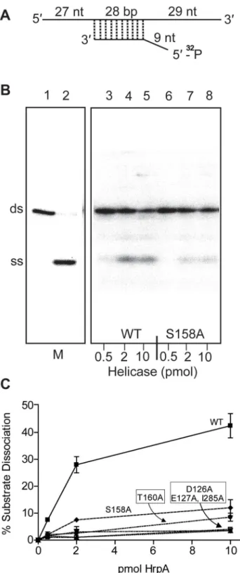
Recombinant wild-type HrpA protein was also assayed for RNA helicase activity using a partially double-stranded RNA substrate (Fig. 3A), which provides both 59and 39single-stranded overhang-ing sequences as previously described [39]. The enzymatic activity (Fig. 3C) was evaluated using a phosphor imager to quantify the signals from native polyacrylamide gels (Fig. 3B). In one hour at 37uC, 10 pmoles of purified HrpA unwound about 40% of the RNA substrate. This activity is similar to that previously reported for NS3 protein from Hepatitis C Virus [39,40]. In conclusion to this point, HrpA displayed the expected RNA-stimulated ATPase activity and RNA unwinding activity expected from its sequence conservation with DEAH-box RNA helicases.
Effect of point mutations in HrpA on ATPase and RNA helicase activity
To further investigate the relationship between HrpA and other DEAH-box RNA helicases, five point mutations were introduced into key motifs (Fig. 1 and Table S1): Motif II (DEAHER), which has a primary function of ATP binding and hydrolysis; Motif III (SAT), which functions as a communication link between the ATP binding and Motif V (TNIAETSITIEN), which functions as an RNA binding site. The purified, recombinant mutant HrpA proteins (Fig. S1) were assayed for ATPase and RNA helicase activity.
Figure 1. Conserved DEAH-box RNA helicase motifs in theB. burgdorferiHrpA protein used for point mutations.Conserved sequence motifs II, III and V of DEAH box RNA helicases are shown, along with their primary functions. The numbers below the motif sequences represent the position of the conserved motifs inBorrelia burgdorferiHrpA. Amino acid residues that are conserved in the mentioned motifs are shown in bold and the residues mutated in this study are indicated.
doi:10.1371/journal.ppat.1003841.g001 Author Summary
ATPase activity in the presence and absence of poly(A) RNA was assayed for all recombinant proteins (Fig. 2). As expected, HrpA with mutations in the ATP binding and hydrolysis motif, D126A and E127A [41], displayed a dramatic loss in activity and hydrolyzed 0.75 and 1.4% of the total ATP, respectively, in the presence of poly(A) and barely detectable and undetectable activity, respectively, in the absence of poly(A). The S158A and T160A mutants hydrolyzed 6 and 7% of the total ATP, respectively, in the presence of poly(A), down about ten-fold from the wild-type protein. However, in the absence of poly(A), S158A and T160A displayed only a two-fold (3.5% substrate hydrolysis) and four-fold (1.5% substrate hydrolysis) reduction in activity, respectively, when compared to the wild-type protein. This differential reduction in activity in the presence or absence of poly(A) is expected for proteins carrying mutations in Motif III [42].The I285A mutant showed a 3.7-fold reduction (18% ATP hydrolysis) in the presence of poly(A) and no reduction in activity (7% ATP hydrolysis) in the absence of poly(A). These results are expected for a mutation that disrupts RNA binding (Motif V) and thereby specifically reduces the RNA-dependent stimulation of ATPase activity.
The mutant HrpA proteins were also assayed for their levels of RNA helicase activity (Fig. 3). HrpA mutants in Motif II (D126A and E127A) and Motif V (I285A) displayed a near complete loss of
helicase activity, as expected for mutations in regions effecting ATP binding and hydrolysis or RNA binding [43,44]. Mutations in the Communication Motif (Motif III, S158A and T160A) also displayed reduced helicase activity, although to a lesser extent than the Motif II and V mutants, in accordance with previously published work on Hepatitis C virus RNA helicase [42]. In summary to this point, HrpA carrying mutations in Motifs II, III and V displayed ATPase and helicase characteristics similar to those of other DEAH-box RNA helicases.
Complementation ofhrpAin murine infection by replacement of the disrupted chromosomal gene
Disruption ofB. burgdorferi hrpAwas previously shown to result in complete loss of infectivity in wild-type C3H/HeN mice [35]. However, in those studies, we were unable to complement thehrpA mutant with a wild-type copy ofhrpAprovidedin transon a shuttle vector. In an alternative attempt to complement hrpA, the disrupted chromosomal gene was replaced with wild-type hrpA by allelic exchange (Fig. 4A). Starting with GCB1164, which carries a gentamicin-resistance cassette withinhrpA, anhrpA+gene with an adjacent kanamycin-resistance cassette was recombined into the chromosome using a suicide vector (pOH62-1). Allelic exchange was confirmed by PCR analysis (Fig. 4B). A total of four mice were infected for each mutant (two mice per clone, two Figure 2. ATP Hydrolysis by wild-type and mutant HrpA proteins.ATPase activity was assayed as described in Materials and Methods. ATPase activity of wild-type HrpA and five different point mutants was measured in the presence and absence of poly adenosine (RNA). The percentage of ATP hydrolyzed was determined by the release of32Pi from 1 mM of [c-32P]ATP after incubation with 500 fmol purified HrpA per microliter of reaction for 1 h at 37uC with and without 1.1 nmol polyadenosine mononucleotide. Experiments were run in duplicate and the standard deviations are represented with error bars. Motifs and assigned functions are shown for the studied mutants.
doi:10.1371/journal.ppat.1003841.g002
RNA Helicase in Infectivity and Tick Transmission
independent clones for each mutation). Tissue samples were cultured weekly for five weeks in BSK-II medium as described in Materials and Methods. All tissue samples from mice infected with thehrpAcomplemented isolate, as well as those infected with the wild-type parent, were positive for spirochetes (Table 1, top three rows).
Effect of point mutations inhrpA on murine infection HrpA is a large (823 amino acid) protein that conceivably may contain functions other than RNA helicase activity. It was, therefore, of interest to test point mutations affecting RNA helicase activity on murine infection to determine whether the inability of anhrpAgene disruption to support infection was a result of a loss of RNA helicase activity. To introduce the point mutations characterized in vitro (Fig. 2 and 3) into the endogenous B. burgdorferi hrpA gene, each mutation allele was first inserted in pPOH62-1 (Fig. 4A), the construct previously used for the complementation of hrpA. The resulting plasmid was then used to transform B. burgdorferi hrpA knockout strain GCB1164. To screen transformants for the presence of the desired point mutation, a PCR strategy was adapted from a method developed to screen for known SNPs between human alleles [45] and used previously to screen for mutants inB. burgdorferi [46]. For each mutation introduced in B. burgdorferi hrpA, a primer with the mutated nucleotide at the 39end was used in conjunction with a primer containing sequence that is conserved between the wild-type and mutant alleles (Fig. S2A). Using this approach, a PCR product was observed only if the mutation was present (Fig. S2B). This strategy was used to screen and recoverhrpA-D126A, E127A, S158A and T160A mutations. Further details can be found in Materials and Methods.
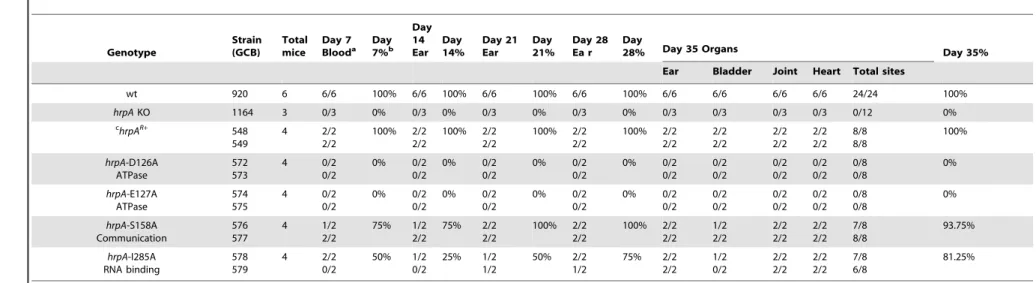
The hrpA-D126A and hrpA-E127A mutations (Motif II, ATP binding and hydrolysis) resulted in a complete loss ofB. burgdorferi infectivity in wild-type mice (Table 1), similar to thehrpAknockout strain (Table 1 and [35]). The loss in infectivity of the HrpA mutants carrying changes in the ATPase domain correlated well with thein vitro loss of both ATPase (Fig. 2) and RNA helicase (Fig. 3) activity exhibited by these mutants.
The mutant in Motif V (RNA binding) displayed a less-dramatic decrease in infectivity. Only 50% of blood samples collected 7 days post-infection were positive for spirochetes. For ear punches collected 14, 21 and 28 days post-infection, a delay in dissemination ofB. burgdorferi hrpA-I285A was observed compared to wild-type B. burgdorferi. Indeed, in contrast to wild-type B. burgdorferi, which were grown from all ear punches collected 14, 21 and 28 days post-infection, spirochetes were observed in samples from only 25, 50 and 75% of the mice, respectively, infected with thehrpA-I285A mutant. By day 35 post-infection thehrpA-I285A mutant had largely caught up to the wild-type in terms of tissue samples positive for spirochetes. The less-dramatic loss of infectivity of the hrpA-I285A mutant correlated with the partial loss of ATPase activity observed for this mutant (Fig. 2). Figure 3. RNA helicase assay. A) Structure of the dsRNA substrate.
Preparation of the helicase substrate is described in Materials and Methods. The long strand was prepared by in vitro transcription of pGEMX-1 that had been cleaved with NheI. The short strand was chemically synthesized and labeled with [c-32P]ATP and polynucleotide kinase at the 59free end. The length of the double strand portion of the substrate is given in base pairs (bp), the single stranded and the overhang portions are given in nucleotides. B) Autoradiograph of a polyacrylamide gel used to assay RNA helicase activity. The helicase assay was carried out for the wild-type and 5 differentB. burgdorferi HrpA mutant proteins as described in the Materials and Methods. The enzymatic reaction products were resolved by 12% native PAGE
followed by autoradiography. The autoradiograph shows wild-type and the S158A mutant HrpA as an example. Lane 1, No enzyme control, lane 2, ds RNA boiled at 95uC for 5 min to generate a marker for the single-stranded (SS) product. Lanes 3–5 contain wild-type HrpA and lines 6–8, the S158A HrpA mutant, with the indicated amounts of purified proteins.C) Quantification of Helicase Activity. Helicase assays were performed in duplicate for three different concentrations of each purified HrpA. The results are plotted as the percentage of double strand RNA substrate converted to single stand as a function of the amount of enzyme per microliter of reaction. Errors bars represent the standard deviation.
Figure 4. Strategy for complementation and insertion of point mutations inB. burgdorferi hrpA. A) Schematics of the strategy used. The B. burgdorferi hrpAKO strain GCB1164 was transformed with a construct carrying either wild-type or mutatedhrpA(hrpA+, a red star indicates the point mutation), aPflgB-driven kanamycin resistance cassette (green) and 500 bp of sequence downstream fromhrpA, to replace the disruptedhrpA gene. The point mutations were transferred in the complementation plasmid pPOH62-1 using PacI and AgeI restriction sites. Allelic exchange was RNA Helicase in Infectivity and Tick Transmission
Finally,hrpA-S158A (Motif III, Communication) resulted in only a slight delay in the infection, where samples from 3 of 4 mice were positive for spirochetes for weeks one and two. At weeks three, four, and five post-infection, all four mice were positive for B. burgdorferi. This mutant was also less severe in vitroand displayed residual ATPase (Fig. 2) and helicase (Fig. 3) activityin vitro. The communication motif is responsible for transmitting allosteric changes in the protein and the lesser effect of this mutantin vivo versusin vitromay result from allosteric protein-protein interactions in vivo, which are absent inin vitroassays.
In conclusion, the data suggest that the ATPase activity may be the most important function of HrpA for murine infection. The less severe infectivity phenotype of the RNA binding motif mutant (I285A) may result from the lack of complete inactivation of the helicase activity in the mutant or from some HrpA function that is independent of helicase activity.
Analysis of RNA levels in the hrpAmutant
In our previous study, we showed that hrpA is involved in controlling the expression of at least 187 proteins inB. burgdorferi [35]. To investigate whether changes in transcription were responsible for the observed changes in protein levels we compared RNA levels of the hrpA mutant versus the wild-type parent by microarray hybridization. Total RNA extracts of the hrpA mutant and the complemented mutant were prepared in duplicate and each run as a separate sample in the microarray analysis. To control biological variation, total RNA was isolated from three separate 50-ml cultures of each clone grown to a cell density of about 96107spirochetes/ml and pooled after the RNA integrity of each sample was verified. RNA samples were then processed for cDNA generation and labeling with fluorescent dyes for hybridization using a NimbleGen 4672K array ofB. burgdorferi B31 genes. A plot depicting the normalized log of hybridization intensities for thehrpAmutant versus the complemented mutant (hrpAR+
) is presented in Fig. 5. Clearly, there were no dramatic changes in mRNA levels in thehrpAmutant; out of 1505 genes examined, only 28 showed$1.5 - fold changes (see Table S4). Of these 28 genes, the largest observed changes were in bb0240-bb0243, which showed downregulation with 1.9 to 2.6 - fold changes. Changes in RNA levels were insufficient to account for the observed changes in expression (4- to 14-fold) of some of the HrpA-regulated proteins, suggesting that HrpA regulation, at least for some genes, was occurring at the post-transcriptional level. The small but significant changes in RNA levels observed in thehrpA mutant versus wild-type may result from small changes in transcription or from post-transcriptional RNA processing.
Changes in RNA processing in thehrpA mutant
The data from microarray hybridization indicated that muta-tion of hrpA does not result in global transcriptional changes. However, expression of about a dozen proteins were up or downregulated by 4- to 14-fold as determined by mass spectrom-etry iTRAQ analysis [35]. If hrpA does not affect major transcriptional changes, then how does it modulate changes in protein expression? To address this question, we used Northern blot hybridization to analyze the processing of RNAs
correspond-ing to some of the more highly regulated proteins or operons: downregulated bb0241 [glpK], bb0242, bb0603, and bba74; and upregulatedbb0502, bbb19, bb0443, bbi39, and bba24[dbpA]. Total RNAs isolated from thehrpAmutant and complement were run on denaturing agarose-formaldehyde gels and then transferred to nitrocellulose. For each transcript, a PCR product targeting the middle of the gene was prepared as a hybridization probe. RNA processing differences between hrpA and hrpA+ spirochetes were detected in five of the 11 transcripts analyzed. As shown in Fig. 6A, a full transcript from bb0603 (encoding the outer membrane protein P66) was present in both strains, but the band was stronger in the mutant. The wild-type strain displayed a smear from 500–1,500 nt that was not present in thehrpAmutant. Moreover, the mutant strain showed a second difference in processing as demonstrated by the new, intense band running just ahead of the 500 nt marker. For bba74 (Fig. 6B) a full length transcript was observed for both the wild-type and the mutant displayed a new intense band at about 500 nt. Changes in RNA processing are also inferred forbb0241[glpK],bb0242andbb0243[glpA] genes, which are part of an operon (Fig. 6C, D and E). RNA levels from each of these genes were lower by two-fold or less in the mutant as determined by microarray hybridization. A similar 2.5-fold decrease was observed for bb0241 by qPCR (data not shown). The large decrease of visible RNA in the mutant in the Northern blots suggests that the RNA is either in small fragments that have run off the bottom of the gel or in larger fragments of heterogeneous length such that they are not visible on the gel.
Northern blots were also performed on the genesdbpA,jag,ospC, rpoAandbbbi39, which are upregulated between 4–12 fold at the protein level in thehrpAmutant [35]. No differences were observed on the Northern blots between the mutant and the complemented mutant.
HrpA-deficientB. burgdorferiare not transmitted to mice via tick bite
We next investigated the contribution of HrpA to the tick phase of the enzootic cycle. Because HrpA-deficient spirochetes are avirulent by syringe-inoculation, we infectedIxodes scapularislarvae with GCB1164 (hrpA) and GCB549 (complemented mutant) using the immersion method developed by Policastro and Schwan [47]. Based on qPCR using a TaqMan assay forflaB, the mutant and complemented isolates survived at similar levels during the larval blood meal (Fig. 7A). Following the molt, we examined the ability of each strain to replicate within feeding nymphs and transmit infection to naı¨ve mice. As shown in Fig. 7B, spirochete burdens increased exponentially in fed versus flat nymphs infected with either the mutant or complement isolates. Using IFA and semi-solid phase plating (Fig. 7C and D, respectively), we confirmed that hrpAmutant organisms survived as well, if not better, than their complemented counterparts during the nymphal blood meal. Despite having high spirochete burdens, unlike nymphs infected with the complement isolate, hrpA-infected nymphs did not transmit infection to naı¨ve C3H/HeJ mice based on serology and tissue culturing performed at 8 weeks post-repletion (Table 2). BecausehrpAmutants are avirulent via syringe-inoculation [35], it is possible that the mutant is transmitted normally by feeding ticks confirmed by PCR using primers indicated by an arrow.B) PCR verification of the allelic exchange. Each construct was confirmed with four PCR analyses.Panel 1) The loss of gentamicin-resistance cassette following allelic exchange was confirmed as shown. The shuttle vector pBSV2G served as the positive control (c+
) for amplification of thegentcassette (lane 5).Panel 2) The replacement of the gentamicin-resistance cassette byhrpAwas confirmed by amplification of the segment ofhrpAthat was deleted in the KO strain GCB1164 (lane 8).Panel 3) The size of thehrpAgene was compared between wild-type (lane 14),hrpAKO (lane 13) and complemented clones (lanes 11 and 12). Lane 10 and 15 was a negative control containing ddH2O as template. A 100 bp and a 1 kb DNA ladder were used (M). The schematic inAof the figure is modified from [74].
Table 1.Effect of complementation and mutations inhrpAonB. burgdorferiinfectivity in C3H/HeN mice. Genotype Strain (GCB) Total mice Day 7 Blooda Day7%b
Day 14 Ear Day 14% Day 21 Ear Day 21% Day 28 Ea r Day
28% Day 35 Organs Day 35%
Ear Bladder Joint Heart Total sites
wt 920 6 6/6 100% 6/6 100% 6/6 100% 6/6 100% 6/6 6/6 6/6 6/6 24/24 100%
hrpAKO 1164 3 0/3 0% 0/3 0% 0/3 0% 0/3 0% 0/3 0/3 0/3 0/3 0/12 0%
c hrpAR+
548 549 4 2/2 2/2 100% 2/2 2/2 100% 2/2 2/2 100% 2/2 2/2 100% 2/2 2/2 2/2 2/2 2/2 2/2 2/2 2/2 8/8 8/8 100% hrpA-D126A ATPase 572 573 4 0/2 0/2 0% 0/2 0/2 0% 0/2 0/2 0% 0/2 0/2 0% 0/2 0/2 0/2 0/2 0/2 0/2 0/2 0/2 0/8 0/8 0% hrpA-E127A ATPase 574 575 4 0/2 0/2 0% 0/2 0/2 0% 0/2 0/2 0% 0/2 0/2 0% 0/2 0/2 0/2 0/2 0/2 0/2 0/2 0/2 0/8 0/8 0% hrpA-S158A Communication 576 577 4 1/2 2/2 75% 1/2 2/2 75% 2/2 2/2 100% 2/2 2/2 100% 2/2 2/2 1/2 2/2 2/2 2/2 2/2 2/2 7/8 8/8 93.75% hrpA-I285A RNA binding 578 579 4 2/2 0/2 50% 1/2 0/2 25% 1/2 1/2 50% 2/2 1/2 75% 2/2 2/2 1/2 0/2 2/2 2/2 2/2 2/2 7/8 6/8 81.25%
aAll fractional values listed correspond to number of cultures positive/number of sites tested. bAll % values listed represent the corresponding number of cultures positive/number of sites tested. cComplemented
but cleared by the host soon after being deposited in the skin. We therefore performed a second experiment using these sameBorrelia burgdorferi-infected nymphs where we cultured the area of skin surrounding the bite site immediately following tick drop-off (,96 h post-placement), a time point that enables one to
distinguish between a defect in tick-to-mammal transmission and clearance by the host [48,49]. As shown in Table 2, none of the skin samples obtained from mice fed on by GCB1164 (hrpA )-infected nymphs were culture-positive, while 21/30 skin samples were positive for the complement (Table 2); it is worth noting that 6 of the 9 culture-negative samples for the complement were from a mouse that was fed on GCB549-infected nymphs that contained substantially lower spirochete burdens (Fig. S3). The absence of HrpA-deficient organisms within the bite site within 24 h of nymphal repletion suggests a defect in their ability to disseminate out of the midgut and/or to penetrate the salivary glands during feeding [48,50].
Discussion
HrpA is an RNA helicase with properties similar to those of other DEAH-box RNA helicases
Borrelia burgdorferihas developed different regulatory pathways that allow it to survive in both ticks and vertebrate animals. We have previously shown that HrpA is involved in the regulation of a large number ofB. burgdorferigene products [35]. Here, for the first time, we have demonstrated that HrpA has both RNA-dependent ATPase and helicase activitiesin vitroand displays activities similar
to those reported for other DEAH-box RNA helicases [39,40,41,51,52,53]. HrpA is the only putative RNA helicase that B. burgdorferipossesses. In contrast, most organisms have multiple RNA helicases: E. coli has over a dozen [54,55] and yeast has about 40 [56]. The paucity of RNA helicases inB. burgdorferiraises the possibility that HrpA is multifunctional and can perform the roles of several different RNA helicases found in other organisms. Alternatively, the level of RNA regulatory roles by RNA helicases may be greatly diminished inB. burgdorfericompared to organisms likeE. coliand yeast.
To gain further insight into the HrpA RNA helicase we generated point mutations in HrpA Motifs II, III and V, which function in ATP binding and hydrolysis, communication between the ATP hydrolysis and RNA binding domains, and RNA binding, respectively (Fig. 1). The effects of these mutations on HrpA enzymatic activityin vitro were similar to those previously reported for other DEAH-box RNA helicases.
The effect of point mutations in HrpA on mouse infectivity
Our previous study showed that disruption of the hrpA gene resulted in a complete loss of infectivity of C3H/HeN mice by needle inoculation [35]. While our previous attempts to comple-menthrpA in transusing a shuttle plasmid were not successful, allelic exchange to replace the disrupted chromosomal gene with a wild-type copy ofhrpA(this work) completely restored infectivity and persistence in mice to a wild–type level. This complementation experiment confirms thathrpAis indeed essential for the infectivity Figure 5. Microarray analysis. Gene level correlation ofhrpA versus complementedhrpA strains ofB. burgdorferiis plotted as the log2of normalized hybridization intensities of thehrpAstrain and the complemented mutant. The green line in the middle shows where both strains have the same intensity and the outer green lines show where the intensities are two-fold apart.
ofB. burgdorferiand that the defect observed in the knockout clones is not the result of secondary mutation(s) in the genome.
The approach used for restoring a wild-typehrpA gene in the chromosome (Fig. 4), coupled with a PCR screening method (Fig. S2), facilitated the construction of point mutations in the B. burgdorferi chromosome. To determine whether loss of RNA helicase and/or ATPase activity has a direct effect on infectivity, point mutations in Motif II, III and V were introduced into B. burgdorferi hrpA.The data suggest that the ATPase activity may be the most important function of HrpA for murine infection. The less severe infectivity phenotype of the RNA binding motif mutant (I285A) may result from the lack of complete inactivation of the helicase activity in the mutant or from some HrpA function that is independent of helicase activity.
The precise mechanism by which the RNA helicase is involved in mouse infectivity remains speculative and is likely through its role in
regulating important genes, such as the integrin-binding protein and porin P66, which is required for mouse infectivity [57]. The full complement of HrpA regulated proteins is currently not known as the iTRAQ proteomics approach used to monitor protein expression in anhrpAmutant [35] identifies only a subset of the expressed proteins. Other proteins whose expression is highly regulated in anhrpAmutant may have escaped identification by the limited proteomics approach and proteins that are expressed in the mouse and/or the tick would not have been identified.
Mutation ofhrpAdoes not result in large changes in transcription
Numerous studies to date have examined the contribution of environmental signals to differential gene expression in B. burgdorferi [1,3,4]. The well characterized RpoN-RpoS pathway, for instance, controls the expression of .100 borrelial genes in Figure 6. Northern blot analysis of p66, oms28, glpK bb0242 and glpA. Purified RNA samples from the hrpA mutant (hrpA) and the complemented strain (hrpA+
) were run on a 1.2% agarose-formaldehyde gel with an RNA marker shown in lane M. Each panel shows a membrane strip that contains 10mg of RNA from the mutant and the complementedhrpAstrain. The blot was hybridized with a32P-labelled gene probe prepared by PCR using the indicated primers. The target transcripts werep66 (bb0603) forPanel A,bba74forPanel B,glpK(bb0241) forPanel C, glpA(bb0243) forPanel Dandbb0242forPanel E.
doi:10.1371/journal.ppat.1003841.g006
RNA Helicase in Infectivity and Tick Transmission
response to increased temperature and/or mammalian host-specific signals [5,6,7,58]. Expression ofhrpAitself does not appear to be controlled by the RpoN-RpoS pathway. The minimal changes observed by microarray analyses comparing the hrpA mutant and complemented isolates contrasted with the signifi-cantly larger changes observed by iTRAQ, which for some proteins were as high as 14-fold downregulation and 12-fold upregulation. Thus, post-transcriptional RNA processing by HrpA represents an additional mechanism by which spirochetes could
modulate their proteome throughout the enzootic cycle. Of note, there appears to only minimal overlap between the RpoS and HrpA regulons. Of the 187 proteins differentially-regulated by HrpA, only 11 are regulated by RpoS [5,6,7]. Interestingly, in each case, the role of HrpA appears to be counter-regulatory to that of RpoS. Of the 11 RpoS-dependent genes within the HrpA regulon, four (mcp1, ospC, dbpA and bb0689) are transcribed by RpoS but were present at higher protein levels in thehrpAmutant. Conversely, transcripts for seven genes (bb0241, bb0243, bb0365, Figure 7. HrpA-deficientB. burgdorferipersist through the molt and survive the nymphal blood meal. A. Spirochete burdens in larvae (3 pools of 15 larvae per isolate) infected with either GCB1164 (hrpA) or GCB549 (Comp) by immersion and then fed to repletion on naı¨ve C3H/HeJ mice. Genome copy numbers were assessed by qPCR using a TaqMan-based assay forB. burgdorferi flaBas previously described [73]. Values represent the averageflaBcopy number per tick6standard error of the mean in each pool.B. Spirochete burdens in flat nymphs (3 pools of 5 per isolate) and nymphs fed to repletion on naı¨ve C3H/HeJ mice (3 pools of 8–10 nymphs per isolate). Genome copies numbers were assessed by qPCR as described for larvae.C. Representative micrographs of nymphs fed to repletion on naı¨ve mice. Spirochetes were detected by immunofluorescence using FITC-conjugateda-Borreliaantibody as previously described [73]. Scale bar, 25mm.D. Viability of GCB1164 (hrpA) and GCB549 (Comp) in nymphs fed to repletion on naı¨ve mice (5 mice per isolate). The average number of CFU per nymph is based on 5 pools (8–10 nymphs per pool) for each isolate (6 standard deviation). Pools were serially-diluted (undiluted, 1021
, and 1022
) in BSK medium and plated in duplicate as previously described [75]. doi:10.1371/journal.ppat.1003841.g007
Table 2.Nymphs infected with HrpA-deficientB. burgdorferiare unable to transmit infection.
Positive cultures
8 wks post-repletiona
,96 hours post-placement
Strain Ear Skin Joint Bladder Serologyb Bite Sitec
GCB1164 (hrpA) 0/3 0/3 0/3 0/3 0/3 0/30
GCB549 (compl) 3/3 3/3 3/3 3/3 3/3 21/30d
a3 mice per isolate.
bBased on immunoblot using whole cell lysates derived from temperature-shifted
B. burgdorferistrain B31 5A4 NP1. cThe area of skin below capsules used to contain feeding nymphs was excised within 24 hours of repletion (
,96 hour post-placement), divided into 6 equal portions for
each mouse (5 mice per isolate), and cultured in BSK II medium containing the appropriate antibiotic as described previously [48].
dOne mouse in this group yielded no positive bite site cultures (0/6), most likely due to significantly lower spirochete burdens in the cohort of nymphs placed in this capsule (See Fig. S1).
bba74,bbi29, andbbk13) that are repressed in an RpoS-dependent manner appear to be stabilized by HrpA (i.e., their protein levels are lower in thehrpAmutant compared to the wild-type).
A role for HrpA in RNA processing and post-transcriptional gene regulation
RNA helicases are known to play a role in all aspects of RNA metabolism and in ribosome biogenesis [24,25,26,27,28,29]. The only known function for an HrpA protein is an involvement in RNA processing of a fimbrial RNA in E. coli, resulting in the generation of a stable mRNA and the upregulation ofdaaE, which encodes a fimbrial adhesin [33]. To further study the mechanism by which HrpA modulates protein expression inB. burgdorferi, we looked for changes in RNA processing in anhrpAmutant versus wild type, in the most highly HrpA-regulated genes. Changes in RNA processing were observed in five downregulated genes: bb0603 [p66], bba74, bb0241 [glpK], bb0242, and bb0243 [glpA] (Fig. 6). In contrast todaaEexpression inE. coli, where HrpA is involved in RNA cleavage to upregulate protein expression, of the five genes that showed changes in RNA processing, all were downregulated. Our data suggest several possible mechanisms by which HrpA may regulate gene expression. In the case of P66, The Northern blot data suggest that HrpA plays a role in the processing of the full-length transcript into shorter transcripts in the wild-type strain. In the mutant strain more of the full-length transcript is observed and the processing is different. The decrease in protein expression in the mutant versus the wild-type may reflect the difference in processing or may result from differences in translational initiation resulting from HrpA mediated RNA remodeling. The case of bbA74 is similar, where a change in processing is observed, but the full length transcript remains a prominent band. Once again, translation may be promoted in the wild-type setting by RNA structural remodeling. The other three downregulated genes we looked at (glpK, bb0242 and glpA) appeared to show a generalized RNA decay in the absence of HrpA, suggesting that HrpA either directly or indirectly protects these transcripts from cellular nucleases. The absence of full-length transcript in these cases correlates well with the decrease in protein expression.
Our work here shows that the DEAH-box protein HrpA displays RNA helicase activityin vitro. It is of noteworthy that four E. coliDEAD-box proteins have recently reported to have ATP-independent RNA annealing, strand displacement and RNA chaperone activityin vitro[59]. These auxillary activities may be important in RNA remodeling and other RNA helicase-mediated processes. It will be of interest to determine if HrpA also carries such auxillary functions.
HrpA-deficientB. burgdorferiare not transmitted to mice via tick bite
Spirochetes lacking HrpA are avirulent in mice infected via needle ([35] and Table 2). Here, we demonstrate that HrpA also is required for tick-to-mammal transmission. HrpA modulates the expression of a large number of borrelial gene products, many of which appear to be involved in spirochete physiology and/or metabolism. Somewhat surprisingly, however, hrpA mutant organisms replicated normally within feeding nymphs, suggesting that the transmission defect is likely due to an inability to disseminate within the vector during the blood meal. Two of the proteins most affected by loss of HrpA are GlpK and GlpA, both of which are involved in glycerol utilization during the tick phase of the enzootic cycle [60]. Unlike the hrpAmutant, glpAand glp operon mutants do not persist through the larval molt and display
modest to severe growth defects, respectively, within feeding nymphs [60,61]. As such, RNA processing of the correspondingglp transcripts does not appear to be responsible for the transmission defect we observed with thehrpAmutant. A survey of the HrpA regulon did not identify any obvious candidate proteins involved in spirochete migration per se. Therefore, further studies will be needed to define more precisely the point(s) during transmission that are controlled by HrpA-regulated gene products.
Materials and Methods
Ethics statement
All animal experimentation for murine infection was carried out in accordance with the principles outlined in the most recent policies andGuide to the Care and Use of Experimental Animalsby the Canadian Council on Animal Care. The animal protocol (AC12-0070) was approved by The Animal Care Committee of the University of Calgary. All animal experimentation for tick transmission studies was conducted following theGuide for the Care and Use of Laboratory Animals(Eighth Edition) and in accordance with protocols reviewed and approved by the University of Connecticut Health Center Institutional Animal Care and Use Committee.
Strains and primers used in this study
All plasmids andE. colistrains used are presented in Table S1. Primers used are listed in Table S2 and allB. burgdorferistrains used are listed in Table S3.
Site-directed mutagenesis and purification of his-tagged HrpA
The point mutations (Fig. 1) D126A, E127A, S158A, T160A and I285A were directly introduced to wild type hrpA in clone pASD1 (Table S1), which contains wild-type hrpA in pET-15b (Novogen). Site-directed mutagenesis and purification of PCR products were performed as described previously [62]. Purified PCR products was digested with DpnI (New England Biolabs) and used to transform chemically competent DH5acells. Mutation of S158A was performed in clone pASD5 and subsequently moved to pET-15b following excision with BamHI and NdeI. Point mutations were confirmed by DNA sequencing. TransformedE. coliRosetta DE3 cells carrying either wild-type or mutant hrpA genes were overexpressed and lysed as described previously [63]. Two-step purification (Ni-NTA Agarose column fallowed by Hydroxyapatite column) was applied to wild-type and mutant HrpA. First, lysates were subjected to His-tag affinity purification as previously described [62] with the following modifications. Affinity columns (1 ml bed volume) were equilibrated with 30 ml equilibration buffer [50 mM NaH2PO4, 0.5 M NaCl, 5 mM
imidazole, 10% glycerol (w/v), pH 8.0]. Samples were diluted in sample preparation buffer [50 mM NaH2PO4, 5 mM imidazole,
10% glycerol (w/v), pH 8.0]. Samples were then loaded on columns and washed with 20 ml of wash buffer [50 mM NaH2PO4, 50 mM NaCl, 10 mM imidazole, 10% glycerol (w/
v), pH 8.0] and 1 ml fractions were collected. Samples were subjected to 7% SDS-PAGE and visualized by Coomassie blue staining. Fractions containing purified His-tagged HrpA were pooled. Salt concentration and pH values of the samples were adjusted to 0.5 M salt concentration through addition of HAP buffer [25 mM MES (pH 6.1), 0.2 mM EDTA, 10% glycerol (w/ v)] and were loaded on a 3 ml Hydroxyapatite (Bio-Gel HTP, BioRad Laboratories) column prepared in HAP buffer containing 0.5 M NaCl. Loaded samples were washed with 10 ml of HAP buffer containing 0.5 M NaCl, followed by 50 mM potassium phosphate, 0.5 M NaCl. Samples were eluted through a gradient RNA Helicase in Infectivity and Tick Transmission
of HAP buffers containing 0.5 M NaCl and 0.6 M potassium phosphate buffer. Once the fractions containing purified HrpA were pooled (Fig. S1), the protein concentrations were determined using the Bradford method [64] and the recombinant proteins were concentrated using Amicon Ultra – 30K centrifugal filters (Millipore).
Enzymatic assays
ATPase assay. Reaction mixtures of 20ml containing 40 mM Tris-HCl (pH 8.0), 2 mM DTT, 2 mM MgCl2, 1 mM
[c-32P]ATP, 0.25 mM of poly(A), and specified concentrations of
HrpA were incubated for 1 h at 37uC. The reactions were ended by addition of 20 mM EDTA. Reaction products were analyzed by spotting onto DE 81 paper (Whatman) strips. Descending chromatography was performed using 0.35 M ammonium formate. After the run, the strips were dried and exposed to a Cyclone phosphor screen for 30 min and ATP hydrolysis quantified using a Cyclone phosphorImager. The plotted results are the average of two separate experiments for each sample.
Helicase assay. Radiolabelled partially double-stranded RNA helicase substrate (Fig. 3A) was prepared as described [39,51] with slight modifications. Briefly, long strand RNA was prepared using pGEM Express Positive control template (Ribop-robe System T7, Promega). Linearized template plasmid was digested with NheI and transcribedin vitroyielding an 84 nt RNA strand, 59-UAAUACGACUCACUAUAGGGAGACCACAACG
GUUUCCCUCUAGAAAUAAUUUUGUUUAACUUUAAGA
AGGAGAUAUACAUAUGG-39. In vitro transcription and re-moval of template DNA were according to manufacturer instructions. The transcript was then extracted with acidified phenol:chloroform:isoamyI alcohol (125:24:1), pH 4.5 (Ambion) and precipitated with 100% ethanol. Precipitated RNA was washed once with 70% ethanol, dried in a Speedvac and dissolved in DEPC-H2O (diethylpyrocarbonate treated water). The 37-mer
short strand, 59-AUCAGACUCACAAAAUUAUUUCUAGAGG GAAACCGUU-39, was synthesized (University of Calgary Core DNA Services) complementary to the middle region of the long strand RNA (bold characters indicate the double stranded portion of the substrate). The short strand RNA was 59end labelled with [c-32P]ATP using T4 polynucleotide kinase (New England Biolabs). Labelled short and long RNA strands were hybridized at a ratio of 1.3:1 (Long strand: Short strand) in 20 mM Hepes-KOH buffer (pH 7.6) containing 0.5 M NaCl, 1 mM EDTA and 0.1% SDS in a 50ml reaction volume. The reaction mixture was heated to 95uC for 5 min followed by a slow cooling to room temperature. Partially double-stranded RNA was cleaned from unbound excess radiolabel by Sephadex G-25 Superfine (GE Healthcare) spin columns. The radiolabelling quality and substrate integrity was confirmed by running the purified substrate on a 12% native polyacrylamide (29:1 acrylamide:bis-acrylamide, 40% solution) gel in TBE buffer at room temperature and visualizing with a Cyclone phosphorImager.
Helicase activity of the samples was assayed in 20ml reaction mixtures containing 20 mM Hepes-KOH (pH 7.6), 2 mM DTT, 50 mM KCl, 2 mM MgCl2, 1 mM ATP, 0.1 mg/ml BSA, 2 units
RNasin, 0.5 pmoles helicase substrate and specified concentrations of purified enzyme. Reaction mixtures were incubated 1 h at 37uC, then stopped by the addition 5ml of 56RNA loading buffer containing 100 mM Tris-HCl, pH 7.5, 50% glycerol, 5 mM EDTA, 0.5% SDS, 0.1% NP-40, 0.1% (w/v) bromophenol blue and xylene cyanol FF. Samples were analyzed by 12% native polyacrylamide (29:1 acrylamide:bis-acrylamide) gel in TBE buffer with running conditions of 8 volts/cm. Double stranded and unwound RNA strands were visualized and quantified using a
Cyclone phosphorImager and ImageQuant software. The back-ground from the no enzyme control was subtracted from the data points to obtain helicase activity levels.
Culture conditions for B. burgdorferi. Except when specified, allB. burgdorferi strains were cultured at 35uC in BSK-II prepared in house [65] and supplemented with 6% rabbit serum (Cedar Lane) in a 1.5% CO2incubator. Cell counts were taken
using a Petroff-Hausser chamber in a dark-field microscope.
Complementation ofB. burgdorferi hrpA. Complementa-tion of hrpA was performed as previously described for the complementation ofB. burgdorferi uvrB[66]. A schematic represen-tation of the strategy used is presented in Fig. 4A. Briefly, overlap extension PCR was used to fuse the wild-typehrpAgene, aPflgB -driven kanamycin-resistance cassette and 500 bp of sequence downstream fromhrpA. The overlap PCR product was cloned into pJET1.2/blunt cloning vector (Fermentas), sequenced and then was used to transformB. burgdorferistrain GCB1164 (hrpAKO) [35] by electroporation, as previously described [67,68]. Allelic exchange was confirmed by PCR for the loss of the gentamicin-resistance cassette, for the presence of the portion ofhrpAthat was deleted in the knockout strain and to confirm that the size of complemented hrpAwas similar to wild-typehrpA(Fig. 4B). The restoration of wild-typehrpAwas finally confirmed by DNA sequencing.
To insert point mutations inB. burgdorferi hrpA, the mutation was inserted in the complementation vector pPOH62-1. The D126A, E127A, S158A and T160A mutations were transferred from their original constructs to pPOH62-1 using PacI and AgeI restriction sites (Fig. 4A). For the I285A mutation, pPOH62-1 was used as the template for site-directed mutagenesis as previously described. The resulting constructs were methylated in vitro with M.SssI CpG methyltransferase (New England Biolabs) as described [69]. Methylated DNA was used to transformB. burgdorferiGCB1164 (hrpA KO) and possible clones were grown in the presence of kanamycin (200mg/ml). A PCR strategy [45] was used to screen for point mutations in B. burgdorferi (Fig. S2). Briefly, a primer containing the mutated base as last nucleotide was used in pair with a primer containing sequence that is conserved between mutated and wild-typeB. burgdorferi hrpA(Fig. S2). Restoration of a completehrpAgene was confirmed by PCR as described forhrpA complementation (Fig. 4) and by DNA sequencing. This strategy was used to screen and recoverhrpA-D126A, E127A, S158A and T160A mutations. However,hrpAT160A could not be recovered and the primers designed for hrpA I285A did not produced a sufficiently clear result to be used for screening B. burgdorferi genomic DNA. Following the transformation, 68 clones for D126A, 93 clones for E127A, 21 clones for S158A, 30 clones for T160A (from 4 transformations) and 41 clones for I285A (from 4 transformations) were recovered. For the D126A mutation, four out of the six clones tested by PCR contained the mutation. From the 12 clones tested for the D127A mutation, nine appeared to contain the mutation by PCR screen. For T160A mutation, 30 clones were recovered over four transformations and all contained only the wild-type hrpA sequence. For I285A, 41 clones were recovered over four transformations and only two contained the expected mutation.
Hayward, CA, USA) and visualized under UV light. All clones used retained all plasmids that were present in the parental GCB1164 strain, which lacks lp28-2 and cp9.
Animal infection. For mouse infection studies, three to four week old C3H/HeNCrl male mice (Charles River Laboratories, St-Constant, QC, Canada) were infected with 26104spirochetes by both intraperitoneal and subcutaneous injection. At 7 days post-infection, 50ml of blood was collected from the saphenous vein and cultured in 1.7 ml of culture medium. At weeks two, three and four, two ear punches per mouse were collected and incubated in 1.5 ml of culture medium. At day 35 post-infection, one ear, the bladder, the heart and one knee joint were recovered. For all tissue sample cultures, 16 Borrelia antibiotic cocktail (20mg/ml phosphomycin, 50mg/ml rifampicin and 2.5mg/ml amphotericin B) was added to the culture medium. Cultures were considered positive for B. burgdorferi when spirochetes could be observed using a dark-field microscope.
B. burgdorfericultures for temperature shift and microarray analysis
Sample preparation for microarray analysis was performed as previously described with slight modifications [71]. Briefly, frozen glycerol stocks of B. burgdorferi strains GCB549 and 1164 were inoculated into 20 ml of BSK-II prepared in-house supplemented with 6% rabbit serum and incubated at 35uC until they reach a density of around 56106spirochetes/ml. The cultures were then diluted to a concentration 16106spirochetes/ml and incubated at 23uC in the dark. At this stage, each culture was prepared in duplicate. When the cultures reached around 26106spirochetes/ ml at 23uC, one replicate was prepared for 35uC shift and the other was kept at 23uC until it reached a density of about 86107 spirochetes/ml and then harvested. The cultures to be shifted to 35uC were diluted to a density of 16106 spirochetes/ml. Following the dilutions, cultures were supplemented with final concentrations of 1% antibiotic cocktail and 5% human blood from which the buffy coat was removed and containing 10% 0.1 M sodium citrate. Blood containing cultures were periodically mixed to keep blood cells in suspension and incubated at 35uC for 48 h where they reached a density of about 86107 spirochetes/ ml. To harvest the Borrelia cells, cultures were centrifuged at 600 rpm for 10 min at 4uC and the culture media containing Borreliacells were pipetted out gently into a new centrifuge tube without disrupting the pelleted blood cells.Borreliawere harvested by centrifugation at 60006 g for 20 min at 4uC after two successive PBS wash steps. An aliquot from each culture was saved to check for expected OspC induction by Western blotting. To control biological variation for the microarray analysis, samples prepared by pooling 4 independent cultures. To prepare whole cell lysates,B. burgdorfericells were lysed using SDS-PAGE loading buffer [72] at 95uC for 5 min. A volume of whole cell lysate generated from 26107spirochetes was loaded per each well of 12% SDS PAGE gels for separation. Samples were then blotted onto nitrocellulose membranes (Amersham Hydrobond-ECL). OspC was detected using rat anti-B31 OspC-His in a dilution of 1/1000 as a primary antibody and peroxidase-conjugated AffiniPure donkey anti-mouse IgG (H+L) (Jackson ImmunoResearch Laboratories) in a dilution of 1/5000 as the secondary antibody. 3,39,5,59-Tetramethylbenzidine (TMB) liq-uid substrate (Sigma) was used as a colorimetric detection reagent according to manufacturer’s instructions. Once the OspC induction was confirmed, total RNA was extracted from hrpA mutant and the complemented mutant clones harvested at their late exponential phase with centrifugation at 60006gfor 20 min at 4uC. RNA samples were extracted using Qiagen RNeasy Total
RNA extraction kit according to the manufacturer’s instructions, Possible remaining contaminant DNA was removed from the samples by incubation with DNase I (BioRad) at 37uC for 2 h. DNase I was inactivated by incubation at 70uC for 20 min. RNA samples were further concentrated with ethanol precipitation and resolubilized in DEPC-H2O at a concentration of 2mg/ml. The
integrity of the RNA samples were checked on 1% Agarose gels. Total RNA extracts of the hrpAmutant and the complemented mutant were prepared in duplicate and each run as a separate sample in the microarray analysis. To control biological variation, total RNA was isolated from three separate 50 ml cultures of each clone grown till their late exponential phase and pooled after RNA integrity was proved. Samples were then sent to the Vancouver Prostate Centre, Laboratory for Advanced Genome Analysis, Vancouver, BC, Canada for microarray (NimbleGen 4672K arrays) and statistical analysis. Gene level t-tests and absolute fold changes with a 1.4-fold change cut off, are given for RNA samples (coded 1164 and 549 for thehrpA mutant and the complemented mutant, respectively) in Supple-mentary Table S4.
B. burgdorferistrains and culture conditions for northern blot analysis
Frozen stock B. burgdorferi strains GCB549 and 1164 were inoculated in 20 ml of BSK-II prepared in-house supplemented with 6% rabbit serum, incubated at 35uC until they reached a density of around 56106spirochetes/ml. The cultures were then diluted to a concentration 16106spirochetes/ml and incubated at 35uC until they reached the density of approximately 86107 spirochetes/ml. To harvest the Borrelia cells, cultures were centrifuged at 60006gfor 20 min at 4uC and washed once with PBS. Resulting pellets were then extracted using an RNeasy Mini Kit (Qiagen) following the manufacturer’s instructions. The RNA quality was checked on a 1% agarose gel run in RML buffer.
Probe preparation. PCR conditions for gene specific Northern probes for selected genes were as follows: 16Phusion High Fidelity (HF) buffer (New England Biolabs), 0.5 mM dNTPs, 0.02 units/ml Phusion DNA polymerase (New England Biolabs), 0.5 mM of each forward and reverse primer (see Table S2 for primers), 50 ng genomic DNA template in a 50ml reaction. Amplification conditions were as follows: 98uC, 30 s followed by 30 cycles of 98uC, 10 sec; 62uC, 30 sec; 68uC, 45 sec and 68uC, for 7 min. PCR products were gel purified with Qiaquick Gel Extraction Kit (Qiagen). Purified probes were labelled using the Random Primers DNA Labelling System (Invitrogen) according to the manufacturer instructions.
For loading of Northern blots the amount of RNA loaded per lane was equivalent as determined by spectrophotometry and verified by agarose gel elctrophoresis and RNA staining with ethidium bromide. For Northern blots ten micrograms of RNA denatured in 50% formamide containing 16 MOPS [3-(N-morpholino)propanesulfonic acid] buffer (20 mM MOPS, 2 mM sodium acetate, 1 mM EDTA [pH 8.0]) by incubation at 85uC for 10 min. Samples were immediately chilled on ice for 10 min followed by a short centrifuge spin to deposit all of the liquid at the bottom of the tubes. For each sample 2ml of 106formaldehyde gel loading buffer (50% glycerol 10 mM EDTA [pH 8.0], 0.25% (w/v) bromophenol blue, and xylene cyanol FF) was added. RNA samples were separated in 2.2 M formaldehyde agarose gel using 16MOPS running buffer for 4 h at 40 V (4 V/cm). Running buffer was refreshed at the half time of the run. The lane containing RNA Millenium molecular weight standards (Ambion) was cut from the visualized gel and the gel was incubated with DEPC-H2O for
10 min, followed by 0.05 N NaOH for 20 min and last, 206SSC RNA Helicase in Infectivity and Tick Transmission
for 40 min. Then, RNAs were transferred onto Hybond-N membranes (Amersham Biosciences) in 206SSC buffer (1.5 M NACl, 0.5 M NaH2PO4, 20 mM EDTA, pH 7.4) by using capillary
transfer overnight at room temperature. RNA was UV cross-linked to the membrane by using a UV Stratalinker 1800 (Stratagene). Membrane strips were incubated for 2 h in prehybridization buffer (0.5 M NaH2PO4, 7% SDS (w/v), 1 mM EDTA) at 85uC, followed
by addition of labelled gene specific probes and overnight hybridization at 68uC. Membranes were washed twice with 16 SSC, 0.1% SDS for 10 min at 68uC. Dried strips were visualized by using a Cyclone Phosporimager. Autoradiography images were produced at different exposure times for each gene. The sizes of the transcripts were estimated by comparison with RNA Millenium molecular weight standards (Ambion).
Tick transmission studies
Pathogen-freeIxodes scapularislarvae (purchased from Oklahoma State University, Stillwater, OK) were infected with GCB1164 (hrpAmutant) and GCB549 (complemented mutant)B. burgdorferi using the immersion method as previously described [47,73]. Briefly, ,200–300 naı¨ve larvae were mixed end-over-end in a
high-density spirochete suspension (26108spirochetes/ml) for 1– 2 h at room temperature. Following immersion, larvae were washed twice with 1 ml of sterile PBS and allowed to recover overnight. Larvae infected by immersion were fed to repletion on a naı¨ve C3H/HeJ mouse housed over water and collected daily. Replete larvae (3 pools of 15 larvae per isolate) were surface-sterilized by successive washes with 1 mL of water, 0.5% bleach, 3% hydrogen peroxide, 70% ethanol and water prior to being processed for qPCR. Total genomic DNA was isolated from replete larvae at drop-off using the Gentra Puregene Yeast/Bact. Kit (Qiagen) according to the manufacturer’s instructions. DNAs were diluted 1:10 in water prior to being analyzed by qPCR using a TaqMan assay forflaBas previously described [73]. Following the molt,,10–12 nymphs were fed to repletion on naı¨ve female
C3H/HeJ mice using the capsule method as previously described [73]. Replete nymphs recovered from each mouse were surface-sterilized and processed for qPCR, immunofluorescence using FITC-conjugated a-Borrelia antibody (Kirkegaard and Perry Laboratories, Gaithersburg, MD), and semi-solid phase plating. Tick-to-mammal transmission was monitored in two separate experiments by (1) culturing the skin surrounding the bite site immediately following drop-off (,96 hours post-placement and/
or within 24 hours of repletion) (5 mice/isolate) [48] and (2) culturing multiple tissues (ear, skin, joint, and bladder) and serology at 8 weeks post-repletion (3 mice/isolate).
Supporting Information
Figure S1 SDS PAGE analysis of purified recombinant HrpA proteins.Proteins were run on a 7% SDS PAGE gel and stained with Coomassie blue. Purification steps are described in Materials and MethodsPanel Ashows a molecular mass marker (M);Panel Bcontains steps for HrpA purification: lane 1, soluble lysate; lane 2, Ni column flow-through; lane 3, Ni column elution;
lane 4, hydroxylapatite column pooled elution fractions;Panel C
contains hydroxylapatite column pooled elution fractions of D126A, E127A, S158A, T160A and I285A HrpA mutants in lanes 5–9, respectively. Ten microliters of each pooled elution fraction was loaded on the gel.
(TIF)
Figure S2 Strategy used to screen for point mutations in
B. burgdorferi. The strategy is adapted from (Newton et al., 1989) A) For each mutant, a primer with a sequence that is conserved between the mutants and the wild-type hrpA (B1220) was used in conjunction with a second primer containing the mutated base as the last nucleotide (B2176 is shown as an example). B) Example of a PCR screen for B. burgdorferi hrpA D126A point mutant (clones 1 and 2). The construct containing the mutatedhrpA(pPOH87) and genomic DNA from wild-typeB. burgdorferiGCB920 strain were used as templates for positive and negative controls, respectively. A PCR reaction lacking template was also included. The presence of point mutations was confirmed by DNA sequencing.
(TIF)
Figure S3 Spirochete burdens in pools ofB. burgdorferi -infected nymphs (8–10 ticks per pool) fed to repletion on individual mice (5 mice per isolate).Genome copy numbers were assessed by qPCR using a TaqMan-based assay for flaB. Values represent the average flaB copy number per nymph 6
standard error of the mean in each pool. Values below each column indicate the number of culture-positive bite site skin samples (out of 6 total per mouse) obtained by culturing the area surrounding the feeding site (e.g., below the capsule used to confine ticks during feeding). Skin samples were collected and cultured within 24 hours of tick drop-off (,96 hours
post-placement). (TIF)
Table S1 Plasmids used in this study.
(PDF)
Table S2 Primers used in this study.
(PDF)
Table S3 B. burgdorferistrains used in this study.
(PDF)
Table S4 Partial list of B. burgdorferi genes regulated based upon microarray analysis.
(PDF)
Acknowledgments
We thank Genevieve Chaconas for technical support and Evelyn Lailey for drawing blood used forB. burgdorferigrowth.
Author Contributions
Conceived and designed the experiments: ASD POH MJC JDR GC. Performed the experiments: ASD POH MJC . Analyzed the data: ASD POH MJC JDR GC. Wrote the paper: ASD POH MJC JDR GC.
References
1. Radolf JD, Caimano MJ, Stevenson B, Hu LT (2012) Of ticks, mice and men: understanding the dual-host lifestyle of Lyme disease spirochaetes. Nat Rev Microbiol.
2. Stanek G, Wormser GP, Gray J, Strle F (2012) Lyme borreliosis. Lancet 379: 461–473.
3. Skare JT, Carroll JA, X.F Y, Samuels DS, Akins DR (2010) Gene regulation, transcriptomics and proteomics. In: Samuels DS, Radolf JD, editors.Borrelia:
Molecular Biology, Host Interaction and Pathogenesis. Norfolk, UK: Caister Academic Press. pp. 67–101.
4. Samuels DS (2011) Gene Regulation inBorrelia burgdorferi. Annu Rev Microbiol. 5. Caimano MJ, Iyer R, Eggers CH, Gonzalez C, Morton EA, et al. (2007) Analysis of the RpoS regulon inBorrelia burgdorferiin response to mammalian host signals provides insight into RpoS function during the enzootic cycle. Mol Microbiol 65: 1193–1217.