ISSN 1546-9239
© 2012 Science Publication
Corresponding Author: Tidarut Vichaidid, Department of Physics, Faculty of Science and Technology, Prince of Songkla University, Pattani Campus, Pattani 94000, Thailand Tel: +66 2872 5253 Fax: +66 2872 5254
Electron Spin Resonance Study of Tertiary
Sediments in Mae Moh Basin, Lampang Province
1
Tidarut Vichaidid,
2Supanee Limsuwan and
2,3Pichet Limsuwan
1
Department of Physics, Faculty of Science and Technology,
Prince of Songkla University, Pattani Campus, Pattani 94000, Thailand
2
Department of Physics, Faculty of Science,
King Mongkut’s University of Technology Thonburi, Bangkok 10140, Thailand
3Thailand Center of Excellence in Physics, CHE,
Ministry of Education, Bangkok 10400, Thailand
Abstract: Electron Spin Resonance (ESR) studies have been carried out on Tertiary sediment collected from Mae Moh basin, well known for its lignite mining in Lampang province of the northern Thailand, a stratigraphic bedding plane in the sediment that formed in geological time of about 13 million years ago. Each individual sedimentary bed has its own unique assemblage and occurs in a stratigraphic succession which allows for close stratigraphic correlations to be made within the Tertiary of Mae Moh Group. The ESR spectra of sediments confined within the basin at the stratigraphic depth between 100 m and 800 m were carried out. The ESR spectra corresponding to Fe3+, Mn2+, SO-3, SO-2, CO-2, CO-3 and E’1 were observed. This result indicated that the sediment consisted of iron oxide (Fe2O3), quartz and calcite (CaCO3).
Key words: Mae moh mine, Electron Spin Resonance (ESR), tertiary sediments
INTRODUCTION
Mae Moh basin is a basin in northern Thailand originated during the early Tertiary that was to become the age of Mammals (Griscom and Beltran-Lopez, 2002). Recently Songtham et al. (2005) report that Mae Moh basin has the largest lignite deposit and is the biggest coalmine in Thailand
Electron Spin Resonance (ESR) is a physical method of observing resonance absorption of microwave power by a solid posseses unpaired electron. The unpaired electrons can be generated by irradiation of a solid with energetic radiations such as X-ray and nuclear radiation. In fact, nearly all substances, if irradiated long enough will show a resonance spectrum. The first successful experiment to observe ESR was performed by Zavoisky (1945). Since then, ESR technique has found wide variety of applications in chemistry, physics, biology and even in the process control and clinical analysis. Zeller et al. (1967) and McDougall (1968) suggested that ESR could also be used in geology and archaeology (Engin et al., 2006). This study presents ESR spectra of Fe3+, Mn2+ and
1
E′ center in iron oxide and quartz and Mn2+, SO3
− ,
2
SO−, CO2
−
and CO3
−
in carbonate from claystone deposits at Mae Moh basin. Each individual sedimentary bed has its own unique assemblage and occurs in a stratigraphic succession, which allows close stratigraphic correlations to be made within the Middle Miocene, Mae Moh Group. This study is of great importance since it is the first ESR study of sediment to be carried out in Neogene’s basin of Thailand and will be used as a reference data and be placed in evidence for the first time.
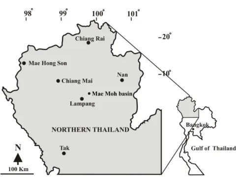
Geology (Songtham et al., 2005): The Mae Moh basin is situated in the Mae Moh District of Lampang Province, which is about 26 km east of Lampang City. It is distributed in the area of about 135 square km, 7 km in east-west and 16 km in north-south as shown in Fig. 1. The basin floor is about 320-340 m above mean sea level.
Fig. 1: Location of the Mae Moh basin, situated in Lampang Province
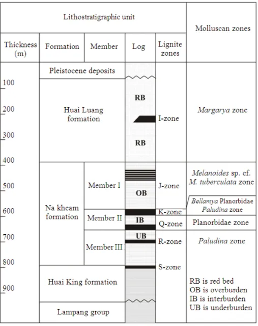
The Tertiary sediments called the Mae Moh Group consist of three formations, namely the Huai King, Na Khaem and Huai Luang formations, in ascending order. The total thickness of three formations is nearly 1,000 meters as shown in Fig. 2.
The Huai King formation unconformably overlies the basement rock, Lampang Group. It consists of a sequence of upward grading conglomerate, sandstone and pebbly sandstone, siltstone and finally fining upwards into interbedded red and gray claystone. The uppermost part of the formation is marked by a thin layer of coal named the S coal zone.
The Na Khaem formation is a coal measure comprising three main coal zones, Q, K and J assigned to Middle Miocene age. This formation has been divided into three members as Member III, Member II and Member I, in ascending order. The Member III, or the so-called Underburden (UB), is a greenish gray to gray claystone with a thin layer of coal, named the R-coal zone. The Member II is composed of two main coal zones, which are the Q coal zone in the lowermost part and the K coal zone in the uppermost part and are intercalated by an Interburden (IB) of claystone. The member I is a thick Overburden (OB) consisting of claystone with a series of coal layers named the J coal zone. The J coal zone is an intercalation between claystone and six main coal layers.
The Huai Luang formation is composed of claystone and siltstone with some sandstone and conglomerate
lenses. A red to brownish red colour is the general characteristic of this formation with some gray layers interbedded in some horizons. A form of gypsum, selenite, is abundant in this formation. There is a coal zone, named I coal zone, in the middle of the formation.
Songtham et al. (2005) also reported that during the Middle Miocene, the Mae Moh basin was a large swamp where living organisms such as fish and various species of aquatic molluscs lived. The molluscs might have appeared and disappeared throughout the time depending on environmental changes that altered the composition of the molluscan assemblages changed from horizon to horizon, resulting in the formation of five molluscan zones (Paludina Molluscan Zone, Planorbidae Molluscan Zone, Bellamya Planorbidae Paludina
Molluscan Zone, Melanoides sp. cf. M. tuberculata
Molluscan Zone and Margarya Molluscan Zone) as shown in Fig. 2.
The purpose of this study is to study the ESR spectra of sediments confined within Red Bed layer (RB) from Huai Luang formation and within three gray claystone layers (overburden, OB; interburden, IB; underburden, UB) from Na Khaem formation.
MATERIALS AND METHODS
were gently ground with a mortar and the grains were sieved in order to separate the size fraction of 75-250 µm. The separated grains were etched by 5% HCl acid for a few minutes in order to remove the surface defect signal at g = 2.0002 caused by the pressure in grinding (Griscom and Beltran-Lopez, 2002). The size fraction was, then washed repeatedly in distilled water and allowed to dry at a temperature of 40°C. All sample preparation procedures were performed in dim red light to avoid the samples being exposed to light. Each sample was divided into three aliquots, approximately 300-500 mg each. These were loaded into fused quartz sample tubes with a nominal inside diameter of 3 mm to
fill a height of 2.5 cm. The sample was placed at the center of microwave cavity for ESR measurement. All ESR measurements were carried out at room temperature. The ESR spectra were recorded using a Bruker spectrometer (E500) operating in the X band of microwave range. The operating conditions adopted during the experiment were as follows: 349 mT central magnetic field; 350, 100 and 10 mT scan ranges; 0.1 mT field modulation amplitude; 100 kHz modulation frequency; 0.63 mW microwave power; 5.12 ms conversion time; 163.84 ms time constant. DPPH with a g-value of 2.0036 was used as an internal standard for g-factor calculations.
RESULTS
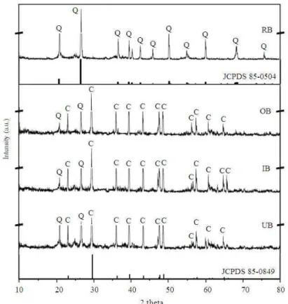
The structure of sediments and claystones was investigated by XRD measurements. Figure 3 shows XRD patterns of sediments confined within Red Bed (RB) layer from Huai Luang formation and OB, IB and UB layers from Na Khaem formation.
ESR spectra, in a scan magnetic field range of 350 mT, of sediments from RB layer and gray claystones from OB, IB and UB layers are shown in Fig. 4.
The ESR signal of sediments from RB layer was further investigated by scanning the magnetic field in narrower ranges of 100 mT and 10 mT and the obtained ESR signals are shown in Fig. 5 and 6, respectively.
Fig. 3: X-ray powder diffraction patterns of sediments collected from Red Bed (RB) layer and gray claystonefrom Overburden (OB), Interburden (IB) and Underburden (UB) layers. C = Calcite, Q = Quartz
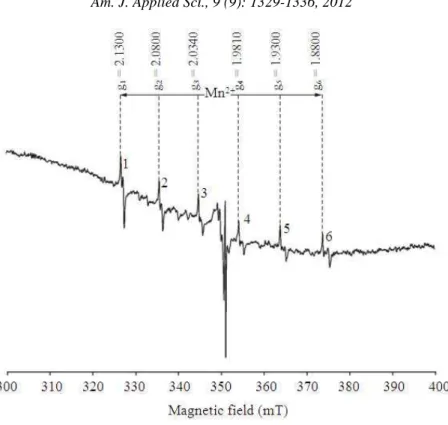
Fig. 5: The sextet hyperfine signals of Mn2+ organic radicals around g = 2.00 of sediments from RB layer. Scan range is 100 mT
Fig. 6: ESR spectra of the E1′ center at g = 2.0018 and g = 2.0003 of sediments from RB layer. Scan range is 10 mT
ESR spectra of sediments from three gray claystone layers (OB, IB and UB) are shown in Fig. 7. The central ESR peaks were further investigated
Fig. 7: The sextet hyperfine signals of Mn2+ organic radicals around g = 2.00 of gray claystonefrom OB, IB and UB layers. Scan range is 100 mT
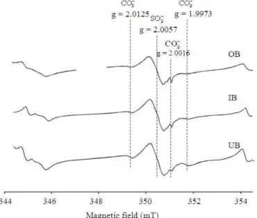
Fig. 8: ESR spectra of gray claystone from OB, IB and UB layers associated with CO3
−
at g = 2.0125, SO2
− at g = 2.0057 and CO2
−
at g= 2.0016 and g= 1.9973. Scan range is 10 mT
DISCUSSION
The diffraction peaks shown in Fig. 3, according to JCPDS file numbers 85-0504 and 85-0849, show that the sediments in RB layer consist mostly of quartz
while those in OB, IB and UB consist mainly of calcite and some fraction of quartz.
radicals around g = 2.0 were observed in samples from all layers as clearly.
As can be seen in Fig. 5, a sharp ESR signal was observed at a magnetic field of about 350 mT. Moreover, the magnified ESR signal in Fig. 6 shows two sharp peaks at g = 2.0018 and g = 2.0003 which are attributed to E1′ center or
3 3
SiO− in quartz (Ikeya, 1993; Ulusoy, 2002; Jani et al., 1983). The ESR results indicate that the sediments in RB layer consist of iron oxide and quartz which agree with XRD results. It should be pointed out that the XRD pattern shown in Fig. 3, no peaks of iron oxide were observed due to its less amount contained in the sediment.
Quartz or silica (SiO2) is a special type of silicate where each oxygen atom is bonded with two Si atoms to form a three-dimensional network in a crystalline form. Quartz is the most common rock-forming mineral next to feldspars and exists in most rocks of sedimentary, igneous and metamorphic origins. A vacancy and interstitial type defects in SiO2 are together called a “Frenkel pair”. The following three pairs occured in SiO2; (i) a paired oxygen vacancy and interstitial O2- which is not stable at room temperature, (ii) a paired Si vacancy and Si interstitial is energetically not stable and (iii) a paired E1′ center (an
electron at an oxygen vacancy) which is associated with ESR signals of sediment sample in Red Bed (RB) layer from Huai Luang formation, is stable and is observed in natural quartz and irradiated amorphous SiO2. The model for E1′ center in quartz is an unpaired electron in
a dangling sp3 hybrid orbital of pyramidal 3 3
SiO− where an oxygen ion, O-, is extracted from a SiO4 tetrahedron (Ikeya, 1993; Jani et al., 1983; Silsbee, 1961).
The selenite-bearing red bed sandy claystone is considered as secondary deposit formed sometimes after the formation of Huai Luang. It is believed that the environment during this secondary formation was in arid condition. The hydrous calcium sulfate crystals, CaSO4.2H2O, are said to be resulting from solution cavities through precipitation of calcium sulfate-bearing groundwater that was saturated under an arid condition. The presence of selenite is in contrast to the massive gypsum, frequently associated with evaporite deposits in playa in the arid environment which formed beds parallel to nearby sedimentary strata. However, the selenite crystals from the Huai Luang formation are randomly scattered like nodules in red bed of sandy claystone without forming beds. The gypsum crystals, selenite and red bed were secondarily formed by some proposed chemical reactions such as ones present below:
CaCO3 → CaO + CO2 (1)
4FeS2 + 11O2 → 2Fe2O3 + 8SO2 (2)
CaO + SO3 + 2H2O → CaSO4 . 2H2O (3)
The source of calcium came from limestone (CaCO3) of the Lampang Group that decayed to become lime (CaO). The lime reacted with sulfur trioxide (SO3) (derived from SO2) and water forming selenite crystals (CaSO4.2H2O) in saturated groundwater under an arid environment according to Eq. 1-3. For the sediments in RB layer, the iron oxide (Fe2O3) produced from the oxidizing of pyrite (FeS2) according to Eq. 2 partially changed the color of original flood plain sediments into the red bed formation containing the selenite crystals. This is why the red bed formation retains some original color of greenish gray sandy claystone occurring in some places (Songtham et al., 2005).
It is seen in Fig. 7 that the sextet hyperfine and the forbidden transitions associated with Mn2+ ions of organic radicals are similar in all three layers.
The ESR signals, as shown in Fig. 8, at g = 2.0125, 2.0057 and 2.0016 and 1.9973 correspond to
3
CO−, SO2
− and
2
CO− molecular ions, respectively (Ikeya, 1993; Serway et al., 1969; Kai and Miki, 1992; Katzenberger et al., 1989; Barabas, 1992).
The results indicated that the sedimentary consisted largely of carbonates. Carbonates are common minerals in nature. The basic constituent unit in all carbonate minerals is the 2
3
CO−molecular ion. Calcium carbonate (CaCO3) has two main crystal structures of calcite and aragonite. Calcite with trigonal (rhombohedral) symmetry is the only thermodynamically stable form of pure CaCO3 at room temperature and under atmospheric pressure (Ikeya, 1993). Elementary defects induced by ionizing radiation are electron centers ( 3
3
CO−) and hole centers (CO3
−
). An oxygen vacancy with an electron, actually the CO2
−
molecular ion, is also formed by radiation (Zavoisky, 1945).
CONCLUSION
layers consist largely of carbonates. In this study, the
1
E′center was also observed in quartz for the sediment collected from RB layer. Recently, E1′ centers have
been used for ESR dating (Ulusoy, 2002; Lee and Yang, 2003; Bartoll et al., 2001). Therefore, the
1
E′centers found in the present study can be used for ESR dating of the formation in the future.
ACKNOWLEDGMENT
The researchers would like to thank EGAT (Electricity Generating Authority of Thailand) and DMR (Department of Mineral Recourses) for providing scientific assistance, access to the mine and company data when staying at the mine. We would like to thank NSRL OAP (National Standard Radioactivity Laboratory, Office of Atoms for Peace) for supporting gamma irradiation exposures and NAA techniques and the Department of Chemistry, Mahidol University for providing ESR facilities. This study was funded by the King Mongkut’s University of Technology Thonburi under The National Research University Project.
REFERENCES
Barabas, M., 1992. The nature of the paramagnetic centres at g = 2.0057 and g = 2.0031 in marine carbonates. Int. J. Radiat. Appli. Instrument Part D. Nucl. Tracks Radiat. Measure., 20: 453-464. DOI: 10.1016/1359-0189(92)90031-P
Bartoll, J., H.P. Schwarcz and W.J. Rink, 2001. ESR studies of E′ centres in geological flint. Radiat. Measure., 33: 893-898. DOI: 10.1016/S1350-4487(01)00091-9
Engin, B., S. Kapan-Yesilyurt, G. Taner, H. Demirtas and M. Eken, 2006. ESR dating of Soma (Manisa, West Anatolia – Turkey) fossil gastropoda shells. Nucl. Instrument. Meth. Phy. Res. Sec. B: Beam Interact. Mater. Atoms, 243: 397-406. DOI: 10.1016/j.nimb.2005.09.008
Griscom, D.L. and V. Beltran-Lopez, 2002. ESR spectra of limestones from the cretaceous-tertiary boundary: Traces of a catastrophe. Adv. ESR Appli., 18: 57-64.
Ikeya, M., 1993. New Applications of Electron Spin Resonance: Dating, Dosimetry and Microscopy. World Scientific Singapore Publishers, London, 1st Edn., ISBN-10: 9810212003, pp: 500.
Jani, M.G., R.B. Bossoli and L.E. Halliburton, 1983. Further characterization of the E'1 center in crystalline SiO2. Phys. Rev. B., 27: 2285-2293.
Kai, A. and T. Miki, 1992. Electron spin resonance of sulfite radicals in irradiated calcite and aragonite. Int. J. Radiat. Appli. Instrument.. Part C. Radiation Phys. Chem., 40: 469-476. DOI: 10.1016/1359-0197(92)90211-W
Katzenberger, O., R. Debuyst, P.D. Canniere, F. Dejehet and D. Apers et al., 1989. Temperature experiments on mollusc samples: An approach to ESR signal identification. Int. J. Radiat. Appli. Instrument. Part A. Applied Radiat. Isotopes, 40: 1113-1118. DOI: 10.1016/0883-2889(89)90048-8 Lee, H.K. and J.S. Yang, 2003. ESR dating of the
Wangsan fault, South Korea. Q. Sci. Rev., 22:
1339-1343. DOI:
10.1016/S0277-3791(03)00018-0
McDougall, D.J., 1968. Thermoluminescence of Geological Materials. 1st Edn., Academic Press, London, New York, pp: 678.
Serway, R.A., S.A. Marshall, J.A. McMillan, R.L. Marshall and W.D. Ohlsen, 1969. Electron spin resonance absorption spectra of hole‐trap centers associated with phosphorous in irradiated single crystal calcite. J. Chem. Phys., 51: 4978-4981. DOI: 10.1063/1.1671892
Silsbee, R.H., 1961. Electron spin resonance in neutron‐irradiated quartz. J. Applied Phys., 32: 1459-1462. DOI: 10.1063/1.1728379
Songtham, W., H. Ugai, S. Imsamut, S. Maranate and W. Tansathien et al., 2005. Middle miocene molluscan assemblages in mae moh basin, lampang province, Northern Thailand. Sci. Asia, 31: 183-191.
Ulusoy, U., 2002. The investigation of ESR dating by using alpha-rays. Adv. ESR Appli., 18: 163-166.
Vichaidid, T., Youngchuay, U. and Limsuwan, P., 2007. Dating of aragonite fossil shell by ESR for paramagnetic species assignment of Mae Moh basin. Nucl. Instrument. Meth. Phy. Res. Sec. B: Beam Interact. Mater. Atoms, 262: 323-328. DOI: 10.1016/j.nimb.2007.05.021
Zavoisky, E.K., 1945. Spin-magnetic resonance in paramagnetics. J. Phys. USSR, 9: 211-245.