www.ambi-agua.net E-mail: [email protected]
This is an Open Access article distributed under the terms of the Creative Commons Attribution License, which permits unrestricted use, distribution, and reproduction in any medium, provided the original work is properly cited.
Electron Spin Resonance (ESR) in detection of aquatic pollution
through host-parasite relationship
ARTICLES doi:10.4136/ambi-agua.2085
Received: 20 Mar. 2017; Accepted: 09 Sep. 2018
Lucas Aparecido Rosa Leite1; Angela Kinoshita1; Oswaldo Baffa2;
Rodney Kozlowiski de Azevedo1; Vanessa Doro Abdallah1*
1Universidade do Sagrado Coração (USC), Bauru, SP, Brasil
Programa de Mestrado em Ciência e Tecnologia Ambiental.
E-mail: [email protected], [email protected], [email protected], [email protected]
2Universidade de São Paulo (USP), Ribeirão Preto, SP, Brasil
Faculdade de Filosofia, Ciências e Letras de Ribeirão Preto (FFCLRP). Departamento de Física (DF). E-mail: [email protected]
*Corresponding author
ABSTRACT
Aquatic environmental pollution due to negative human activities remains a major problem. Bioindicators that primarily describe the total concentration of the respective pollutant are very useful tools to aid in the chemical analysis of water in order to obtain knowledge about the levels of pollutants in the environment. This study therefore used Electron Spin Resonance Spectroscopy (ESR) to detect the presence of transition metals (copper, iron and manganese) and possible radicals present in samples of Neoechinorhynchus curemai and its host tissues Prochilodus lineatus, as well in water and sediment of the Batalha River, at the same sample
site where fish were collected. Spectral analysis of samples showed the presence of three metals (Cu2+, Fe3+ and Mn2+), in addition to nitric oxide (NO) and humic acid (HA). Quantification of
the elements in the samples was possible only for Cu detected in the spectrum of parasites, which was equivalent to 2 ppm. ESR proved to be efficient in the detection of transition-metal ions (Cu2+, Fe3+ and Mn2+), in addition to NO and HA. However, the low concentration values
of these compounds in P. lineatus tissues (liver, muscle and intestine) and in the water and
sediment samples collected did not allow their quantification, as they were below the limit of detection. It can be concluded that N. curemai had the capacity to accumulate these ions,
especially copper.
Keywords: environmental monitoring, free radicals, Neoechinorhynchus curemai, Prochilodus lineatus, transition metals.
Ressonância do Spin Eletrônico (ESR) na detecção de poluição
aquática através da relação parasito-hospedeiro
RESUMO
Rev. Ambient. Água vol. 13 n. 6, e2085 - Taubaté 2018
auxílio das análises químicas da água que primariamente descrevem a total concentração do respectivo poluente. Nesse contexto, o objetivo desse trabalho foi o de empregar a técnica de Espectroscopia de Ressonância do Spin Eletrônico (ESR) para detectar a presença de poluentes, diretamente em espécimes de Neoechinorhynchus curemai e nos tecidos do seu hospedeiro Prochilodus lineatus, assim também como na água e sedimento do rio Batalha, no mesmo ponto
onde os peixes foram coletados. A análise do espectro das amostras identificou a presença de três metais (Cu2+, Fe3+ e Mn2+), além do óxido nítrico (NO) e do ácido húmico (AH). A
quantificação dos elementos nas amostras só foi possível para o Cu, detectado no espectro dos parasitos, que foi de 2ppm. A ESR se mostrou eficiente na detecção de íons metálicos de transição (Cu2+, Fe3+ e Mn2+), além do NO e do AH. Entretanto, a baixa concentração desses
compostos nos tecidos (fígado, músculo e intestino) de P. lineatus. não possibilitaram sua
quantificação, já que as concentrações dos mesmos estão abaixo do limite de detecção do aparelho. É possível concluir que N. curemai teve a capacidade de acumular esses íons,
especialmente cobre.
Palavras-chave: metais de transição, monitoramento ambiental, Neoechinorhynchus curemai, Prochilodus lineatus, radicais livres.
1. INTRODUCTION
Pollution of aquatic ecosystems as a result of anthropogenic activities remains a major problem in these environments, causing disastrous effects on the health of living organisms and becoming subject of many discussions (Khan and Thulin, 1991; Sures, 2008). In freshwater ecosystems, both organic and inorganic pollution (especially metals) are global issues and deserve attention since they directly and indirectly affect organisms, causing them irreparable damage (Wen et al., 2017; Schmeller et al., 2018; Väänänen et al., 2018).
Bioindicators that primarily describe the total concentration of a respective pollutant are very useful tools in the chemical analyses of water in order to obtain knowledge about levels of pollutants in the environment. Recent studies have shown that, in addition to already established indicators, certain parasites and their hosts can be used as biomonitoring tools, since the pollution also effects their health and, consequently, their occurrence and distribution. Another important point to consider is that, just like pollutants, parasites affect the health of organisms, and some of these organisms’ responses to pollutants and parasites are very similar (Lafferty, 2008; Lafferty and Kuris, 2005; Sures, 2008; Thielen et al., 2004).
Studies evaluating the role of parasites as accumulators and pollution indicators have been carried out for decades, using a vast host-parasite combination (Sures et al., 1999; 2017) where fish are the most frequently used hosts (Vidal-Martínez and Wunderlich, 2017). In addition, the high potential of parasites for metal accumulation (especially heavy metals) and their high effectiveness in assessing aquatic pollution compared to free-living organisms (such as mussels, for example) has been clearly demonstrated (Sures et al., 1997; 1999;).
Different fish endoparasites have been suggested for use in the detection of pollution in aquatic environments. Among them, acanthocephalans are highlighted as presenting a high capacity for heavy metal accumulation (Vidal-Martínez et al., 2009; Nachev et al., 2013). Laboratory studies have shown that fish parasitized by acanthocephalans have lower levels of heavy metal contamination in their tissues when compared to non-parasitized specimens. However, the results of the analyses are difficult to predict, and there is a need for further studies in this field to contextualize information already obtained in different types of environments with different levels of degradation (Thielen et al., 2004; Sures, 2008).
Rev. Ambient. Água vol. 13 n. 6, e2085 - Taubaté 2018 al., 2017; Nachev et al., 2010; 2013; Thielen et al., 2004) or Atomic absorption spectrometry
(AAS) (Baruš et al., 2001; Bayoumy et al., 2015; Baruš et al., 2007; Dural et al., 2011; Morsy et al., 2012). Eletron Spin Resonance (ESR) is a spectroscopy designed to detect non-zero electronic spin systems, such as free radicals and some transition metals. In biological systems, it can be used to detect the presence of some ions, including copper, manganese and iron, directly in tissues without biochemical manipulations, thus reducing the risk of structural disturbances. This makes the technique a powerful tool in research involving biological and environmental systems (Wertz and Bolton, 2012).
ESR has already been used to detect pollutants (both organic and inorganic) in several systems. In aquatic ecosystems, ESR has been used to detect Reactive Oxygen Species (ROS)
in amphibians (D’Errico et al., 2018) and to detect and quantify Mn2+in fish otoliths (Di
Beneditto and Franco, 2018). In both cases the technique proved to be efficient, and could be used to aid other spectrometric techniques. In the host-parasite system, however, there are still no published studies where ESR was used. In this context, the objective of this study was to use the ESR technique to detect transition metals (copper, iron and manganese) and possible radicals present in parasites and host tissues (muscle, liver and intestine).
2. MATERIAL AND METHODS
2.1. STUDY AREA
This study was conducted in a stretch of the Batalha River, the Tietê-Batalha River Basin,
located in the municipality of Reginópolis, State of São Paulo (21º53’17” S and 49º13’31” W).
It is an area with severe anthropic influences and predominantly lotic characteristics. Limnologic and structure variables of the stretch are plotted in Table 1.
The river area in this stretch, even though surrounded by native riparian forest for most of its extension, is used predominantly for agricultural and livestock purposes, with emphasis on cattle raising, sugar cane, corn and eucalyptus plantations, increasing the amount of organic matter and leachate pollutants. In addition, the municipality does not treat sewage, and so sewage is thrown in natura into a tributary of the river (São Paulo, 2010; Santos and Heubel,
2008; Sistema Nacional De Informações Sobre Saneamento, 2016). Table 1. Mean values of physicochemical parameters: pH, temperature, dissolved oxygen (DO) and conductivity (CD); and structural parameters: width of the stretch, and width of the permanent preservation area (APP) surrounding the sampling point, located in the municipality of Reginópolis.
Parameter Value ± SD
pH 7.7 ± 0.3
Temperature (ºC) 26.5 ± 1.5
DO (mg/L) 4.9 ± 1.6
CD (µS/cm) 68 ± 14.7
Width (m) 13.2 ± 1.7
APP width (m) 83.1 ± 7.9
2.2. SAMPLING AND PROCESSING OF PARASITES AND HOSTS
Thirty-nine specimens of Prochilodus lineatus (Valenciennes, 1837) (Characiformes:
Rev. Ambient. Água vol. 13 n. 6, e2085 - Taubaté 2018
stereomicroscope to collect parasites. In this study, only fish specimens that showed positive results for Neoechinorhynchus curemai Noronha, 1973 (Acanthocephala:
Neoechinorhynchidae) parasitism were used, totaling 19 P. lineatus specimens analyzed.
After collection, the parasites were placed in a recepticle containing distilled water and then washed with a vortex mixer. Subsequently, they were placed in glass flasks and frozen at -20ºC until the ESR analysis. Samples of muscle, liver and intestine were also taken from hosts. The washing and storage of the tissue samples followed the same methodology used for parasites.
2.3. WATER AND SEDIMENT SAMPLING
Water and sediment samples were collected from the Batalha River at the same point of the fish collection. The sampling followed the procedures recommended by Gomes and Filizola (2006) for sediment and Pires et al. (2006) for water. The samples were stored in 20 mL plastic tubes and frozen at -20ºC until ESR analysis.
2.4. ESR ANALYSIS
Samples of parasites and host tissues were externally cleaned with ultrapure water (Milli Q) and then dehydrated in absolute alcohol by immersion for 10 minutes. These materials and sediment were then oven-dried at 40ºC (Table 2). After drying, the samples were carefully crushed with agate mortar and pestle for homogenization under low impact, in order to avoid induction of other radicals by mechanical action. Subsequently, an aliquot of each sample was transferred to an ESR quartz tube, with a 3mm internal diameter. The mass used to record the spectrum was determined (Table 2). The water sample was placed in a capillary tube and sealed prior to insertion in the quartz tube. The tube containing the sample was inserted into a quartz dewar for freezing with liquid nitrogen for spectrum recording (77K).
A JEOL Band-X spectrometer was used to record the spectra, some of them with a scan width of 500mT and others with 80mT, centered on the g~2 spectral region. Some spectra were recorded simultaneously with a Mn2+ standard, present in the spectrometer, that consists of
MgO crystal doped with Mn2+. A 0.5 mM solution of CuSO
4 complexed with C3H7NO2 was
used for the quantification of copper (Cu2+) present in the sample. The spectra of the parasite
and the copper solution were acquired under the same spectrometer conditions of microwave power, modulation and gain. Copper can be complexed with several compounds producing different spectral symmetries. Alanine was used as a ligand due to the same symmetry of the complex found in the parasites samples. The volume of the standard solution used was 20µL.
Table 2. Mass values (mg) of parasites, host tissues and sediment samples before and after drying, and the amount used in ESR analysis.
Mass Parasite Intestine Liver Muscle Sediment Wet 102 mg 189.5 mg 690 mg 674.4 mg --
Dry 17.1 mg 20.21 mg 77.44 mg 114 mg --
ESR 5.68 mg 6.08 mg 24.45 mg 5.8 mg 77.6 mg
3. RESULTS AND DISCUSSION
The specimens of P. lineatus showed mean standard length and weight of 27.44 ± 6.37 cm
and 568.38 ± 318.14 g, respectively. The specimens of N. curemai were all adults and had mean
abundance of 2.83 ± 0.12 parasites per fish and mean intensity of 5.52 ± 0.13 parasites.
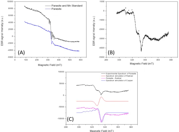
The analysis of the parasite samples’ spectrum in the 500mT scan was performed under
Rev. Ambient. Água vol. 13 n. 6, e2085 - Taubaté 2018 the obtained signals showed that there is no Mn2+ in the parasite samples (Figure 1A). In the
spectrum of the central region g~2 it was possible to notice characteristic lines of copper (Figure 1B). To confirm the presence of Cu2+ in the sample, spectral simulation of the Cu2+ present in
the parasites was carried out using Simfonia-Bruker software. Through the simulation, it was possible to observe the agreement between the simulated Cu2+ signal and the radical present in
the parasites. Double integration of the Cu2+ simulated signal was compared to element pattern
spectrum. The Cu2+ mass of the total mass of the parasite was obtained from the atomic mass
of the ion (63.6g), resulting in a concentration of 2 ppm of Cu2+ in the parasites (Figure 1C).
Figure 1. ESR spectrum of Neoechinorhynchus curemai specimens with and without the simultaneous register of manganese (A), centered on the g~2 region, where the signals indicated by asterisks represent the lines belonging to the copper (B)(g⊥: 2.0032 ± 0.0001; g//: 2.1160 ± 0.0001; A⊥: 1.1 ± 0.1 mT; A//: 4.8 ± 0.1 mT) and spectral simulation of the
radical (g-factor: 2.0084 ± 0.0001) (C). Acquisition parameters are: Center Field 336 mT, Modulation Frequency 100 kHz, Modulation amplitude 0.1 mT, gain 2000, microwave frequency 9131.4 MHz, Microwave Power 5 mW. In (A) sweep width 500mT and (B) 80 mT.
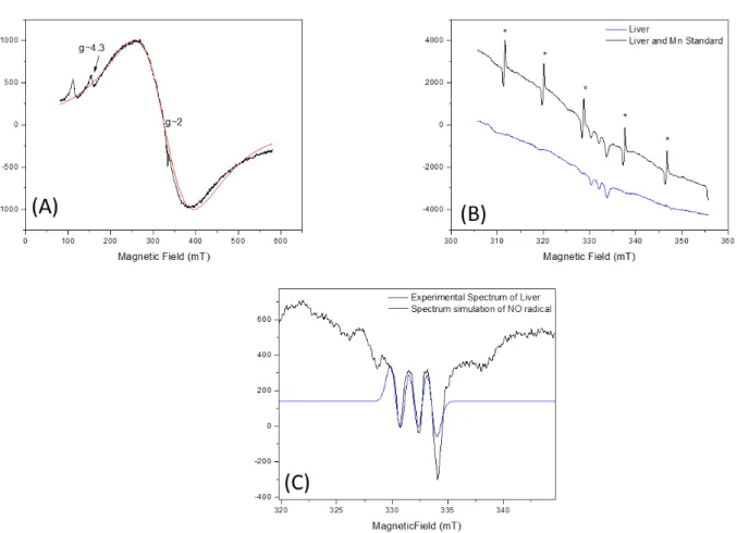
The liver sample spectrum was recorded with 500mT scanning width. The spectrum was dominated by iron, as shown in the simulation (Figure 2A). In the g~2 region of the spectrum it was possible to identify other radicals. By performing a more detailed recording of the spectrum in the g~2 region it was possible to observe a structure of lines. The spectrum was recorded with the manganese pattern for investigation, where very weak signals of this ion in this region can be noted (Figure 2B), indicating a small concentration of Mn2+ in the tissue. A
more detailed scan at the g~2 region showed that the lines’ structure in the central region corresponds to nitric oxide (NO), bound to Fe3+, leading to the three-line structure observed.
Rev. Ambient. Água vol. 13 n. 6, e2085 - Taubaté 2018
Figure 2. ESR spectrum in the 500mT scan of the fish liver samples and iron spectral simulation in red (A) (g-factor = 2.0453± 0.0001). The spectrum centered on the g~2 region with and without the manganese (*) pattern (B). Spectrum of the sample also centered on the g~2 region and the simulation of the NO (g-factor = 2.0040 ± 0.0001, A=1.82 ± 0.01mT) present in the sample (C). Acquisition parameters are: Center Field 330 mT, Modulation Frequency 100 kHz, Modulation amplitude 0.1 mT, gain 300, microwave frequency 9133.4 MHz, Microwave Power 5 mW. In (A) sweep width 500mT and (B) 100 mT.
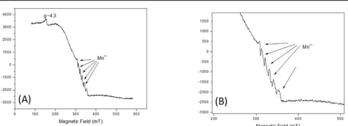
In the spectrum of fish intestine samples, it was possible to notice the presence of Fe3+ and
Mn2+ (Figure 3A). The spectrum was recorded with Mn2+standard to confirm the presence of
this element in the sample (Figure 3B). Incorporation of manganese ions in different matrices have been studied with multifrequency ESR, and this can give more information about the complex formed; but these techniques are not easily found, and these studies have corroborated the experiments done at X band (Murzakhanov et al., 2017).
In the fish muscle spectrum, it was also possible to notice the presence of the Fe3+ element
and Mn2+ traces (Figure 4), suggesting that this element, although present in the sample, was in
Rev. Ambient. Água vol. 13 n. 6, e2085 - Taubaté 2018 Figure 3. ESR spectrum of the fish intestine samples centered in the g~2 region where the presence of iron is noted, with a peak at g~4.3 and manganese, as indicated (A). Spectrum in the Mn2+ region, recorded with manganese standard, for confirmation (B). Acquisition
parameters are: Center Field 330 mT, Modulation Frequency 100 kHz, Modulation amplitude 0.1 mT, gain 300, microwave frequency 9133.4 MHz, Microwave Power 5 mW, sweep width 500mT.
Figure 4. ESR spectrum of the fish muscle samples, where the presence of iron is observed and traces suggesting the presence of manganese (*). Acquisition parameters are: Center Field 330 mT, Modulation Frequency 100 kHz, Modulation amplitude 0.1 mT, gain 300, microwave frequency 9133.4 MHz, Microwave Power 5 mW, sweep width 500mT.
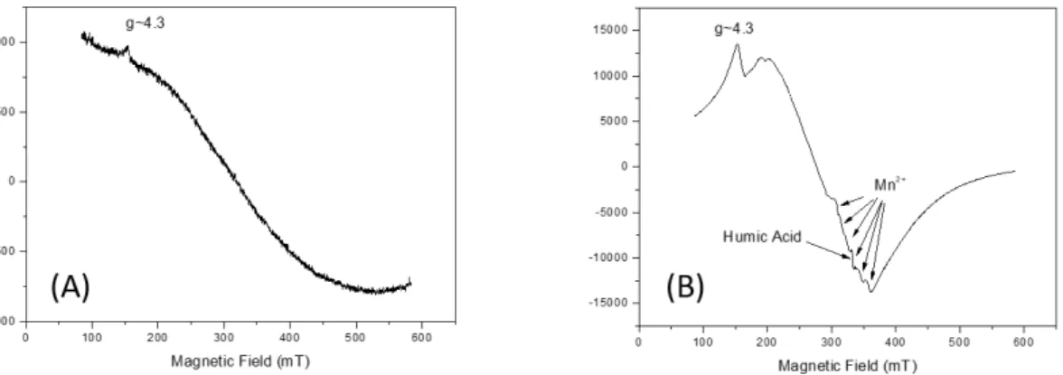
In the spectrum of the sediment samples analyzed, the presence of Fe3+ and Mn2+ can be
observed. In addition to these elements, another central radical was recorded in the g~2 region, attributed to humic acid (AH) (Figure 5B). In the water sample, the spectrum showed only the presence of Fe3+ (Figure 5A).
Low concentrations of elements considered essential, that is, those necessary for the development of organisms and that occur naturally in the environment (Merian et al., 2004), are expected in acanthocephalans fish parasites, which are more susceptible to accumulate high concentrations of toxic elements (Nachev et al., 2013). Although copper is considered essential
Rev. Ambient. Água vol. 13 n. 6, e2085 - Taubaté 2018
depend on water physico-chemical characteristics, such as alkalinity, hardness and pH (Carvalho and Fernandes, 2006). Generally, concentrations of this element in water are lower than those found in fish and parasites (Porto and Ethur, 2009; Brázová et al., 2012), and the maximum concentration of copper allowed for Class 2 waters, such as the Batalha River, is 0.009 mg/L (Conama, 2005). In this case, as the element was not detected in fish-tissue spectrums nor in the water or sediment collected from the river, it was assumed that the concentrations are below the ESR detection limit.
Figure 5. ESR spectrum of the water sample, where the presence of iron (A) is noted. The ESR spectrum of sediment sample shows the presence of iron, traces of manganese, as indicated, and the presence of humic acid in the g~2 region (g-factor = 1.9976 ± 0.0001) (B). Acquisition parameters are: Center Field 330 mT, Modulation Frequency 100 kHz, Modulation amplitude 0.1 mT, gain 300, microwave frequency 9133.4 MHz, Microwave Power 5 mW, sweep width 500mT.
The presence of iron and manganese ions in the spectrum of the tissues (liver, muscle and intestine) of P. lineatus and also in the samples of water and sediment of the Batalha River was
already expected due to the high natural occurrence of these elements, which are the two most abundant metals in the environment (Förstner and Wittmann, 1983). Both elements in water and sediment are considered essential for both flora and fauna (Howe et al., 2004; Schümann and Elsenhans, 2004). Iron in fish acts mainly in oxidation-reduction and oxygen-transport processes, occurring as a component of the respiratory pigment (heme), as hemoglobin and myoglobin, as well as the heme enzymes (peroxidase, catalase and cytochromes) being absorbed from the water through the gills (Lim et al., 2001). Manganese plays a key role in the metabolism of amino acids, lipids, proteins and carbohydrates, besides being involved in functions of the immune system, blood glucose regulation, reproduction and also in defense mechanisms against free radicals (Keen et al., 1999).
In aquatic vertebrates, iron excess can cause several effects such as breathing problems, reduced growth, feeding difficulties, high mortality and histopathological changes in liver cells (Bury et al., 2003; Lim et al., 2001), in addition to the production of oxygen free radicals, which may be toxic to cells (Bury et al., 2003). Negative effects of excess Mn on fish include anemia, leukocytosis, disruption of sodium balance, impaired calcium absorption and impacts on metabolism (Agrawal and Srivastava, 1980; Barnhoorn et al., 1999; Gonzalez et al., 1990; Nath and Kumar, 1987; Reader et al., 1988).
The presence of NO detected in the spectrum of fish liver samples may also be associated with Fe3+ domain in the spectrum of the same tissue, since the molecule exerts its physiological
functions through the binding with Fe3+ present in heme (Denninger and Marletta, 1999). In
Rev. Ambient. Água vol. 13 n. 6, e2085 - Taubaté 2018 on tissue concentration or clearance (Hansen and Jensen, 2010). In addition, NO production may be directly associated with hypoxias situations, where the increase in NO causes fish and other aquatic organisms to become more resistant to situations of low oxygen availability in water (Jensen, 2009; McNeill and Perry, 2006). Hansen and Jensen (2010), evaluating the production of NO in goldenfish (Carassius auratus Linnaeus, 1758), observed that in situations
of severe hypoxia, NO production by fish was increased as well as its resistance capacity, since the vasodilatation caused by NO increased the oxygen delivery rate in the organs. Thus, it is possible to verify that presence of NO in the tissues of P. lineatus can be an indicative of the
lack of oxygen in the water, which in the study site of the Batalha River is around 4.9 mg/L, below the established limit by CONAMA for Class 2 waters (Conama, 2005). However, it would be necessary to quantify NO in the samples in order to obtain safer and more accurate results. Another factor that should be considered is the fact that NO production is also directly associated with production of nitrosamines, which are potentially carcinogenic organic compounds (Al Bulushi et al., 2009). In humans, the highest rate of exposure to this compound occurs through food, which includes water and fish (Dutra et al., 2007).
Humic acids (HA), derived from humic substances (HS), which are part of organic matter, belong to the class of natural products most abundant in the biosphere, being precursors of fossil fuels. They are the most-studied fraction of HS, since they can contain voids of different sizes in their molecular structures, and can accommodate a wide range of pollutants (Mangrich and Vugman, 1988). HA act as binders for the complexation of ions, mainly Mn2+, Fe3+ and Cu2+,
where the formation and transport of these complexes, and their deposition, can be important mechanisms in the accumulation of metals in the sedimentary deposits (Saab, 1999). It is known that in HA the content of certain ions, such as Fe3+, in concentrate domain sites increases with
decreases in the degree of environmental pollution (Silva, 2001). However, the mere presence of HA in the sediment spectrum cannot be considered an indicator of pollution, and a more robust analysis of its structure is necessary to establish relationships with environmental impacts such as forest-pasture conversion (Araújo et al., 2011) or pollution by heavy metals, pesticides and other substances of anthropogenic origin (Toscano, 1999).
4. CONCLUSION
In this study, the Electron Spin Resonance technique proved to be efficient in detecting transition metals (manganese, iron and copper) and nitric oxide and humic acid. However, low concentration values of these compounds in P. lineatus tissues (liver, muscle and intestine) and
in water and sediment samples collected did not allow their quantification, as the concentration of metals in the tissues is at a level below the limit of detection. It can be concluded that N. curemai had the capacity to accumulate these ions, especially copper.
5. ACKNOWLEDGMENTS
We thank the Fundação de Amparo à Pesquisa do Estado de São Paulo (FAPESP) (process nº 2014/12682-0 and 07/06720-4) and the Coordenação de Aperfeiçoamento de Pessoal de Nível Superior - Brasil (CAPES) - Finance Code 001, for the financial support.
6. REFERENCES
Rev. Ambient. Água vol. 13 n. 6, e2085 - Taubaté 2018
AL BULUSHI, I.; POOLE, S.; DEETH, H. C.; DYKES, G. A. Biogenic Amines in Fish: Roles in Intoxication, Spoilage, and Nitrosamine Formation – A Review. Critical Reviews in Food Science and Nutrition, v. 49, p. 369-377, 2009. https://doi.org/10.1080/10408390802067514
ARAÚJO, E.; KER, J. C.; MENDONÇA, E. S.; SILVA, I. R; OLIVEIRA, E. K. Impacto da conversão floresta-pastagem nos estoques na dinâmica do carbono e substâncias húmicas do solo no bioma Amazônico. Acta Amazônica, v. 41, n. 1, p. 103-114, 2011.
BARNHOORN, I.; VAN VUREN, J. H. J.; PREEZ, H. H. Sublethal effects of manganese on the carbohydrate metabolism of Oreochromis mossambicus after acute and chronic
exposure. African Zoology, v. 34, p. 102-107, 1999.
BARUŠ, V.; TENORA, F.; KRÁCMAR, S.; PROKES, M. Cadmium and lead concentrations
in Contracaecum rudolphii (Nematoda) and its host, the cormorant Phalacrocorax carbo
(Aves). Folia Parasitologica, v. 48, p. 77-78, 2001.
BARUŠ, V.; JARKOVSKÝ, J.; PROKEŠ, M. Philometra ovata (Nematoda: Philometroidea):
a potential sentinel species of heavy metal accumulation. Parasitology Researh, v. 100, p. 929-933, 2007. https://doi.org/10.1007/s00436-006-0384-8
BAYOUMY, E. M.; ABOU-EL-DOBAL, S. K. A.; HASSANAIN, M. A. Assessment of Heavy Metal Pollution and Fish Parasites as Biological Indicators at Arabian Gulf off Dammam Coast, Saudi Arabia. International Journal of Zoological Research, v. 11, n. 5, p. 198-206, 2015. http://dx.doi.org/10.3923/ijzr.2015.198.206
BRÁZOVÁ, T.; TORRES, J.; EIRA, C.; HANZELOVÁ, V.; MIKLISOVÁ, D.; ŠALAMÚN,
P. Perch and Its Parasites as Heavy Metal Biomonitors in a Freshwater Environment: The
Case Study of the Ružín Water Reservoir, Slovakia. Sensors, v. 12, p. 3068-3081, 2012. https://doi.org/10.3390/s120303068
BRÁZOVÁ, T.; HANZELOVÁ, V.; MIKLISOVÁ, D.; ŠALAMÚN, P.; VIDAL-MARTÍNEZ, V. M. Host-parasite relationships as determinants of heavy metal concentrations in perch (Perca fluviatilis) and its intestinal parasite infection. Ecotoxicology and
Environmental Safe, n. 122, p. 551-556, 2015. https://doi.org/10.1016/j.ecoenv.2015.09.032
BURY, N. R.; WALKER, P. A.; GLOVER, C. N. Nutritive metal uptake by teleost fish.
Journal of Experimental Biology, v. 206, p. 11-23, 2003. https://dx.doi.org/10.1242/jeb.00068
CARVALHO, C. S.; FERNANDES, M. N. Effect of temperature on copper toxicity and hematological responses in the neotropical fish Prochilodus scrofa at low and high pH.
Aquaculture, v. 251, p. 109-117, 2006. https://doi.org/10.1016/j.aquaculture.2005.05.018
CONSELHO NACIONAL DO MEIO AMBIENTE – CONAMA. Resolução nº 357, de 17 de março de 2005. Dispõe sobre a classificação dos corpos de água e diretrizes ambientais para o seu enquadramento, bem como estabelece as condições e padrões de lançamento de efluentes, e dá outras providências. Diário Oficial [da] União, n. 53, p. 58-63, 18 mar. 2015.
Rev. Ambient. Água vol. 13 n. 6, e2085 - Taubaté 2018
D’ERRICO, G.; VITIELLO, G.; TOMMASO, G.; ABDEL-GAWAD, F. K.; BRUNDO, M. V.;
FERRANTE, M. et al. Electron Spin Resonance (ESR) for the study of Reactive Oxygen Species (ROS) on the isolated frog skin (Pelophylax bergeri): A non-invasive method for
environmental monitoring. Environmental Research, v. 165, p. 11-18, 2018. https://doi.org/10.1016/j.envres.2018.03.044
DI BENETITTO, A. P. M. D.; FRANCO, R. W. A. Mn2+ concentrations in coastal fish otoliths:
understanding environmental and biological influences from EPR. Journal of Biological Physics, 2018. https://doi.org/10.1007/s10867-018-9502-y
DURAL, M.; GENC, E.; SANGUN, M. K.; GÜNER, O. Accumulation of some heavy metals in Hysterothylacium aduncum (Nematoda) and its host sea bream, Sparus aurata
(Sparidae) from North-Eastern Mediterranean Sea (Iskenderun Bay). Environmental Monitoring and Assessment, v. 174, p. 147-155, 2011. https://doi.org/10.1007/s10661-010-1445-0
DUTRA, C. B.; RATH, S.; REYES, F. G. Nitrosaminas voláteis em alimentos. Alimentos e Nutrição, v.18, p. 111-120, 2007.
EDDY, F. B. Role of nitric oxide in larval and juvenile fish. Comparative Biochemistry and Physiology Part A: Molecular & Interative Physiology, v. 142, p. 221-230, 2005. https://doi.org/10.1016/j.cbpb.2005.05.038
FÖRSTNER, U.; WITMANN, G. T. W. Metal Pollution in Aquatic Environment. 2. ed. Berlin: Spinger-Verlag, 1983. 485 p.
GOMES, M. A. F.; FILIZOLA, H. F. Amostragem de sedimento para análise de metais pesados. In: FILIZOLA, H. F.; GOMES, M. A. F.; SOUZA, M. D. (Eds.). Manual de procedimentos de coleta de amostras em áreas agrícolas para análise da qualidade ambiental: solo, água e sedimentos. Jaguariúna: Embrapa Meio Ambiente, 2006. p. 119-124.
GONZALEZ, R. J.; GRIPPO, R. S.; DUNSON, W. A. The disruption of sodium balance inbrook charr, Salvelinus fontinalis (Mitchell), by manganese and iron. Journal of Fish
Biology, v. 37, p. 765-774, 1990. https://doi.org/10.1111/j.1095-8649.1990.tb02540.x HANSEN, M. N.; JENSEN, F. B. Nitric oxide metabolites in goldfish under normoxic and
hypoxic conditions. Journal of Experimental Biology, v. 213, p. 3593-3602, 2010. Htttps://dx.doi.org/10.1242/jeb.048140
HOWE, P.; MALCOLM, H.; DOBSON, S. Manganese and its compounds: Environmental aspects. Geneva: World Health Organization, 2004, 63 p.
JENSEN, F. B. The role of nitrite in nitric oxide homeostasis: a comparative perspective.
Biochimica et Biophysica. Acta (BBA) - Bionergetics, v. 1787, p. 841-848, 2009. https://doi.org/10.1016/j.bbabio.2009.02.010
KEEN, C. L.; ENSUNSA, J. L.; WATSON, M. H.; BALY, D. L.; DONOVAN, S. M.; MONACO, M. H. et al. Nutritional aspects of manganese from experimental studies.
Neurotoxicology, v. 20, p. 213-223, 1999.
KHAN, R. A.; THULIN, J. Influence of pollution on parasites of aquatic animals. Advances in Parasitology, v. 30, p. 201-238, 1991. https://doi.org/10.1016/S0065-308X(08)60309-7 LAFFERTY, K. D. Ecosystem consequences of fish parasites. Journal of Fish Biology, n. 73,
Rev. Ambient. Água vol. 13 n. 6, e2085 - Taubaté 2018
LAFFERTY, K. D.; KURIS, A. M. Parasitism and environmental disturbances. In: THOMAS, F.; RENAUD, F. L.; GUÉGAN, J. (Org.). Parasitism and Ecosystems. Oxford: Oxford University Press., 2005. p. 113-123.
LEITE, L. A. R.; PEDRO, N. H. O. P.; AZEVEDO, R. K.; KINOSHITA, A.; GENNARI, R. F.; WATANABE, S. et al. Contracaecum sp. parasitizing Acestrorhynchus lacustris as a
bioindicator for metal pollution in the Batalha River, southeast Brazil. Science of the Total Environment, v. 575, p. 836-840, 2017. https://doi.org/10.1016/j.scitotenv.2016.09.132
LIM, C.; KLEUSIUS, P. H.; SHOEMAKER, C. A. Dietary Iron and Fish Health. In: LIM, C.; WEBSTER, C. D. Nutrition and Fish Health. New York: Food Products Press, 2001. p. 216-226.
MANGRICH, A. S.; VUGMAN, N. V. Bonding Parameters of Vanadyl ion in humic acid from the Jucu river estuarine Region, Brazil. Science of the Total Environment, v. 75, p. 235-241, 1988. https://doi.org/10.1016/0048-9697(88)90036-8
MCNEILL, B.; PERRY, S. F. The interactive effects of hypoxia and nitric oxide on catecholamine secretion in rainbow trout (Oncorhynchus mykiss). Journal of
Experimental Biology, v. 209, p. 4214-4223, 2006. https://dx.doi.org/10.1242/jeb.02519 MERIAN, E.; ANKE, M.; IHNAT, M.; STOEPPLER, M. (Eds.). Elements and their compounds in the environment: occurrence, analysis and biological relevance. 2. ed. Weinheim: WILEY-VCH Verlag GmbH & Co. KGaA, 2004. 1806 p.
MOMČILOVIĆ, B. The Copper Group. In: MERIAN, E.; ANKE, M.; IHNAT, M.;
STOEPPLER, M. (Eds.). Elements and their compounds in the environment: occurrence, analysis and biological relevance. 2. ed. Weinheim: WILEY-VCH Verlag GmbH & Co. KGaA, 2004. p. 731-750.
MONCADA, S.; HIGGS, E. A. The Discovery of nitric oxide and its role in vascular biology.
British Journal of Pharmacology, n. 147, p. S193-S201, 2006. https://doi.org/10.1038/sj.bjp.0706458
MORSY, K.; MORSY, K.; BASHTAR, A. R.; ABDEL-GHAFFAR, F.; MEHLHORN, H.; QURAISHY, S. A. et al. First record of anisakid juveniles (Nematoda) in the European seabass Dicentrarchus labrax (family: Moronidae), and their role as bio-indicators of
heavy metal pollution. Parasitology Research, v. 110, p. 1131-1138, 2012. https://doi.org/10.1007/s00436-011-2600-4
MURZAKHANOV, F.; MAMIN, G.; VOLOSHIN, A.; KLIMASHINA, E.; PUTLYAEV, V.; DORONIN, V. et al. Conventional electron paramagnetic resonance of Mn 2+ in synthetic
hydroxyapatite at different concentrations of the doped manganese. IOP Conference Series: Earth and Environmental Science, v. 155, p. 1-5, 2017. https://dx.doi.org/10.1088/1755-1315/155/1/012006
NACHEV, M.; ZIMMERMANN, S.; RIGAUD, T; SURES, B. Is metal accumulation in
Pomphorhynchus laevis dependent on parasite sex or infrapopulation size? Parasitology,
v. 137, 1239-1248, 2010. https://doi.org/10.1017/S0031182010000065
NACHEV, M.; SCHERTZINGER, G.; SURES, B. Comparison of the metal accumulation capacity between the acanthocephalan Pomphorhynchus laevis and larvae nematodes of
the genus Eustrongylides sp. infecting barbell (Barbus barbus). Parasites & Vectors, v.
Rev. Ambient. Água vol. 13 n. 6, e2085 - Taubaté 2018 NATH, K; KUMAR, N. Toxicity of manganese and its impact on some aspects of Carbohydrate
metabolism of a freshwater teleost, Colisa fasciatus. Science of the Total Environment,
v. 67, p. 257-262, 1987. https://doi.org/10.1016/0048-9697(87)90216-6
PAPAGIANNIS, I.; KAGALOU, I.; LEONARDOS, J. PETRIDIS, D.; KALFAKAKOU, V. Copper and zinc in four freshwater fish species from Lake Pamvotis (Greece).
Environment International, v. 30, p. 357-362, 2004. https://doi.org/10.1016/j.envint.2003.08.002
PIRES, A. M. M.; SOUZA, M. D.; LIGO, M. A. V. Amostragem de água para análise de nitrato e de metais pesados. In: FILIZOLA, H. F.; GOMES, M. A. F.; SOUZA, M. D. (Eds.).
Manual de Procedimentos de Coleta de Amostras em Áreas Agrícolas para Análise da Qualidade Ambiental: Solo, Água e Sedimentos. Jaguariúna: Embrapa Meio Ambiente, 2006. p. 103-106.
PORTO, L. C. S.; ETHUR, E. M. Elementos traço na água e em vísceras de peixes da Bacia Hidrográfica Butuí-Icamaquã, Rio Grande do Sul, Brasil. Ciência Rural, v. 39, n. 9, p. 2512-2518, 2009.
READER, J. P.; DALZIEL, T. R. K.; MORRIS, R. Groth, mineral uptake and skeletal calcium deposition in brown trout, Salmo trutta L., yolk-sac fry exposed to aluminium and
manganese in soft acid water. Journal of Fish Biology, v. 32, p. 607-624, 1988. https://doi.org/10.1111/j.1095-8649.1988.tb05399.x
RUDNICK, J.; PÜTTMANN, B.; TESCH, P. A.; ALKNER, B.; SCHOSER, B. G.; SALANOVA, M. et al. Differential expression of nitric oxide synthases (NOS 1-3) in human skeletal muscle following exercise countermeasure during 12 weeks. FASEB J., v. 18, p. 1228-1230, 2004. https://doi.org/10.1096/fj.03-0792fje
SAAB, S. C. Caracterização da matéria orgânica em gleissolos por espectroscopias de EPR, RMN, IV e UV-Visível. 1999. Tese (Doutorado em Ciências) – Universidade de São Paulo, São Carlos, 1999.
SANTOS, F. S.; HEUBEL, M. T. C. D. Composição da comunidade ictiológica e biometria taxológica na lagoa de captação de água do DAE no rio Batalha (Bauru-SP). Salusvita, v. 27, n. 1, p. 29-44, 2008.
SÃO PAULO (Estado). Comitê da bacia hidrográfica do Tietê Batalha: fundamentos para implantação da cobrança pelo uso dos recursos hídricos. 2010. Disponível em: https://goo.gl/F4ienM. Acesso em: 23 set. 2016.
SCHMELLER, D. S.; LOYAU, A.; BAO, K.; BRACK, W.; CHATZINOTAS, A.; VLEESCHOUWER, F. et al. People, pollution and pathogens – Global change impacts in mountain freshwater ecosystems. Science of the Total Environment, v. 622-623, p. 756-763, 2018. https://doi.org/10.1016/j.scitotenv.2017.12.006
SCHÜMANN, K.; ELSENHANS, B. The Iron Group. In: MERIAN, E.; ANKE, M.; IHNAT, M.; STOEPPLER, M. (Eds.). Elements and their compounds in the environment: occurrence, analysis and biological relevance. 2. ed. Weinheim: WILEY-VCH Verlag GmbH & Co. KGaA, 2004. p. 811-824.
Rev. Ambient. Água vol. 13 n. 6, e2085 - Taubaté 2018
SISTEMA NACIONAL DE INFORMAÇÕES SOBRE SANEAMENTO. Série histórica: informações sobre água e esgoto dos municípios brasileiros. 2016. Disponível em: http://app.cidades.gov.br/serieHistorica/#. Acesso em: 23 set. 2016.
SURES, B. Environmental Parasitology: Interactions between parasites and pollutants in the aquatic environment. Parasite, v. 15, p. 434-438, 2008. https://doi.org/10.1051/parasite/2008153434
SURES, B.; NACHEV, M.; SELBACH, C.; MARCOGLIESE, D. J. Parasites responses to
pollution: what we know and where we go in ‘Environmental Parasitology’. Parasites & Vectors, v. 10, p. 1-19, 2017. https://doi.org/10.1186/s13071-017-2001-3
SURES, B.; SIDALL, R.; TARASCHEWSKI, H. Parasites as accumulation indicators of heavy metal pollution. Parasitology Today, v. 15, p. 16-21, 1999. https://doi.org/10.1016/S0169-4758(98)01358-1
SURES, B.; TARACHEWSKI, H.; RYDLO, M. Intestinal fish parasites as heavy metal bioindicators: a comparison between Acanthocephalus lucii (Palaeacanthocephala) and
the Zebra Mussel, Dreissena polymorpha. Bulletin of Environmental Contamination
and Toxicology, v. 59, p. 14-21, 1997. https://doi.org/10.1007/s001289900437
THIELEN, F.; ZIMMERMAN, S.; BASKA, F.; TARASCHEWSKI, H.; SURES, B. The intestinal parasite Pomphorhynchus laevis (Acanthocephala) from barbel as a
bioindicator for metal pollution in the Danube River near Budapest, Hungary.
Environmental Pollution, v. 121, p. 421-429, 2004. https://doi.org/10.1016/j.envpol.2003.11.011
TOSCANO, I. A. S. Influência das substâncias húmicas aquáticas na determinação de atrazina por imunoensaio (elisa). 1999. Tese (Doutorado em Química) – Universidade
Estadual Paulista “Júlio de Mesquita Filho”, Araraquara, 1999.
VÄÄNÄNEN, K.; LEPPÄNEN, M. T.; CHEN, X.; AKKANEN, J. Metal bioavailability in ecological risk assessment of freshwater ecosystems: From science to environmental management. Ecotoxicology and Environmental Safety, v. 147, p. 430-446, 2018. https://doi.org/10.1016/j.ecoenv.2017.08.064
VIDAL-MARTÍNEZ, V. M.; PECH, D.; SURES, B.; PURUCKER, S. T.; POULIN, R. Can parasites really reveal environmental impact? Trends in Parasitology, v. 26, n. 1, p. 44-51, 2009. https://doi.org/10.1016/j.pt.2009.11.001
VIDAL-MARTÍNEZ, V. M.; WUNDERLICH, A. C. Parasites as bioindicators of environmental degradation in Latin America: a meta-analysis. Journal of Helminthology, v. 91, n. 2, p. 165-173, 2017. https://doi.org/10.1017/S0022149X16000432
WEN, Y.; SCHOUPS, G.; van de GIESEN, N. Organic pollution of rivers: Combined threats of urbanization, livestock farming and global climate change. Scientific Reports, v. 7, p. 43289, 2017. https://doi.org/10.1038/srep43289