Clinical and pathological factors influencing
the survival of breast cancer patients with
malignant pleural effusion*
,**
Fatores clínicos e anatomopatológicos que influenciam a sobrevida de pacientes com câncer de mama e derrame pleural neoplásico
Giovana Tavares dos Santos, João Carlos Prolla, Natália Dressler Camillo, Lisiane Silveira Zavalhia, Alana Durayski Ranzi, Claudia Giuliano Bica
Abstract
Objective: The objective of this study was to identify the clinical and pathological factors that can influence the prognosis of breast cancer patients with clinical symptoms of malignant pleural effusion. Methods: This was a clinical cohort study, in which we analyzed the medical charts of patients diagnosed with malignant pleural effusion between 2006 and 2010. By examining the charts, we identified the female patients with a history of breast cancer. For those patients, we collected pathology data related to the primary tumor and cytopathology data related to the pleural metastasis. Results: We evaluated 145 patients, 87 (60%) of whom had tested positive for malignant cells in the pleural fluid. Ductal histology was observed in 119 (82%). The triple-negative breast cancer phenotype was seen in 25 cases (17%). Those patients had the worst prognosis (with a sharp decline in the survival curve), and 20 of the 25 (80%) died during the follow-up period (through June of 2011). The mean survival after the identification of malignant pleural effusion was 6 months. Conclusions: In patients with triple-negative breast cancer who test positive for malignant cells in the pleural fluid, the prognosis is poor and survival is reduced.
Keywords: Pleural effusion, malignant/mortality; Breast neoplasms/mortality; Breast neoplasms/genetics.
Resumo
Objetivo: O objetivo deste estudo foi identificar os fatores clínicos e anatomopatológicos que possam influenciar o prognóstico de pacientes com câncer de mama e sintomas clínicos de derrame pleural neoplásico. Métodos: Trata-se de um estudo clínico de coorte, no qual foram analisados os prontuários médicos de pacientes que receberam diagnóstico de derrame pleural neoplásico entre 2006 e 2010. Por meio da análise dos prontuários, identificamos as pacientes com história de câncer de mama. Para essas pacientes, coletamos dados anatomopatológicos relacionados ao tumor primário e dados citopatológicos relacionados à metástase pleural. Resultados: Das 145 pacientes avaliadas, 87 (60%) apresentaram, no exame citológico, resultado positivo para células neoplásicas no líquido pleural; além disso, 119 (82%) apresentaram tipo histológico ductal. O fenótipo triplo-negativo foi observado em 25 pacientes (17%), as quais apresentaram o pior prognóstico, com queda acentuada na curva de sobrevida. Das 25 pacientes, 20 (80%) evoluíram a óbito durante o período de seguimento (até junho de 2011). A sobrevida média após a identificação de derrame pleural neoplásico foi de 6 meses. Conclusões: Em pacientes com câncer de mama triplo-negativo e exame citológico com resultado positivo para células neoplásicas no líquido pleural, o prognóstico é ruim e a sobrevida é menor.
Descritores: Derrame pleural maligno/mortalidade; Neoplasias da mama/mortalidade; Neoplasias da mama/ genética.
* Study conducted at the Federal University of Health Sciences of Porto Alegre, Porto Alegre, Brazil.
Correspondence to: Giovana Tavares dos Santos. Departamento de Patologia, Universidade Federal de Ciências da Saúde de Porto Alegre, Rua Sarmento Leite, 245, Sala 304, CEP 90050-170, Porto Alegre, RS, Brasil.
Tel. 55 51 3303-8760. E-mail: giovanat@ufcspa.edu.br
Financial support: This study received financial support in the form of grants: Giovana Tavares dos Santos is the recipient of a Graduate Support Program grant from the Coordenação de Aperfeiçoamento de Pessoal de Nível Superior (CAPES, Office for the Advancement of Higher Education); and Natália Dressler Camillo is the recipient of a Young Investigator Program grant from the Fundação de Amparo à Pesquisa do Rio Grande do Sul (FAPERGS, Foundation for the Support of Research in the state of Rio Grande do Sul).
Submitted: 23 February 2012. Accepted, after review: 13 June 2012.
cancer who develop pleural effusion. To that end, we evaluated factors related to the diagnosis, the histological type of the primary tumor, the identification of malignant cells in the pleural fluid, the expression of immunohistochemical markers of breast cancer, and outcomes.
Methods
This was a hospital-based, historical cohort study, conducted at the Santa Casa Sisters of Mercy Hospital, a referral hospital located in the city of Porto Alegre, Brazil. We analyzed the medical charts of patients diagnosed with malignant pleural effusion at the hospital between 2006 and 2010. The inclusion criteria were being female, having been diagnosed with pleural effusion, and having a history of breast cancer. Patients diagnosed with a primary tumor at a site other than the breast were excluded, as were those for whom cytology or pathology data were missing from the medical charts.
We investigated the following variables: age at diagnosis of breast cancer; age at diagnosis of malignant pleural effusion; age at death (the primary outcome measure); the histological type of the primary tumor (ductal, lobular, or other); the expression of immunohistochemical markers of breast cancer, including the estrogen receptor (ER), progesterone receptor (PR), Ki-67 protein, p53 protein, and c-erbB-2 protein; and the results of the pleural fluid cytology. All of the data were obtained from medical carts or from pathology, cytopathology, and immunohistochemistry reports issued by the hospital laboratory. The results of all laboratory tests had been evaluated by qualified professionals who were experienced in the areas of cytopathology and anatomopathology.
We conducted this study in accordance with Brazilian National Health Council Resolution 196/96, respecting the ethical and legal anonymity and confidentiality of information. The study was approved by the Research Ethics Committees of the Federal University of Health Sciences of Porto Alegre and the Santa Casa Sisters of Mercy Hospital.
Survival time is defined as the interval between the onset or diagnosis of a disease (the starting point of the follow-up period) and death. In the present study, the date on which the cytopathology report showed malignant cells in the pleural fluid was taken as the starting point of follow-up, and the occurrence of death was the primary
Introduction
In recent decades, the incidence of breast cancer has increased worldwide. This is probably due to improvements in screening and diagnosis, as well as to changes in the lifestyle and habits of women, including, for instance, reproductive behavior.(1,2) In Brazil, the number of new cases
breast cancer is estimated at 52,689 for 2012. In the state of Rio Grande do Sul, the crude incidence of primary breast cancer is 81.97/100,000 population, making it the Brazilian state with the second highest incidence of breast cancer.(3)
Breast cancer is considered a public health problem because of its high incidence, morbidity, and mortality, as well as the high cost of treatment. In 2008, the total number of breast cancer deaths in Brazil was 12,098, women accounting for 11,969 of those deaths.(4)
Many breast cancer patients have complications triggered by distant metastases, which primarily affect the brain, lungs, liver, and bones.(5) In
this context, the lack of concrete data about breast cancer metastases is a concern.(2) However,
studies have shown that 20-80% of patients with primary breast tumor will develop metastatic disease, the proportion varying depending on the biology of the tumor, the stage at diagnosis, and the subsequent treatment.(2,6) The incidence
of lung metastases in breast cancer patients with a primary tumor depends on the stage at diagnosis; when the diagnosis is made at an early stage, the probability of pulmonary metastases is lower.(7) In breast cancer patients, pleural and
pericardial effusions are common and, when present, occur frequently over the course of the disease.(8,9) Such pulmonary metastases have been
associated with a poor prognosis.(10,11)
Because the lungs are common sites of metastatic disease,(12) the identification of
malignant cells in the pleural fluid, together with the results of a biopsy of the parietal pleura, can provide evidence that the primary disease has spread or progressed, and such progression has been associated with decreased life expectancy.(13)
Therefore, we stress the importance of identifying organ-specific characteristics of metastatic breast cancer, because such data can further the understanding of the natural history of the disease.(5,14)
pleural effusion, the mean age at death was 57.8 ± 12.0 years. Therefore, the mean time from the diagnosis of the primary tumor to the identification of malignant pleural effusion was 20 months, and the mean survival after the identification of malignant pleural effusion was 6 months.
On the basis of the histological analysis, the primary tumor was classified as ductal in 119 patients (91%), lobular in 8 (6%), and other in 4 (3%). Of the 119 patients with a ductal tumor, 75 (63%) tested positive for malignant pleural effusion (Table 1).
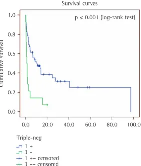
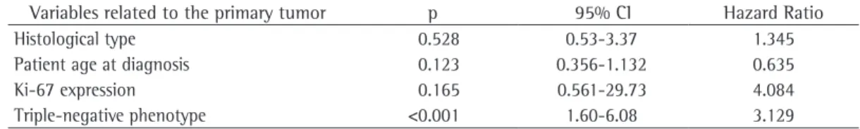
In the Cox univariate analysis, mortality was not found to be significantly associated with the identification of malignant cells in the pleural fluid, with the histological type of the primary tumor, with the age at diagnosis of the primary tumor, or with Ki-67 expression. However, the Cox univariate analysis and the Kaplan-Meier curves both revealed a statistically significant association between triple-negative breast cancer and mortality (Table 2 and Figure 1, respectively), the latter showing a strong log-rank test association (p < 0.001), with a hazard ratio of 3.12.
Of the 140 patients for whom the outcome was known, 91 (65%) died. Of the 25 patients with triple-negative breast cancer, 20 (80%) died during the follow-up period.
outcome measure. Patients were followed until June of 2011. Data were censored for patients who were lost to follow-up, as well as for those who were still alive at the end of the follow-up period.
The data were processed with Microsoft Excel 2007 and analyzed with the Statistical Package for the Social Sciences, version 18.0 (SPSS Inc., Chicago, IL, USA). Survival curves were calculated using the Kaplan-Meier method, and Cox univariate analysis was applied to identify independent factors associated with survival. The log-rank test was used in order to determine whether there were any statistical differences between the distributions of the curves. We used the Student’s t-test to compare the mean values obtained for malignant pleural effusion and Ki-67 protein expression. The level of statistical significance was set at p < 0.05.
Results
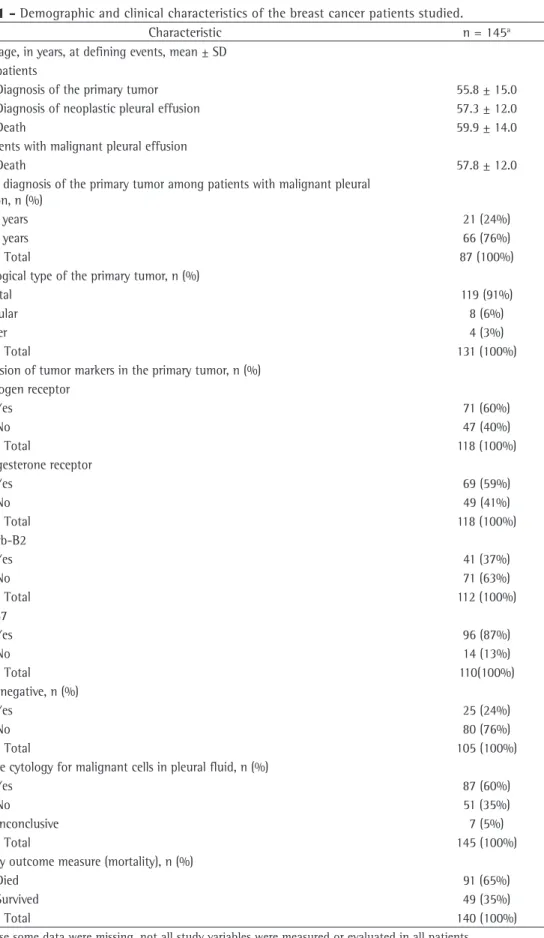
We identified and evaluated 145 female breast cancer patients who were referred to our hospital for investigation of malignant pleural effusion. Table 1 shows the main demographic and clinical characteristics of the study sample.
There were some missing data. Five patients were lost to follow-up, and their survival status data was therefore unknown. For another 6 patients, the cytological analysis was unsatisfactory, because there was hemorrhagic material in the pleural fluid. In addition, the immunohistochemical panel applied to the primary tumor comprised only 3 markers in some cases, whereas it comprised 5 markers in others, and, in some cases, there were no immunohistochemical data available. Therefore, such data were available in only 118 cases for ER and PR; 110 cases for p53 and Ki-67; 112 cases for c-erbB-2; and 105 cases for the triple-negative phenotype.
In the study sample as a whole, the age at diagnosis of the primary tumor ranged from 20 and 90 years. Of the patients with malignant pleural effusion, 21 (24%) had been diagnosed with a primary tumor before 50 years of age (Table 1).
As can be seen in Table 1, the mean age of the patients at each defining event was as follows: 55.8 ± 15.0 years at diagnosis of the primary tumor; 57.3 ± 12.0 years at identification of malignant pleural effusion; and 59.9 ± 14.0 years at death. Among the patients with malignant
Table 1 - Demographic and clinical characteristics of the breast cancer patients studied.
Characteristic n = 145a
Mean age, in years, at defining events, mean ± SD All patients
Diagnosis of the primary tumor 55.8 ± 15.0
Diagnosis of neoplastic pleural effusion 57.3 ± 12.0
Death 59.9 ± 14.0
Patients with malignant pleural effusion
Death 57.8 ± 12.0
Age at diagnosis of the primary tumor among patients with malignant pleural effusion, n (%)
≤50 years 21 (24%)
>50 years 66 (76%)
Total 87 (100%)
Histological type of the primary tumor, n (%)
Ductal 119 (91%)
Lobular 8 (6%)
Other 4 (3%)
Total 131 (100%)
Expression of tumor markers in the primary tumor, n (%) Estrogen receptor
Yes 71 (60%)
No 47 (40%)
Total 118 (100%)
Progesterone receptor
Yes 69 (59%)
No 49 (41%)
Total 118 (100%)
C-erb-B2
Yes 41 (37%)
No 71 (63%)
Total 112 (100%)
Ki-67
Yes 96 (87%)
No 14 (13%)
Total 110(100%)
Triple-negative, n (%)
Yes 25 (24%)
No 80 (76%)
Total 105 (100%)
Positive cytology for malignant cells in pleural fluid, n (%)
Yes 87 (60%)
No 51 (35%)
Inconclusive 7 (5%)
Total 145 (100%)
Primary outcome measure (mortality), n (%)
Died 91 (65%)
Survived 49 (35%)
Total 140 (100%)
Such variation might be attributed to the calendar year studied, the region of the country in which the patients were treated, the study methodology, or the population studied.
Fentiman et al.(8) found that the mean interval
between the diagnosis of the primary tumor and the presentation of pleural effusion was 41.5 months, and that the mean survival after the presentation of pleural effusion was 15.7 months. However, van Galen et al.(11) reported a longer
mean time from the diagnosis of breast cancer to the detection of pleural effusion (63.6 months) and a shorter mean survival (9.3 months), findings that are in stark contrast with those of our study (20 months and 6 months, respectively).
Studies of the spread of breast cancer have shown that a longer interval free of pleural metastases is an indicator of a worse prognosis,(8,10,11) and this poor prognosis has been
associated with factors such as the site of the metastasis and the identification of malignant cells in the pleural fluid(23); the expression of
c-erbB-2(21); the triple-negative phenotype(11,15-17);
and Ki-67 protein expression, which has been shown to be present in 63% of patients with malignant pleural effusion.(18,24,25)
The worst breast cancer survival rates are in developing countries.(26,27) Survival in malignant
pleural effusion has also been widely studied.(8,10,11)
In the present study, mean survival was shorter than that reported in studies conducted in developed countries, which might be attributable to more aggressive forms of the disease, limited access to appropriate treatment,(21) or even to delayed
diagnosis of the primary tumor (i.e., diagnosis at an advanced stage), which could account for the shorter interval free of pleural metastases and for the shorter survival.
According to the literature, the site of metastasis is an important prognostic factor in breast cancer. Visceral metastases, such as those affecting lungs, are predictive of a poor prognosis and shorter survival.(23,28) In addition, the clinical
Data regarding the expression of Ki-67, an immunohistochemical marker of cell proliferation, were available for 103 patients, 64 (63%) of whom tested positive for Ki-67 expression. The association between Ki-67 expression and the identification of malignant cells in the pleural fluid was found to be statistically significant (p = 0.0041).
The immunohistochemical panel of the primary tumor showed that there was expression of Ki-67, ER, PR, p53, and c-erbB-2 in 87%, 60%, 59%, 38%, and 37% of the patients, respectively, comparable to the 93%, 64%, 56%, 44%, and 35%, respectively, found when we considered only the patients who were diagnosed with malignant pleural effusion. The triple-negative phenotype (characterized by the lack of expression of ER, PR, and c-erbB-2) was identified in 14 (21%) of the 66 such patients for whom data on all three markers were available.
Discussion
In the present study, the prognosis was worse for patients with triple-negative breast cancer than for those with other breast cancer phenotypes. This is in agreement with data in the literature, which show that, in such patients, the tumor is more aggressive, the incidence of distant metastases, particularly in visceral organs such as the lung, is higher, and survival is lower.(5,11,15-18)
We found that there was a tendency toward a worse prognosis in patients who had been diagnosed with a primary tumor before 50 years of age, perhaps suggesting that diagnosis after the age of 50 is a protective factor. The prognosis for breast cancer is relatively good if it is diagnosed in the early stages.(19) The cumulative median
overall five-year survival is approximately 65% in developed countries, 56% in developing countries, and 61% worldwide.(20) Epidemiological studies
conducted in Brazil have provided inconsistent data on the five-year survival of breast cancer patients, which ranges from 61% to 87.7%.(21,22)
Table 2 - Cox univariate analysis of possible risk factors for mortality in the breast cancer patients studied.
Variables related to the primary tumor p 95% CI Hazard Ratio
Histological type 0.528 0.53-3.37 1.345
Patient age at diagnosis 0.123 0.356-1.132 0.635
Ki-67 expression 0.165 0.561-29.73 4.084
8. Fentiman IS, Millis R, Sexton S, Hayward JL. Pleural effusion in breast cancer: a review of 105 cases. Cancer. 1981;47(8):2087-92. http://dx.doi. org/10.1002/1097-0142(19810415)47:8<2087::AID-CNCR2820470830>3.0.CO;2-9
9. Pokieser W, Cassik P, Fischer G, Vesely M, Ulrich W, Peters-Engl C. Malignant pleural and pericardial effusion in invasive breast cancer: impact of the site of the primary tumor. Breast Cancer Res Treat. 2004;83(2):139-42. PMid:14997044. http:// dx.doi.org/10.1023/B:BREA.0000010706.24181.b6 10. Singer TS, Sulkes A, Biran S. Pleural effusion in breast
cancer: influence upon clinical course and survival. Chemioterapia. 1986;5(1):66-9. PMid:3955787. 11. van Galen KP, Visser HP, van der Ploeg T, Smorenburg
CH. Prognostic factors in patients with breast cancer and malignant pleural effusion. Breast J. 2010;16(6):675-7. PMid:21070453. http://dx.doi. org/10.1111/j.1524-4741.2010.00986.x
12. Yhim HY, Han SW, Oh DY, Han W, Im SA, Kim TY, et al. Prognostic factors for recurrent breast cancer patients with an isolated, limited number of lung metastases and implications for pulmonary metastasectomy. Cancer. 2010;116(12):2890-901. PMid:20564396. http:// dx.doi.org/10.1002/cncr.25054
13. Teixeira LR, Pinto JA, Marchi E. Derrame pleural neoplásico. J Bras Pneumol. 2006;32(Suppl 4):182-9.
14. Fidler IJ. Review: biologic heterogeneity of cancer metastases. Breast Cancer Res Treat. 1987;9(1):17-26. PMid:3297212. http://dx.doi.org/10.1007/BF01806690 15. Rakha EA, Chan S. Metastatic triple-negative breast
cancer. Clin Oncol (R Coll Radiol). 2011;23(9):587-600.
PMid:21524569. http://dx.doi.org/10.1016/j.
clon.2011.03.013
16. Tischkowitz M, Brunet JS, Bégin LR, Huntsman DG, Cheang MC, Akslen LA, et al. Use of immunohistochemical markers can refine prognosis in triple negative breast cancer. BMC Cancer. 2007;7:134. PMid:17650314 PMCid:1948892. http://dx.doi.org/10.1186/1471-2407-7-134
17. Dent R, Hanna WM, Trudeau M, Rawlinson E, Sun P, Narod SA. Pattern of metastatic spread in triple-negative breast cancer. Breast Cancer Res Treat. 2009;115(2):423-8.
PMid:18543098. http://dx.doi.org/10.1007/
s10549-008-0086-2
18. Keam B, Im SA, Lee KH, Han SW, Oh DY, Kim JH, et al. Ki-67 can be used for further classification of triple negative breast cancer into two subtypes with different response and prognosis. Breast Cancer Res. 2011;13(2):R22. PMid:21366896 PMCid:3219180. http://dx.doi.org/10.1186/ bcr2834
19. Brewster AM, Do KA, Thompson PA, Hahn KM, Sahin AA, Cao Y, et al. Relationship between epidemiologic risk factors and breast cancer recurrence. J Clin Oncol. 2007;25(28):4438-44. PMid:17785707. http:// dx.doi.org/10.1200/JCO.2007.10.6815
20. Cadaval Gonçalves AT, Costa Jobim PF, Vanacor R, Nunes LN, Martins de Albuquerque I, Bozzetti MC. Increase in breast cancer mortality in Southern Brazil from 1980 to 2002 [Article in Portuguese]. Cad Saude Publica. 2007;23(8):1785-90. PMid:17653396. 21. de Moraes AB, Zanini RR, Turchiello MS, Riboldi J, de
Medeiros LR. Survival study of breast cancer patients treated at the hospital of the Federal University in Santa Maria, Rio Grande do Sul, Brazil [Article in Portuguese].
evolution of patients with isolated pulmonary metastatic disease remains a concern.(12)
Expression of Ki-67, a tumor marker closely related to the process of cell proliferation, might represent a negative prognostic factor.(24,25) The
breast cancer subtypes in which there is high Ki-67 expression have been shown to respond better to the initial chemotherapy but have also been associated with a poor long-term prognosis.(18)
In conclusion, we found that the triple-negative breast cancer phenotype is an unfavorable characteristic in patients with malignant pleural effusion, worsening the prognosis and reducing life expectancy. In addition, Ki-67 protein expression was found to be associated with the development of malignant pleural effusion.
The burden imposed by breast cancer calls for expanding our knowledge of its prognostic factors, not only to inform decisions regarding it treatment but also to increase our understand of its evolution. Certain prognostic factors are well established and can be safely evaluated in clinical practice. However, some are still the targets of studies attempting to provide further information about the evolution of cancer and to identify new predictors of its prognosis.
References
1. Paulinelli RR, Freitas Jr R, Curado MP, Souza AA. A situação do câncer de mama em Goiás, no Brasil e no mundo: tendências atuais para a incidência e a mortalidade. Rev Bras Saude Mater Infant. 2003;3(1):17-24. http://dx.doi. org/10.1590/S1519-38292003000100004
2. Dewis R, Gribbin J. Breast Cancer: Diagnosis and treatment. An assessment of need. Cardiff: National Collaborating Centre for Cancer (UK); 2009.
3. Instituto Nacional de Câncer - INCA [homepage on the Internet]. Brasília: Ministério da Saúde. [cited 2011 Dec 1]. Estimativa 2012: incidência de câncer no Brasil. Available from: http://www.inca.gov.br/estimativa/2012 4. Instituto Nacional de Câncer - INCA [homepage on
the Internet]. Brasília: Ministério da Saúde. [cited 2011 Dec 1]. Tipos de câncer: Mama. Available from: http:// www2.inca.gov.br/wps/wcm/connect/tiposdecancer/ site/home/mama
5. Koo JS, Jung W, Jeong J. Metastatic breast cancer shows different immunohistochemical phenotype according to metastatic site. Tumori. 2010;96(3):424-32. PMid:20845803.
6. Jung SY, Rosenzweig M, Sereika SM, Linkov F, Brufsky A, Weissfeld JL. Factors associated with mortality after breast cancer metastasis. Cancer Causes Control. 2012;23(1):103-12. http://dx.doi.org/10.1007/ s10552-011-9859-8
25. de Azambuja E, Cardoso F, de Castro G Jr, Colozza M, Mano MS, Durbecq V, et al. Ki-67 as prognostic marker in early breast cancer: a meta-analysis of published studies involving 12,155 patients. Br J Cancer. 2007;96(10):1504-13. PMid:17453008 PMCid:2359936. http://dx.doi.org/10.1038/sj.bjc.6603756 26. Schneider IJ, d’Orsi E. Five-year survival and prognostic
factors in women with breast cancer in Santa Catarina State, Brazil [Article in Portuguese]. Cad Saude Publica. 2009;25(6):1285-96. http://dx.doi.org/10.1590/ S0102-311X2009000600011
27. Parkin DM, Bray F, Ferlay J, Pisani P. Global cancer statistics, 2002. CA Cancer J Clin. 2005;55(2):74-108. http://dx.doi.org/10.3322/canjclin.55.2.74
28. Cutler SJ, Ardyce JA, Taylor SG 3rd. Classification of patients with disseminated cancer of the breast. Cancer. 1969;24(5):861-9. http://dx.doi.org/10.1002/1097-0142(196911)24:5<861::AID-CNCR2820240502>3.0.CO;2-3 Cad Saude Publica. 2006;22(10):2219-28. http://dx.doi.
org/10.1590/S0102-311X2006001000028
22. Guerra MR, Mendonça GA, Teixeira MT, Cintra JR, Carvalho LM, Magalhães LM. Five-year survival and prognostic factors in a cohort of breast cancer patients treated in Juiz de Fora, Minas Gerais State, Brazil [Article in Portuguese]. Cad Saude Publica. 2009;25(11):2455-66. http://dx.doi.org/10.1590/S0102-311X2009001100015 23. Largillier R, Ferrero JM, Doyen J, Barriere J, Namer M,
Mari V, et al. Prognostic factors in 1,038 women with metastatic breast cancer. Ann Oncol. 2008;19(12):2012-9. PMid:18641006 PMCid:2733115. http://dx.doi.org/10.1093/ annonc/mdn424
24. Mohsenifar J, Almassi-Aghdam M, Mohammad-Taheri Z, Zare K, Jafari B, Atri M, et al. Prognostic values of proliferative markers ki-67 and repp86 in breast cancer. Arch Iran Med. 2007;10(1):27-31. PMid:17198450.
About the authors
Giovana Tavares dos Santos
Biologist, Graduate Program in Pathology, Federal University of Health Sciences of Porto Alegre, Porto Alegre, Brazil.
João Carlos Prolla
Cytopathologist, Laboratory of Pathology, Santa Casa Sisters of Mercy Hospital, Porto Alegre, Brazil.
Natália Dressler Camillo
Medical Student, Federal University of Health Sciences of Porto Alegre, Porto Alegre, Brazil.
Lisiane Silveira Zavalhia
Medical Biologist, Graduate Program in Pathology, Federal University of Health Sciences of Porto Alegre, Porto Alegre, Brazil.
Alana Durayski Ranzi
Medical Biologist, Graduate Program in Pathology, Federal University of Health Sciences of Porto Alegre, Porto Alegre, Brazil.
Claudia Giuliano Bica