Toxicity of a cyanobacterial extract
containing microcystins to mouse lungs
Laboratórios de 1Fisiologia da Respiração, 2Ecofisiologia e Toxicologia da Cianobacteria, and
3Investigação Pulmonar, Instituto de Biofísica Carlos Chagas Filho,
Universidade Federal do Rio de Janeiro, Rio de Janeiro, RJ, Brasil M.R. Picanço1, R.M. Soares2,
V.R. Cagido1,
S.M.F.O. Azevedo2,
P.R.M. Rocco3 and W.A. Zin1
Abstract
Toxic cyanobacteria in drinking water supplies can cause serious public health problems. In the present study we analyzed the time course of changes in lung histology in young and adult male Swiss mice injected intraperitoneally (ip) with a cyanobacterial extract con-taining the hepatotoxic microcystins. Microcystins are cyclical hep-tapeptides quantified by ELISA method. Ninety mice were divided into two groups. Group C received an injection of saline (300 µl, ip) and group Ci received a sublethal dose of microcystins (48.2 µg/kg,
ip). Mice of the Ci group were further divided into young (4 weeks old) and adult (12 weeks old) animals. At 2 and 8 h and at 1, 2, 3, and 4 days after the injection of the toxic cyanobacterial extract, the mice were anesthetized and the trachea was occluded at end-expiration. The lungs were removed en bloc, fixed, sectioned, and stained with hematoxylin-eosin. The percentage of the area of alveolar collapse and the number of polymorphonuclear (PMN) and mononuclear cell infil-trations were determined by point counting. Alveolar collapse in-creased from C to all Ci groups (123 to 262%) independently of time, reaching a maximum value earlier in young than in adult animals. The amount of PMN cells increased with time of the lesion (52 to 161%). The inflammatory response also reached the highest level earlier in young than in adult mice. After 2 days, PMN levels remained un-changed in adult mice, while in young mice the maximum number was observed at day 1 and was similar at days 2, 3, and 4. We conclude that the toxins and/or other cyanobacterial compounds probably exert these effects by reaching the lung through the blood stream after ip
injection.
Correspondence
W.A. Zin
Laboratório de Fisiologia da Respiração
Instituto de Biofísica, UFRJ 21949-900 Rio de Janeiro, RJ Brasil
Fax: +55-21-2564-1578 E-mail: [email protected] Research supported by PRONEX-MCT, CNPq, FINEP, and FAPERJ.
Received November 10, 2003 Accepted May 6, 2004
Key words
•Microcystins
•Lung
•Inflammation •Alveolar collapse
Toxic cyanobacteria in drinking water supplies have raised public health concerns in many countries due to the frequent occur-rence of cyanotoxins produced by these mi-croorganisms. Microcystins are the cyano-toxins most commonly found in these blooms (1). These toxins are cyclic heptapeptides
dam-age to the tissue, which may lead to death or, in a chronic intoxication, may promote he-patic tumors (3). Microcystins in the liver can also induce hepatocytes to produce ara-chidonic acid metabolites such as prostacy-clin (6-Keto F1α) and thromboxane (TXB2)
by stimulation of the cyclooxygenase path-way (4). This observation suggests that mi-crocystins can promote inflammation, which may contribute to the hepatic shock that leads to death.
There are some reports from different regions of the world of human intoxication due to toxic cyanobacteria ingestion (5,6). In Brazil, more than 100 chronic renal patients were intoxicated in the town of Caruaru in 1996 and 52 died due to intravenous expo-sure to water containing microcystins during dialysis treatment (7-9).
In addition to the oral and intravenous routes of intoxication, human beings can be exposed to cyanotoxins through inhalation. This may occur in recreational water con-taining cyanobacteria or their toxins. Fitz-george et al. (10) observed extensive necro-sis of the epithelium of both olfactory and respiratory zones in mice that received a purified microcystin-LR (MCYST-LR) by the intranasal route. Through this route, the LD50 of this toxin was the same as that
observed after administration by the intra-peritoneal (ip) route. These investigators sug-gested that the phenomenon results from the necrosis of the nasal epithelium, which fa-cilitates the absorption of the toxin into the blood stream through the nasal capillaries.
However, there are few reports analyzing the effects of microcystins on the lungs. Turner et al. (11) reported a case of two recruits in England who developed severe pneumonia after contact with water contain-ing toxic Microcystis aeruginosa, and Slatkin et al. (12) detected pulmonary thrombosis in mice injected with lethal doses of microcys-tins. It has been already demonstrated that this toxin can reach the lung, and its distribu-tion after oral and intratracheal
administra-tions has been well described by Ito et al. (13,14).
The aim of the present study was to ana-lyze the time course of changes in lung his-tology in young and adult mice injected ip
with a cyanobacterial extract containing mi-crocystins.
A toxic M. aeruginosa strain (NPJB-1) was cultured in ASM-1 medium as described by Ferrão-Filho and Azevedo (15). The cul-ture was not axenic, but the concentration of bacteria was considered to be very low. At the exponential growth phase (between 15 and 20 days) the cells were harvested, con-centrated with a Pellicon Cassette System (Millipore, Billerica, MA, USA) that uses a tangential flow multiple filter technique, re-suspended in deionized water, and disrupted by cycles of freeze-thawing. Particulate or-ganic matter was removed from the solution by filtration through fiberglass filters and the extract containing dissolved microcystins was used in the experiments. This strain has al-ready been described as a producer of two types of microcystins: MCYST-LR and MCYST-LF (16). For this study, the total microcystins present in the strain extract were quantified by ELISA using the Envirol-ogix Inc. (Portland, ME, USA) commercial plate kits, according to the manufacturer’s protocol.
Ninety male Swiss mice were divided into two groups. The control group (C, N = 30 adult animals) received an ip injection of 0.9% NaCl (300 µl) and the test group (Ci) received an ip injection of a sublethal dose of cyanobacterial extract containing micro-cystins (48.2 µg/kg body weight). The ani-mals of the test group were equally divided into two groups according to age, i.e., young (4 weeks old) and adult (12 weeks old). At 2 and 8 h and at 1, 2, 3, and 4 days after the ip
lungs were removed en bloc.
The right lung was quick-frozen by im-mersion in liquid nitrogen and fixed with Carnoy’s solution (ethanol:chloroform:acetic acid, 70:20:10) at -70ºC for 24 h (17). The Carnoy’s solution was replaced with pro-gressively increasing ethanol concentrations at -20ºC up to 100% ethanol. The tissue was maintained at -20ºC for 4 h, warmed to 4ºC for 12 h, and then allowed to reach and remain at room temperature for 2 h. After fixation, the tissue was embedded in paraf-fin, blocks were cut into 4-µm thick sections with a microtome and the slices were stained with hematoxylin-eosin. Each slide received a code and two investigators who were un-aware of the origin of the material performed microscopic examination.
Morphometric analysis was performed using an integrating eyepiece and a 100-point grid consisting of 50 lines of known length coupled to a conventional light mi-croscope (Axioplan, Zeiss, Oberkochen, Germany) (18). The fraction of the area of collapsed alveoli was determined by the point-counting technique at 200X magnifi-cation across 10 random non-coincident mi-croscopic fields. Points falling on collapsed alveolar spaces were counted and divided by the total number of points in each micro-scopic field. Polymorphonuclear (PMN) and mononuclear cells and pulmonary tissue were evaluated at 1000X magnification. Points falling on the tissue area were counted and divided by the total number of points in each microscopic field. Thus, data were reported as the fractional area of pulmonary tissue. The same method was applied to determine the amount of PMN cells. All morphometric parameters determined for all groups were compared by one-way ANOVA and the level of significance was set at 5% in all analyses. Statistical analyses were performed using the SigmaStat software (Jandel Scientific, San Rafael, CA, USA).
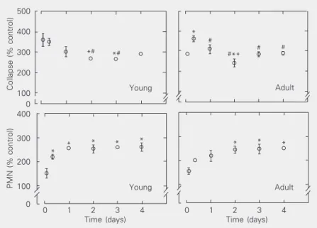
The percentages of alveolar collapse and PMN cells are illustrated in Figure 1. The
fraction of alveolar collapse was higher in all test groups compared to control, independ-ently of the time of analysis, reaching a maximum value earlier in young than in adult animals. The amount of PMN cells increased with the temporal evolution of the lesion. It is interesting to observe that in young mice the inflammatory response also reached its highest level earlier than in adult animals. After two days, PMN levels re-mained unchanged in adult mice, while in young mice the maximum level was ob-served at day 1 and was similar at days 2, 3 and 4 (Figure 2). Additionally, young ani-mals responded with an earlier increase of PMN, followed by collapse whereas adult mice exhibited an inverse profile. These dif-ferences could probably be attributed to dis-crepancies in inflammatory mechanisms, i.e., in young mice, together with PMN infiltra-tion, type II pneumocytes may also be al-tered very early in the course of lung injury, leading to surfactant dysfunction and
alveo-Collapse (% control)
500
400
300
200
100
400
300
200
100
0 0
Young Adult
*# *#
* #
#**
# #
* *
* * * * * *
0 1 2 3 4
Time (days)
0 1 2 3 4
Time (days)
Figure 1. Effect of a cyanobacterial extract on the fractional area of alveolar collapse and the amount of polymorphonuclear (PMN) cells compared to the control group. The left panels provide the data for the young mice who received a sublethal dose of cyanobacte-rial extract containing microcystins (48.2 µg/kg, ip). The right panels contain the data for adult mice who received the same dose. Data are reported as means ± SEM for 5 lungs in each group (10 random non-coincident microscopic fields were analyzed in each lung). *P < 0.05 compared to the 2-h period; #P < 0.05 compared to the 8-h period; **P < 0.05 compared to the 24-h period (one-way ANOVA).
lar collapse, while in adult mice alveolar collapse peaked at 8 h.
Therefore, we observed that the M. aeru-ginosa extract containing microcystins gen-erated a rapid inflammatory process in mouse lungs, with interstitial edema and
recruit-ment of inflammatory cells that remained stable until day 4, starting earlier in young animals. There was 100% survival until the end of the experiment.
Ito et al. (14) administered a similar sub-lethal dose of MCYST-LR intratracheally to mice and, using an immunostaining method, observed the presence of this toxin in the lungs until 7 h after injection. After this time, the tissue no longer showed any staining. They also detected a few stained macro-phages in the lung and no lesion until the end of the experiment, 2 weeks later. Our results showed that cyanobacterial extracts contain-ing microcystins lead to a rapid inflamma-tory response in the lung, which continued until the fourth and last day of the experi-ment. In addition, we observed alveolar col-lapse that also started rapidly and reached a maximum level earlier in young animals (Fig-ure 1).
According to Naseem et al. (19), MCYST-LR can stimulate alveolar macrophages to produce inflammatory mediators. Nakano et al. (20) observed that TNF-α production by cultured macrophages from mouse perito-neal exudates is stimulated by purified mi-crocystins and toxic and non-toxic extracts of M. aeruginosa (a non-lipopolysaccharide producer). They also observed that the toxic extract was more effective than purified mi-crocystins. Therefore, it is important to con-sider that other cyanobacterial secondary metabolites present in the extract may in-duce inflammatory responses in lung or act synergistically with microcystins to promote inflammation.
We cannot rule out the presence of bacte-ria in the extract, which might contribute to those inflammatory effects. However, bacte-rial contamination of the M. aeruginosa cul-ture was found to be very low and therefore negligible in the final volume of M. aerugi-nosa after the concentration process.
In the present study, these toxins and/or other cyanobacterial compounds may have caused the effects described above by reach-Figure 2. Photomicrographs of lung parenchyma stained with hematoxylin-eosin. A, Control
ing the lung through the blood stream after ip
injection. Therefore, intravenous exposure to microcystins can represent a risk to the lungs in addition to the known targets, i.e., liver and kidney. Thus, whenever human health depends on the quality of water for
direct consumption and recreational or medi-cal use, such as dialysis treatment, the in-crease of cyanobacterial blooms producing microcystins in the water supplies ought to be carefully considered.
References
1. Carmichael WW (1994). The toxins of cyanobacteria. Scientific American, 270: 78-86.
2. Chorus I & Bartram J (1999). Toxic Cyanobacteria in Water - A Guide to Their Public Health Consequences, Monitoring and Manage-ment. E & FN Spon, London, UK.
3. Falconer I & Humpage A (1996). Tumour promotion by cyanobacte-rial toxins. Phycologia, 35: 74-79.
4. Naseem SM, Hines HB & Creasia DA (1990). Inhibition of microcys-tin-induced release of cyclooxygenase products from rat hepato-cytes by anti-inflammatory steroids. Proceedings of the Society for Experimental Biology and Medicine, 195: 345-349.
5. Falconer IR, Beresford AM & Runnegar MTC (1983). Evidence of liver damage by toxin from a bloom of the blue-green algae, Micro-cystis aeruginosa. Medical Journal of Australia, 1: 511-514. 6. Teixeira MdaG, Costa MdaC, Carvalho VLP, Pereira MS & Hage E
(1993). Gastroenteritis epidemic in the area of the Itaparica dam, Bahia, Brazil. Bulletin of the Pan American Health Organization, 27: 244-253.
7. Jochimsen EM, Carmichael WW, An J et al. (1998). Liver failure and death after exposure to microcystins at a haemodialysis center in Brazil. New England Journal of Medicine, 33: 873-878.
8. Carmichael WW, Azevedo SMFO, An J, Molica RJR, Jochimsen EM, Lau S, Rinehart KL, Shaw GR & Eaglesham GK (2001). Human fatalities from cyanobacteria: chemical and biological evidence for cyanotoxins. Environmental Health Perspectives,109: 663-668. 9. Azevedo SMFO, Carmichael WW, Jochimsen EM, Rinehart KL, Lau
S, Shaw GR & Eaglesham GK (2002). Human intoxication by micro-cystins during renal dialysis treatment in Caruaru - Brazil. Toxicol-ogy, 181: 441-446.
10. Fitzgeorge RB, Clark SA & Kelvin CW (1994). Routes of intoxication. In: Codd GA, Jeffries TM, Kelvin CW & Potter E (Editors), Detection Methods for Cyanobacterial (Blue-Green Algae) Toxins. The Royal Society of Chemistry, Cambridge, UK.
11. Turner PC, Gammie AJ, Hollinrake K & Codd GA (1990). Pneumonia associated with cyanobacteria. British Medical Journal, 300: 1400-1441.
12. Slatkin DN, Stoner RD, Adams WH, Kycia JH & Siegelman HW (1983). Atypical pulmonary thrombosis caused by a toxic cyanobac-terial peptide. Science,220: 1383-1385.
13. Ito E, Kondo F & Harada K-I (2000). First report on the distribution of orally administered microcystin-LR in mouse tissue using an immu-nostaining method. Toxicon,38: 37-48.
14. Ito E, Kondo F & Harada K-I (2001). Intratracheal administration of microcystin-LR, and its distribution. Toxicon,39: 265-271. 15. Ferrão-Filho AS & Azevedo SMFO (2002). Effects of unicellular and
colonial forms of toxic Microcystis aeruginosa from laboratory cul-tures and natural populations on tropical cladocerans. Aquatic Ecol-ogy, 37: 23-35.
16. Azevedo SMFO, Evans WR, Carmichael WW & Namikoshi M (1994). First report of microcystins from a Brazilian isolate of the cyanobac-terium Microcystis aeruginosa. Journal of Applied Phycology, 6: 261-265.
17. Nagase T, Lei M, Robatto FM, Eidelman DH & Ludwig MS (1992). Tissue viscance during induced constriction in rabbit lung: morpho-logical-physiological correlation. Journal of Applied Physiology, 73: 1900-1907.
18. Weibel ER (1990). Morphometry: stereological theory and practical methods. In: Gil J (Editor), Models of Lung Disease - Microscopy and Structural Methods. Marcel Dekker, New York.
19. Naseem SM, Hines HB & Creasia DA (1989). Effect of toxins on arachidonic acid metabolism in rat cultured pulmonary alveolar mac-rophages. Biochemistry International, 19: 583-592.