Acute oral toxicity of
Celtis iguanaea
(Jacq.) Sargent leaf extract (Ulmaceae) in rats
and mice
GONÇALVES, N.Z.1; LINO JÚNIOR, R.S.1; RODRIGUES, C.R.1; RODRIGUES, A.R.1; CUNHA, L.C.1
1Universidade Federal de Goiás. Núcleo de Estudos e Pesquisas Tóxico-Farmacológicas, Faculdade de Farmácia.
Av.Universitária Q 62/2°/sala 36, Setor Universitário. Goiânia GO. 74605-010. *Autor para correspondência: [email protected]
Recebido para publicação em 06/10/2014 Aceito para publicação em 22/07/2015
ABSTRACT: Celtis iguanaea (Jacq.) Sargent is popularly used to treat urinary infections, kidneys, breast, body aches, rheumatism, asthma, cramps, poor digestion and as a diuretic medicine. This study aims to determine the acute toxicity of the aqueous leaf extract of Celtis iguanaea (Jacq.) Sargent in rodents. After the collection processes, identification, drying and grinding, the lyophilized powder of the leaves produced, by infusion, the aqueous extract and it was dissolved in saline 0.9%. The administration was made by gavage at a dose of 2000 mg kg-1 to rats and mice of both genders. The oral toxicity was determined according to the OECD
423 guide. Signs of toxicity were observed for 15 days and classified from 0 to 4 respectively as missing, rare, mild, moderate and severe. The weight of the animals and the physiological parameters such as food intake and excrements production were observed. All animal tissue samples were collected for histological analysis. The extract was included in Type 5 (substance with LD50 higher than 2000 mg kg-1 and less than 5000 mg kg-1), being considered of low toxicity,
but the histopathologycal findings suggested nephrotoxicity and cardiotoxicity. The absolute weight of the kidneys and the heart of the male rats and mice increased, but there was no significant raise in the relative weight of the animals’ organs.
Keywords: medicinal plants, Celtis iguanaea, acute oral toxicity.
RESUMO: Toxicidade oral aguda do extrato de folhas de Celtis iguanaea (Jacq.) Sargent em ratos e camundongos. Celtis iguanaea (Jacq.) Sargent é uma planta usada popularmente para tratar infecções do trato urinário, rim, mama, dores no corpo, reumatismo, asma, cólicas, má digestão e também é usada como diurético. Este trabalho objetivou determinar a toxicidade aguda do extrato aquoso de folhas de Celtis iguanaea (Jacq.) Sargent em roedores. Após os processos de coleta, identificação, secagem e moagem, o pó liofilizado das folhas da planta foi utilizado para produzir o seu extrato aquoso por infusão e então dissolvido em solução salina a 0.9 %. A administração foi feita por gavagem na dose de 2000 mg kg-1 em ratos e camundongos
de ambos os sexos. A toxicidade oral foi determinada de acordo com o guia 423 da OECD. Sinais de toxicidade foram observados por 15 dias e tabulados de 0 a 4, respectivamente, como ausentes, raros, leves, moderados e graves. Foi acompanhado o peso dos animais e parâmetros fisiológicos tais como alimentação e excreções. Amostras do tecido de todo o animal foram coletadas para análise histológica. A toxicidade encontrada para o extrato foi incluída na classe 5 (substâncias com DL50 superior a 2000 mg kg-1 e menor que 5000 mg kg-1) sendo considerada
baixa, porém, as observações histopatológicas sugerem nefrotoxicidade e cardiotoxicidade. O peso absoluto dos rins e coração de ratos e camundongos machos aumentou, porém, não houve aumento significativo no peso relativo dos órgãos dos animais.
Palavras-chave: plantas medicinais, Celtis iguanaea, toxicidade oral aguda.
10.1590/1983-084X/14_128 INTRODUCTION
Popular tradition in the state of Goiás indicates the use of the species Celtis iguanaea as tea for the treatment of various complaints such as body aches, rheumatism, chest pain, asthma,
in the treatment of urinary tract infections. Ortêncio (1994) found an indication of Celtis iguanaea for the treatment of renal calculi and pyelonephritis in the state of Goiás. Paula (2009) and Martins et al. (2011) demonstrated antiulcer and gastric acid antisecretory effects of aqueous extract and consequently Sousa et al. (2013) found an antiulcerogenic activity of crude ethanolic extract of Celtis iguanaea leaves.
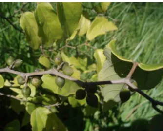
Celtis iguanaea is a shrubby species commonly known as ‘esporão-de-galo’, ‘tela’, ‘taleira’, ‘sarã’, ‘gurrupiá’ or ‘gumbixava’, depending on the region of Brazil where it is found (Giehl & Jarenkow, 2008). This plant belongs to the family Ulmaceae and it is a dicotyledonous angiosperm classified in the sub-division Magnoliophyta inside the Rosales order (Corrêa, 1978). Ulmaceae is a family of trees and shrubs comprising 16 genera and approximately 2000 species, occurring both in temperate regions and in those with tropical climates (Heywood, 1993). Celtis iguanaea leaves are shown in Figure 1.
According to Paula et al. (2010) the leaves of Celtis iguanaea (Jacq.) Sargent have mucilage, coumarin and flavonoids. In their study the aqueous extract of the plant given orally was able to protect mice against gastric mucosal lesions induced by indomethacin, ethanol and stress, and reduce the volume and acidity of gastric acid secretion in mice and significantly alter intestinal motility by increasing the traffic.
Considering the popular use of Celtis iguanaea (Jacq.) Sargent and the absence of toxicological studies on this matter, it was proposed to determine the experimental acute toxicity of its aqueous extract.
MATERIALS AND METHODS Bothanical material
Celtisiguanaea leaves were collected from an adult plant located in riparian vegetation in the city of Campestre - Goiás, Brazil (612 m, 16 º 46 ‘01.7 “S, 49 ° 42’ 00.6” W) in February 2010. The botanical material was identified by Prof. Dr. José Realino de Paula, Faculty of Pharmacy, Federal University of Goiás (UFG) and a voucher specimen was deposited at UFG’s Herbarium under number 40110.
Preparation of the aqueous extract of Celtis iguanaea (AECI)
Leaves of the Celtis iguanaea were air-dried in an oven at 40.0 ± 2 ºC and then the dried plant was cut and sprayed. From 120 g dried powdered plant material, the aqueous extracts (infusion) were prepared at 80 ºC for 30 min. The solvent was then eliminated by a rotary vacuum evaporator at 60 ºC under 650 mmHg pressure and lyophilized for 20 h,
representing a yield of 21.86% of the dry material extracted. The aqueous extracts obtained was dissolved in 0.9% saline just before administration, and administered at 2000 mg kg-1 (Farmacopeia
Brasileira, 2010).
Animals
Male and female (nulliparous and non-pregnant) Wistar rats (200–250 g) and male and female (nulliparous and non-pregnant) Swiss mice (25-30 g) supplied by the Center for Animal Resources (UFG), were housed in a temperature and light-controlled room (24.0 ± 2 ºC; 12 h light/dark cycle) and acclimatized in the laboratory for a period of at least 10 days before any experimental studies, with free access to water and food (Purina Labina®).
All procedures were approved by the Institutional Ethics Committee under protocol number 232/10.
Acute Oral Toxicity Testing
The acute oral toxicity test was performed using methods described in guide 423 of the Organization for Economic Cooperation and Development (OECD, 2001). Table 1 details the animals used in the experiment:
It was orally administered 2000 mg kg-1 of
AECI dissolved in saline solution 0.9 % by gavage Figure 1. Celtis iguanaea (Jacq.) leaves. Camprestre, Goiás, February 2007.
TABLE 1. Number of animals used in determination of acute oral toxicity of Celtis iguanaea (Jacq.) Sargent leaf extract (Ulmaceae) in rats and mice.
Control Group Test Group Total
Rats 6 male 6 male 24
6 female 6 female
Mice 3 male 3 male 12
to the test group of rats and to the test group of mice. Both control groups (rats and mices) were treated only with saline solution 0.9%. Behavioral observations were conducted systematically at times 15 min, 30 min, 1h, 2h, 4h, and 8h after administration and thereafter daily, until the fourteenth day, through the Hippocratic screening for all groups (test and control groups of mice and rats). Signs of toxicity, onset time, intensity, duration and progression of these were recorded, tabulating them on a scale of 0 to 4 (absent, rare, mild, moderate, severe) for further analysis (Malone & Robichaud, 1962, Malone, 1977). The body weight of each animal was given on the first day of experiment and weekly thereafter, and the body weight gain was calculated using the mean difference between weight per week. Physiological parameters (food intake and excreta production) were determined weekly. At the end of the observation period all surviving animals were euthanized and autopsied (xylazin-ketamin (2:8) 0.2 mL 100g-1) (Kohn, 1997).
T h e p r o c e d u r e o f p r e p a r a t i o n a n d histopathological evaluation was performed at the Instituto de Patologia Tropical e Saúde Pública - UFG (IPTSP-UFG) in Patologia Geral Sector, using double-blind method. Tissue samples were collected from the whole animal, specially the organs liver, lung, kidney, heart, spleen, pancreas, colon and small intestine. Tissues were then fixed for at least for 24 h in formol buffer (100 mL of 37% formaldehyde: 900 mL of distilled water, 4.0 g sodium phosphate monobasic, 6.5 g of 10% sodium phosphate dibasic anhydrous). The proportion was 20 times the volume of fixative solution to volume ratio of the parts. After fixation, the fragments were washed in running water for five minutes, thus starting up the process of dehydration in ethanol, in ascending series from 70% to absolute alcohol. After that, it was performed the clarifying process with xylene, and wax infiltration with paraffin for the block confection. The 5 mm sections were placed in glass slides and stained with hematoxylin & eosin (HE) (Junqueira & Junqueira, 1983).
The analyses were performed by a researcher who did not know which were the treated groups. The general pathologic processes analyzed within the tissues were: degeneration, necrosis, interstitial modifications, such as fibrosis and deposits, inflammation and hyperemia. These processes were described and classified in a semi-quantitative form as follows: absent or score 0, discrete with up to 25% of area commitment or score 1, moderate with 26 to 50% of area commitment or score 2 and accentuated with more than 50% of area commitment or score 3. The statistical analysis was performed with non-parametric tests (Kruskal-Wallis) through the Sigma Stat®
software. All general pathologic processes were registered in photomicrographs performed with a photomicroscope attached to a digital camera. Another results were submitted to statistical tests, especially Student’s t Test.
RESULTS AND DISCUSSION
Regarding to the mice, there was one death of a female mouse on the fifth day of the experiment. That animal presented moderate general activity, tremors, ataxia and intense response for clamping tail within 24 h. The experiment was repeated with 3 other mice and no deaths were observed. Other mice and rats showed no abnormal behavioral changes during the study and no macroscopic alterations were found in the organs during the necropsy.
According to guide 423 (OECD, 2001) in an acute toxicity test in which no death occurred in more than one of the six animals given a dose of 2000 mg kg-1, the LD
50 value can be considered greater than
2000 mg kg-1 and less than 5000 mg kg-1. Trevisan et
al. (2012) tested bark extract of this plant for toxicity against Artemia salina living organisms and found no toxic effect as well.
Guide 423 (OECD, 2001) states that only one species is used and usually the female, on the other hand the ‘Guide for the conduct of studies Preclinical Toxicity of Herbal Medicines’ recommends that both genders must be tested with a number of six animals per gender and only one animal species (Brasil, 2004). The use of two animal species is justified by the possible difference in results between species, although both are rodents. The use of only three animals per gender allowed the estimation of LD50 from guide 423, and the use of alternative
methods to estimate the LD50 of herbal medicines in
Brazil is not restricted.
Male rats treated with AECI (2000 mg kg-1)
had absolute kidney and heart weights increased in relation to the control group (treated group -3.12 ± 0.15 g and 1.19 ± 0.072 g; control group- 2.50 ± 0.07 g and 0.97 ± 0.02 g, respectively) (Table 2).
Male mice treated with AECI (2000 mg kg -1, given orally) also had kidney and heart absolute
weights increased in the control group (saline 1 ml kg-1), with values of 0.64 ± 0.05 g and 0.29 ± 0.009
TABLE 2. Absolute organ weights and weight gain (mean ± SEM) of Wistar rats (n=6) and Swiss mice (n = 12) treated with Celtis iguanaea extract.
Absolute weights of organs of rats and body gain
GROUP Liver
(g)
Kidneys
(g)
Heart
(g)
Spleen
(g)
Body weight
gain (X ± SEM)
(g)
I 12.73 ± 1.16 2.50 ± 0.07 0.97 ± 0.2 0.67 ± 0.03 13.17 ± 0.88
II 13.57±1.250 3.12 ± 0.15 (a)
1.19 ± 0.07 (b)
0.87 ± 0.08 21.50 ± 3.75
III 9.33 ± 1.05 1.90 ± 0.06 0.80 ± 0.02 0.58 ± 0.07 1.83 ± 1.45 IV 8.04 ± 0.29 1.95 ± 0.04 0.83 ± 0.03 0.64 ± 0.05 8.00 ± 0.76 (c)
Absolute weights of the organs of mice and body gain
GROUP Liver
(g)
Kidneys
(g)
Heart
(g)
Spleen
(g)
Body weight
gain (X ± SEM)
(g)
I 1.92 ± 0.11 0.48 ± 0.03 0.2 ± 0.02 0.08 ± 0.01 -1.17 ± 2.40
II 1.87 ± 0.15 0.64 ± 0.05 (d)
0.29 ± 0.009 (e)
0.10 ± 0.01 5.17 ± 2.95
III 1.60 ± 0.04 0.32 ± 0.11 0.25 ± 0.09 0.15 ± 0.02 4.17 ± 1.96
IV 1.44 ± 0.14 0.37 ± 0.15 0.18 ± 0 0.11 ± 0.06 1.50 ± 0.50
Caption: Group I (male animals treated with saline, 1 mL kg-1 - control); Group II (male treated with AECI 2000 mg kg-1); Group III (female,
treated with saline, 1 ml kg-1 - control) and Group IV (female, treated with AECI 2000 mg kg-1). The words a, b, c, d and e represents statistical
significance related to Group I, as follows: (a) p = 0.0053, (b) p = 0.0082, (c) p = 0.0198, (d) p = 0.049 and (e) 0.0179 (Student’s t Test).
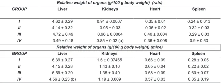
TABLE 3. Organs’ weights in relation to body (mean ± SEM) of Wistar rats (n=6) and Swiss mice (n = 12) treated with Celtis iguanaea extract.
Relative weight of organs (g/100 g body weight) (rats)
GROUP Liver Kidneys Heart Spleen
I 4.62 ± 0.29 0.91 ± 0.0007 0.35 ± 0.01 0.24 ± 0.013
II 4.14 ± 0.32 0.95 ± 0.03 0.36 ± 0.02 0.32 ± 0.03
III 4.72 ± 0.49 0.96 ± 0.0004 0.40 ± 0.004 0.29 ± 0.03 IV 3.49 ± 0.18 0.85 ± 0.02 (a) 0.36 ± 0.008 0.9 ± 0.60
Relative weight of organs (g/100 g body weight)(mice)
GROUP Liver Kidneys Heart Spleen
I 6.39 ± 0.27 1.6 ± 0.07465 0.66 ± 0.09 0.28 ± 0.05
II 4.15 ± 0.28 1.43 ± 0.10 0.65 ± 0.04 0.22 ± 0.02
III 6.59 ± 0.29 1.35 ± 0.49 0.58 ± 0.09 0.60 ± 0.07
IV 4.56 ± 0.23 (b) 1.19 ± 0.009 0.57 ± 0.03 0.35 ± 0.19
Caption: Group I (male animals treated with saline, 1 mL kg-1 - control); Group II (male treated with AECI 2000 mg kg-1); Group III (female,
treated with saline, 1 ml kg-1 - control) and Group IV (female, treated with AECI 2000 mg kg-1). The words a and b represents statistical
significance related to Group I, as follows: (a) p = 0.0059 and (b) p = 0.0160. (Student’s t Test).
and Table 3). Table 3 shows organs weights relative to body of Wistar rats and Swiss mice.
The monitoring of physiological parameters on the last day of each week of the experiment suggested no change in the animals’ behavior (Table 4). The low values found for the group of female rats and male mice treated with the negative control
(0.9% saline) can be explained by issues of handling the animals, which may have caused them stress, changing the physiological parameters.
intestine. However, animals in the group treated with 2000 mg kg-1 of AECI had pathological changes in
the kidney of male rats after microscopic analysis, with hyperemia, inflammatory infiltration and hyaline casts in the glomerulus, with special attention to this casts, although this does not present a statistically significant difference according to the rating scale used (p = 0.067, Student’s t Test) (Figure 2 and Table 5).
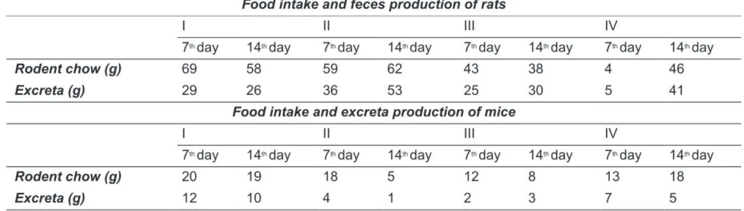
The glomerulus is the initial site of exposure to chemicals in the nephron, and various nephrotoxic substances produce lesions on this location. In some cases, the chemical change glomerular permeability to proteins by altering the size and charge-selective functions. Circulating immune complexes can be trapped within the glomerulus which may result in complement activation, attraction of neutrophils and phagocytosis. Neutrophils and macrophages are TABLE 4. Food consumption and excreta production, measured on the 7th and 14th days of confinement (mean
± SEM) of Wistar rats (n=6) and Swiss mice (n = 12) treated with Celtis iguanaea extract.
Food intake and feces production of rats
I II III IV
7th day 14th day 7th day 14th day 7th day 14th day 7th day 14th day
Rodent chow (g) 69 58 59 62 43 38 4 46
Excreta (g) 29 26 36 53 25 30 5 41
Food intake and excreta production of mice
I II III IV
7th day 14th day 7th day 14th day 7th day 14th day 7th day 14th day
Rodent chow (g) 20 19 18 5 12 8 13 18
Excreta (g) 12 10 4 1 2 3 7 5
Caption: Group I (male treated with AECI 2000 mg kg-1); Group II (male treated with saline, 1 ml kg-1 - control); Group III (female, treated
with saline, 1 ml kg-1 - control) and Group IV (female, treated with AECI 2000 mg kg-1).
FIGURE 2. Kidney photomicrograph of male Wistar rats subjected to treatment with AECI 2000 mg kg-1 and
saline 1 ml kg-1. In (A) Control rat’s Kidney with normal appearance (H & E, scale = 100 μm) (B) previous figure
commonly seen in the glomerulus in membranous glomerulonephritis, and the local release of cytokines and reactive oxygen species may contribute to glomerular injury. Antibody reactions with cell surface antigens may lead to formation of immune deposits in the glomerulus and subsequently the tissue glomerular injury. Glomerulonephritis is found in numerous cylinders, kidney cells and some leukocytes, and frank hematuria and albuminuria (Lima et al., 2001, Klaassen, 2013).
T h e p r e s e n t s t u d y p e r f o r m e d t h e histopathological investigation and organs did not show macroscopic alterations in order to increase the possibility of detecting any toxicity.
CONCLUSIONS
The aqueous extract of Celtis iguanaea was classified in class 5 (substance with LD50 higher than
2000 mg kg-1 and less than 5000 mg kg-1), being
considered of low toxicity and presented discrete signals of nephrotoxicity and cardiotoxicity in males rats and mice at a dose of 2000 mg kg-1, demanding,
thus, specific and more detailed studies.
ACKNOWLEDGMENTS
The authors are grateful to CNPq (Conselho Nacional de Ciência e Tecnologia, Brasil), FAPEG (Fundação de Amparo à Pesquisa do Estado de Goiás, Brasil), FUNAPE (Fundação de Apoio à Pesquisa da Universidade Federal de Goiás, Brasil) for their financial support and to the Center for Animal Resources of Universidade Federal de Goiás.
REFERENCES
BRASIL. Ministério da Saúde. Agência Nacional de Vigilância Sanitária. Resolução nº 90 de 16 de Março
de 2004. Determina a publicação de padrões para
estudos toxicológicos de produtos de origem vegetal. DOU. Poder Executivo, Brasília, DF, 2004.
CORRÊA, MP. Dicionário das plantas úteis do Brasil
e das exóticas cultivadas. Volume 6. Rio de Janeiro:
Ministério da Agricultura, Instituto Brasileiro de Desenvolvimento Florestal, 1978. 646p.
KOHN, DF; WIXSON S; WHITE W; BENSON G.
Anesthesia and analgesia in laboratory animals. 1.
ed. New York: Academic Press. 1997. 426p.
ANVISA. AGENCIA NACIONAL DE VIGILÂNCIA SANITÁRIA. Farmacopeia Brasileira, volume 1. 5ª Ed. Brasilia, 2010.
GIEHL, ELH; JARENKOW, JA. Structural gradient of the tree component and relationship with flooding in a riverine forest, Rio Uruguai, southern Brazil. Brasilica
Botanica Acta, v.22, n.03, 2008.
HEYWOOD, VH. Flowering Plants of the World. 1. ed. Updated edition. London: Batsford. 1993. 336p. KLAASSEN, CD. Casarett & Doull’s Toxicology: The
Basic Science of Poisons. 7. ed. Portugal: Mc
Graw-Hill, 2013. 1309p.
JUNQUEIRA, LCU; JUNQUEIRA, LMMS. Técnicas
básicas de citologia e histologia. 1. ed. Rio de
Janeiro: Guanabara Koogan, 1983. 506p.
LIMA, AO; SOARES JB; GRECO JB; GALIZZI J; CANÇADO JR. Métodos de laboratório aplicados à
clínica técnica e interpretação. 8. ed. Rio de Janeiro:
Guanabara Koogan. 2001. 688 p.
MALONE, MH; ROBICHAUD, RC. A hippocratic screening for pure or drug materials. Lloydia, v. 25, p. 23-53, 1962. MALONE, MH. Pharmacological approaches to natural
product, screening and evaluation. In: WAGNER H;
WOLFF P. New natural products and plant drugs with pharmacological, biological or therapeutical activity.
1. ed. Berlin: Wolf Springer–Verlag, 1977. p. 23-53. MARTINS, JLR; COSTA, EA; PAULA, JR; SOUSA,
FB; FAJEMIROYE, JO; BRITO, AF; COUTO, RO; FLORENTINO, IF. Efeitos do extrato hexânico de
Celtis iguanaea em modelos de lesões gástricas
induzidas em camundongos. In: REUNIÃO ANUAL
DA SBPC, 63ª, 2011, Goiânia.
OECD Guidelines for the Testing of Chemicals (2001). OECD 423. Acute Oral Toxicity—Acute Toxic Class
Method. Organisation for Economic Cooperation and
Development, Paris.
ORTÊNCIO, WB. Medicina popular do Centro-Oeste.
2. ed. Brasilia: Thesaurus. 1994. 373p.
PAULA, MA et. al. Caracterização farmacognóstica da
Celtis iguanaea (Jacq.) Sargent. Latin American
Journal of Pharmacy, v. 29, p. 526–533, 2010.
PAULA, MA. Caracterização farmacognóstica e atividade gastroprotetora do extrato aquoso de
folhas de Celtis iguanaea (Jacq.) Sargent. 2009. 90p.
Dissertação (Mestrado em Ciências Farmacêuticas) - Faculdade de Farmácia, Universidade Federal de Goiás, Goiânia.
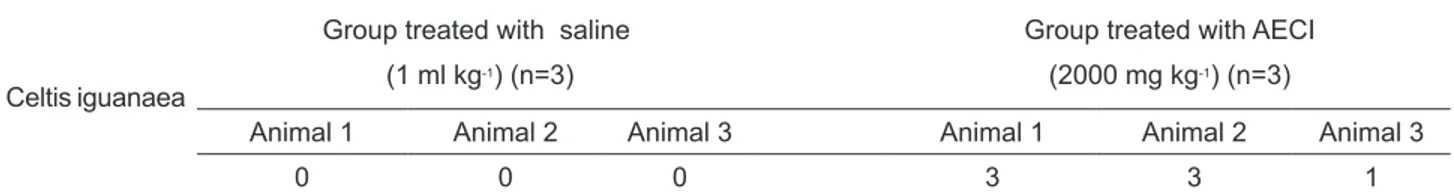
SILVA, CSP; PROENÇA, CEB. Use and availability of medical resources in Ouro Verde de Goiás, Goiás State, Brazil. Brasilica Botanica Acta, v. 22, n. 02, 2008. TABLE 5. Intensity values injury casts assigned to the parameter of kidneys from Wistar rats. Scale of 0 to 4 means absent, rare, mild, moderate and severe respectively.
Celtis iguanaea
Group treated with saline (1 ml kg-1) (n=3)
Group treated with AECI (2000 mg kg-1) (n=3)
Animal 1 Animal 2 Animal 3 Animal 1 Animal 2 Animal 3
SOUSA, FB et. al. Preliminary studies of gastroprotective effect of Celtis iguanaea (Jacq.) Sargent leaves (Ulmaceae). Natural Product Research: Formerly
Natural Product Letters. v. 27, n. 12, p. 1102-1107,
2013.
TREVISAN, RR et. al. Avaliação da atividade fitotóxica com enfoque alelopático do extrato das cascas de
Celtis iguanaea (Jacq.) Sargent Ulmaceae e purificação
de dois triterpenos. Revista Brasileira de Plantas