ISSN 0100-879X
BIOMEDICAL SCIENCES
AND
CLINICAL INVESTIGATION
www.bjournal.com.br
www.bjournal.com.br
Volume 42 (11) 993-1118 November 2009
Institutional Sponsors
The Brazilian Journal of Medical and Biological Research is partially financed by
Braz J Med Biol Res, November 2009, Volume 42(11) 1015-1019
Hydrogen peroxide induces a specific DNA base change profile
in the presence of the iron chelator 2,2' dipyridyl in
Escherichia
coli
Hydrogen peroxide induces a specific DNA base
change profile in the presence of the iron chelator
2,2’ dipyridyl in
Escherichia coli
D.L. Felício
1, C.E.B. Almeida
2, A.B. Silva
1and A.C. Leitão
11Laboratório de Radiobiologia Molecular, Instituto de Biofísica Carlos Chagas Filho,
Universidade Federal do Rio de Janeiro, Rio de Janeiro, RJ, Brasil
2Laboratório de Radiobiologia, Instituto de Radioproteção e Dosimetria,
Comissão Nacional de Energia Nuclear, Rio de Janeiro, RJ, Brasil
Abstract
Pretreatment of Escherichia coli cultures with the iron chelator 2,2’-dipyridyl (1 mM) protects against the lethal effects of low
concentrations of hydrogen peroxide (<15 mM). However, at H2O2 concentrations equal to or greater than 15 mM, dipyridyl
pretreatment increases lethality and mutagenesis, which is attributed to the formation of different types of DNA lesions. We
show here that pretreatment with dipyridyl (1 mM) prior to challenge with high H2O2 concentrations (≥15 mM) induced mainly
G:C→A:T transitions (more than 100X with 15 mM and more than 250X with 20 mM over the spontaneous mutagenesis rate)
in E. coli. In contrast, high H2O2 concentrations in the absence of dipyridyl preferentially induced A:T→T:A transversions (more
than 1800X and more than 300X over spontaneous mutagenesis for 15 and 20 mM, respectively). We also show that in the
fpg nth double mutant, the rpoB gene mutation (RifS-RifR) induced by 20 mM H2O2 alone (20X higher) was increased in 20 mM H2O2 and dipyridyl-treated cultures (110X higher), suggesting additional and/or different lesions in cells treated with H2O2
under iron deprivation. It is suggested that, upon iron deprivation, cytosine may be the main damaged base and the origin of
the pre-mutagenic lesions induced by H2O2.
Key words: Hydrogen peroxide; Dipyridyl; Mutagenesis; Iron; Fenton reaction
Introduction
Correspondence: A.C. Leitão, Laboratório de Radiobiologia Molecular, Instituto de Biofísica Carlos Chagas Filho, UFRJ, CCS Bloco G,
21941-902 Rio de Janeiro, RJ, Brasil. E-mail: acleitao@biof.ufrj.br
Research supported by CNPq, FAPERJ and CAPES.
Received May 13, 2009. Accepted August 21, 2009. Available online October 26, 2009. In Escherichia coli, it has been demonstrated that H2O2
induces cell death by two different modes of action as a func-tion of H2O2 concentration, both of which are accompanied
by enhanced mutagenesis (1,2). Oxidative DNA damage induced by hydrogen peroxide is thought to occur through the Fenton reaction in the Haber-Weiss cycle. Our group and others have demonstrated (2-4) that pretreatment with the iron chelators 2-2’dipyridyl, 1-10 phenanthrolene or de-feroxamine protects E. coli cells against the lethal effect of H2O2, suggesting that ionic iron is the main transition metal
that mediates its genotoxicity (5). Dipyridyl also protects the cells against the oxidative lesions produced by UV-B radiation (6). Asad and Leitão (3) have proposed a differ-ent pathway for DNA damage induced by H2O2 when cells
are depleted of iron because DNA lesions produced under these conditions were repaired even in an exonuclease III
(xthA)-deficient mutant (3). It was thought that this pathway would occur through the Fenton reaction mediated by transi-tion metals other than iron. Indeed, copper ions have been implicated as candidates to mediate this pathway. It was suggested that copper plus H2O2 would generate different
types of DNA lesions or a higher number of the same le-sions that are generated through the iron-mediated Fenton reaction (7). Following this characterization, Asad et al. (8) detected the increased sensitivity of fpg and uvrA mutants to H2O2 challenge when iron deprived. Based on the fact that
oxidation involving copper generates a significant amount of 8oxo-G and that Fpg and UvrA play an important role in repairing this lesion, Almeida et al. (7) have suggested that 8oxo-G should be the most important DNA lesion produced by H2O2 challenge in iron-depleted cultures (9,10). The SOS
1016 D.L. Felício et al.
survival after treatment with H2O2 in the
presence of iron chelators (9).
In the present study, we investigated H2O2-induced mutagenesis under iron
deprivation using a reversion test with a series of E. coli mutant strains (11) as well as a forward mutation test (12) using base excision repair mutant strains.
Material and Methods
Bacterial strains
The bacterial strains used are de-rived from E. coli K-12 and are listed in Table 1.
Growth conditions
Bacterial cultures were grown over-night in M9 minimal medium (13) contain-ing glucose (4 g/L) supplemented with 2.5 mg/mL casamino acids and 1 µg/ mL thiamine at 37°C under shaking. The supplemented medium was designated M9S. A starting inoculum (0.25 mL, ≈108
cells) was taken from these cultures and the cells were grown in 10 mL fresh M9S medium until the mid-exponential phase (2 x 10⁸ cells/mL).
Survival experiments
Cultures in the mid-exponential phase of growth (pre-treated or not with 1 mM dipyridyl) were challenged with 20 mM H2O2 (30% perhydrol; Merck, 7722-84-1, Brazil)
for 20 min (CC strains) or 5 min (AB1157 and base exci-sion repair mutant strains) in M9S medium at 37°C under shaking. Residual H2O2 was inactivated by the addition of
excess catalase (5 µg/mL; EC 1.11.1.6 Sigma 9001-05-2, USA). Samples were collected at the end of the incubation time, diluted in M9 salt solution, and spread on lysogeny broth (LB) medium (13) solidified with 1.5% agar (Difco). The colony forming units (CFU) were scored after overnight incubation at 37°C.
Pretreatment with dipyridyl
Cultures in the mid-exponential growth phase were incu-bated with the iron chelator dipyridyl (1 mM; 2,2’- bipyridine, Sigma, 366-18-7) for 20 min in M9S medium at 37°C, with shaking. Treatment with the metal ion chelator alone did not affect cell viability (data not shown). Cultures treated with 1 mM dipyridyl are referred to as iron-depleted culture.
Analysis of the Lac-
→
Lac+ mutagenesis induced by H 2O2Mutagenesis assays for transitions and transversions are based upon the Lac-→Lac+ reversion of specific
muta-tions in the lacZ gene located on an F’-plasmid described by Coulondre and Miller (14) and Cupples and Miller (11).
Reversion is examined in a set of strains (CC101 to 106) in which the Lac+ phenotype is recovered after specific base
sub-stitution restores codon 461 of the lacZ gene to Gln. We have screened base changes using the CC strains after treatment with 15 and 20 mM H2O2 for 20 min with or without dipyridyl
(1 mM) pretreatment. Cultures in the mid-exponential growth phase were challenged as indicated in the section Survival experiments. Aliquots (0.8 mL) were taken for survival and mutagenesis experiments, centrifuged for 4 min at 9500 g and resuspended in 0.4 mL. An aliquot of 0.2 mL was added to 2 mL LB and incubated for 20 h at 37°C with shaking (150 rpm). Next, a 0.1-mL portion was spread on minimal medium containing 0.4% lactose as a carbon source (11) and incubated for 72 h at 37°C to allow growth of Lac+ revertants. Viable cell numbers in
the 20-h culture were counted on LB plates after 24 h at 37°C. The mutation frequency is reported as number of mutant cells per 108 viable cells. Data are reported as means ± SEM of
three experiments, as indicated in the tables. Fold increase in mutation frequency (base substitution) was compared between strains by ANOVA and the Tukey-Kramer multiple comparison test (GraphPad InsTat, GraphPad Software Inc., USA). The level of significance was set at P < 0.01 in all analyses.
As a control we measured the sensitivity of CC strains to challenge with 20 mM H2O2 and observed that the
sur-vival was about 10% and for dipyridyl-pretreated cells the survival was 2 to 5% (data not shown).
Analysis of the RifS
→
RifR mutagenesis induced byH2O2
Mutagenesis experiments were performed as described by Sedgwick and Goodwin (12). Cells in the mid-exponential
Table 1. Strain description.
Escherichia coli strains
Designation* Relevant genotype Reversion event
CC101 ara-. ∆(lac - proB)XIII [F’ lacI
-Z-.proB+] A:T→C:G CC102 ara-. ∆(lac - proB)XIII [F’ lacI-Z-.proB+] G:C→A:T
CC103 ara-. ∆(lac - proB)XIII [F’ lacI
-Z-.proB+] G:C→C:G CC104 ara-. ∆(lac - proB)XIII [F’ lacI-Z-.proB+] G:C→T:A
CC105 ara-. ∆(lac - proB)
XIII [F’ lacI
-Z-.proB+] A:T→T:A CC106 ara-. ∆(lac - proB)XIII [F’ lacI
-Z-.proB+] A:T→G:C
AB1157 F
- thr-1, leuB6, thi-1, argE3, his-4, proA2, lacY1,
galK2, xyl-5, ara-14, rspL13, tsx-33, supE44
BW375 As AB 1157, but nth
BH20 As AB 1157, but fpg
BH160 As AB 1157, but nth fpg
*The CC strains were a gift from Prof. J.H. Miller (University of California, Los An
phase of growth, pretreated with 1 mM dipyridyl or not (20 min), were treated with 20 mM H2O2 for 5 min. All strains showed
at least 60% survival after the H2O2 challenge. Aliquots were
taken for survival experiments, and 0.1-mL samples were added to 3 mL of melted LB containing 0.75% agar, which was then layered on 15-mL LB plates. An additional 3-mL layer was added to the plates, which were then incubated at 37°C for 5 h to allow cell division and mutation fixation. After this period, a final 3-mL layer of melted LB containing 800 μg/mL rifampicin (Sigma 13292-46-1) was added and plates were then incubated for 16 h. Diffusion of the antibiotic through the medium led to a final concentration of 100 μg/mL rifampicin. Rifampicin-resistant CFU represented cells with a mutated rpoB gene and mutation frequency was expressed as the ratio between the number of rifampicin-resistant CFU and the number of viable cells after treatment detected in the corresponding survival experiment.
Results and Discussion
To analyze the nature of the lesions produced when bacte-rial cultures are pretreated with 2-2’dipyridyl and challenged
with H2O2 at high concentrations, we conducted mutagenesis
assays for transitions and transversions based upon the Lac
-→Lac+ reversion of specific mutations in the lacZ gene located
on an F’-plasmid described by Coulondre and Miller (14) and Cupples and Miller (11).
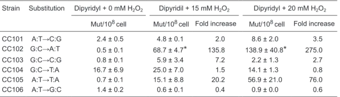
A clearly predominant A:T→T:A (CC105) transversion was seen when strains were treated with H2O2 concentrations ≥15 mM (Table 2). On the other hand, these concentrations induced a completely different pattern of base changes in cultures pretreated with 2-2'dipyridyl. In this case there was a clear preference for G:C→A:T (CC102) transition instead of the massive A:T→T:A (CC105) transversion induced by H2O2 alone (Tables 2 and 3). It should be noted that A:T→T:A
(CC105) base substitution appears as the second highest mutation frequency of base substitution when H2O2 treatment
is performed under iron deprivation (Table 3).
By our characterization of DNA base substitution induced by treatment with high concentrations of H2O2 upon iron
deprivation, we obtained a clear difference in the profile of DNA lesions induced in the two situations. A:T→T:A transversion induced by H2O2 can be related to translesion
synthesis at AP sites that is strongly dependent on SOS
Table 2. Mutagenesis of CC culture strains exposed to H2O2.
Strain Substitution 0 mM H2O2 15 mM H2O2 20 mM H2O2
Mut/108cell Mut/108cell Fold increase Mut/108cell Fold increase
CC101 A:T→C:G 1.7 ± 0.3 12.5 ± 1.5 7.4 55.3 ± 30.9 32.5
CC102 G:C→A:T 0.5 ± 0.3 0.0 0.0 0.0 0.0
CC103 G:C→C:G 0.6 ± 0.2 6.0 ± 4.2 9.8 18.4 ± 10.8 30.2
CC104 G:C→T:A 3.3 ± 1.4 33.7 ± 5.7 10.2 50.0 ± 11.5 15.2
CC105 A:T→T:A 0.5 ± 0.2 978.8 ± 242.0* 1856.9 162.9 ± 6.9* 309.0
CC106 A:T→G:C 0.3 ± 0.1 0.5 ± 0.1 1.7 0.1 ± 0.0 0.2
Cultures in the mid-exponential phase of growth in M9S medium at 37°C under shaking were exposed to H2O2
treatment for 20 min. The data represent the mean of at least 3 experiments and are used in Table 3 and Figure
1.
*P < 0.01 compared to all other strains (Tukey-Kramer test).
Table 3. Mutagenesis of CC culture strains exposed to dipyridyl and H2O2.
Strain Substitution Dipyridyl + 0 mM H2O2 Dipyridil + 15 mM H2O2 Dipyridyl + 20 mM H2O2
Mut/108cell Mut/108cell Fold increase Mut/108cell Fold increase
CC101 A:T→C:G 2.4 ± 0.5 4.8 ± 0.1 2.0 8.6 ± 2.0 3.5
CC102 G:C→A:T 0.5 ± 0.1 68.7 ± 4.7* 135.8 138.9 ± 40.8* 275.0
CC103 G:C→C:G 0.8 ± 0.1 5.9 ± 3.4 7.2 2.2 ± 1.3 2.7
CC104 G:C→T:A 16.7 ± 6.9 25.0 ± 7.0 1.5 14.1 ± 1.3 0.8
CC105 A:T→T:A 0.7 ± 0.1 15.1 ± 8.8 20.2 56.9 ± 21.0 76.0
CC106 A:T→G:C 1.4 ± 0.2 0.6 ± 0.1 0.4 0.9 ± 0.0 0.6
Cultures in the mid-exponential phase of growth in M9S medium at 37°C under shaking were pretreated with 1 mM dipyridyl for 20 min and then exposed to H2O2 treatment for 20 min. The data represent the mean of at least
1018 D.L. Felício et al.
response (15).
The appearance of A:T→T:A is consistent with a pref -erence for adenine (A rule) insertion in the bypass at AP sites (16). Therefore, we assume that our observation is consistent with the production of AP sites, a well-known type of oxidative DNA damage (17), after exposure to high H2O2 concentrations. However, we cannot exclude
the possibility of another oxidative base damage, such as 2-hydroxyadenine, that can also induce A:T→T:A trans-version by mispairing with adenine. The accumulation of 2-hydroxyadenine is reported to occur after H2O2 treatment
in human cells (18,19).
The clear preferential induction of G:C→A:T transition by high H2O2 upon iron deprivation indicates that the nature
of DNA base damage induced by this challenge may be different from that induced by H2O2 alone. The occurrence
of 8oxo-G as well as AP sites due to damaged guanine in DNA does not support the appearance of this transition. Both lesions would produce G:C→T:A as a result of mispair-ing between 8oxo-G with A or by preferential insertion of adenine at an AP site (20). This led us to focus on cytosine as the main target for DNA lesion induced by H2O2 upon
iron ion deprivation.
Hydroxyl ion reacts with cytosine by adding to the C5-C6 double bond leading to the formation of cytosine glycol, which is unstable and can break down further to 5-hydroxy-2’deoxycytosine OHdC), 5-hydroxy-2’deoxyuracil (5-OHdU) and uracil glycol (21,22). The major stable oxidation product of cytosine is 5-OHdU that preferentially pairs with
adenine (13). 5-Hydroxy-2’deoxyuracil shares this same property and it would also result in the G:C→A:T transi -tion (19,23). Whenever 5-OHdC is present in DNA, it is removed by the action of endonuclease III (Nth) via the N-glycosylase/β elimination reaction, by the formamidopy-rimidine-DNA glycosylase (Fpg) via the N-glycosylase/β,δ
elimination reaction (24,25) in E. coli or by their homologs in eukaryotes (26,27). Nth and Fpg can remove 5-OHdU from DNA through the cited mechanisms and additionally by uracil DNA N-glycosylase (Ung) generating AP sites (25). Mutagenesis induced by 20 mM H2O2 upon iron
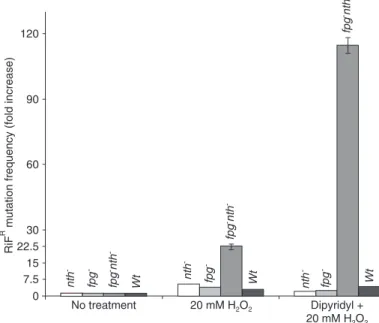
depriva-tion in the rpoB gene, performed as described by Sedgwick and Goodwin (12), was evaluated using either nth and fpg single mutants or double mutants. A higher mutation frequency was detected in the double mutant (Figure 1) for both conditions. We detected a 110-fold increase in muta-tion frequency in dipyridyl-pretreated cells and a 20-fold increase in cultures not treated with the metal chelator, suggesting the appearance of additional and/or different lesions in cells challenged with H2O2 upon iron deprivation.
In both cases the sum of the mutation frequency observed in the single mutants did not correspond to that observed in the double mutant. This behavior may indicate that both nth and fpg share a role in preventing the appearance of mutagenic lesions. We suggest that 5-OHdC and 5-OHdU are the premutagenic DNA lesions produced by the chal-lenge of H2O2 in iron-depleted cultures. We assume that
the G:C→A:T transition would be due to the mispairing of damaged cytosine, in this case 5-OHdC, with adenine. In the case of 5-OHdU we can speculate that it would be converted to an AP site, since Ung is active in nth fpg double mutants, leading to base substitu -tion of C to T as a consequence of the preference for adenine insertion during translesion synthesis at an AP site. The nature of base substitution in the nth, fpg and nth fpg background is presently under investigation by our group.
We suggest that lethal and mutagenic pathways produced by high concentrations (more than 15 mM) of H2O2 in E. coli under iron deprivation might be a
result of cytosine oxidation, yielding their mutagenic products 5-OHdC and/or 5-OHdU. Taken together, our data support the hypothesis of two different path-ways for H2O2-induced lesions upon iron deprivation,
which depend on H2O2 concentration.
Acknowledgments
We are grateful to J.S. Cardoso for expert techni-cal assistance and to D.P. Carvalho and M. Pádula for help and discussion during the preparation of the manuscript.
Figure 1. Mutagenesis of Escherichia coli cultures exposed to dipyridyl
and H2O2. Cultures in the mid-exponential phase of growth in M9S me
References
1. Imlay JA, Linn S. Bimodal pattern of killing of
DNA-repair-defective or anoxically grown Escherichia coli by hydrogen
peroxide. J Bacteriol 1986; 166: 519-527.
2. Imlay JA, Chin SM, Linn S. Toxic DNA damage by hydrogen peroxide through the Fenton reaction in vivo and in vitro.
Science 1988; 240: 640-642.
3. Asad NR, Leitao AC. Effects of metal ion chelators on DNA
strand breaks and inactivation produced by hydrogen perox
-ide in Escherichia coli: detection of iron-independent lesions. J Bacteriol 1991; 173: 2562-2568.
4. Asad NR, Asad LMBO, Almeida CE, Felzenszwalb I,
Cabral-Neto JB, Leitão AC. Several pathways of hydrogen peroxide action that damage the E. coli genome. Gen Mol Biol 2004;
27: 291-303.
5. Luo Y, Han Z, Chin SM, Linn S. Three chemically distinct types of oxidants formed by iron-mediated Fenton reactions
in the presence of DNA. Proc Natl Acad Sci U S A 1994; 91:
12438-12442.
6. Souza LL, Eduardo IR, Padula M, Leitao AC. Endonuclease
IV and exonuclease III are involved in the repair and muta
-genesis of DNA lesions induced by UVB in Escherichia coli.
Mutagenesis 2006; 21: 125-130.
7. Almeida CE, Galhardo RS, Felicio DL, Cabral-Neto JB, Leitao AC. Copper ions mediate the lethality induced by hydrogen peroxide in low iron conditions in Escherichia coli.
Mutat Res 2000; 460: 61-67.
8. Asad NR, de Almeida CE, Asad LM, Felzenszwalb I, Leitao AC. Fpg and UvrA proteins participate in the repair of DNA lesions induced by hydrogen peroxide in low iron level in
Escherichia coli. Biochimie 1995; 77: 262-264.
9. Asad LM, Asad NR, Silva AB, de Almeida CE, Leitao AC.
Role of SOS and OxyR systems in the repair of Escherichia
coli submitted to hydrogen peroxide under low iron condi
-tions. Biochimie 1997; 79: 359-364.
10. Galhardo RS, Almeida CE, Leitao AC, Cabral-Neto JB. Repair of DNA lesions induced by hydrogen peroxide in the presence of iron chelators in Escherichia coli: participation
of endonuclease IV and Fpg. J Bacteriol 2000; 182:
1964-1968.
11. Cupples CG, Miller JH. A set of lacZ mutations in Escherichia coli that allow rapid detection of each of the six base substi
-tutions. Proc Natl Acad Sci U S A 1989; 86: 5345-5349.
12. Sedgwick SG, Goodwin PA. Differences in mutagenic and recombinational DNA repair in enterobacteria. Proc Natl Acad Sci U S A 1985; 82: 4172-4176.
13. Miller JH. A short course in bacterial genetics: a laboratory manual and handbook for E. coli and related bacteria. Cold
Spring Harbor: Cold Spring Harbor Lab. Press; 1992. 14. Coulondre C, Miller JH. Genetic studies of the lac repressor.
III. Additional correlation of mutational sites with specific
amino acid residues. J Mol Biol 1977; 117: 525-567.
15. Schaaper RM, Loeb LA. Depurination causes mutations
in SOS-induced cells. Proc Natl Acad Sci U S A 1981; 78:
1773-1777.
16. Kunkel TA. Mutational specificity of depurination. Proc Natl Acad Sci U S A 1984; 81: 1494-1498.
17. Wallace SS. Biological consequences of free radical-dam
-aged DNA bases. Free Radic Biol Med 2002; 33: 1-14.
18. Jaruga P, Dizdaroglu M. Repair of products of oxidative DNA
base damage in human cells. Nucleic Acids Res 1996; 24:
1389-1394.
19. Kamiya H. Mutagenic potentials of damaged nucleic acids produced by reactive oxygen/nitrogen species: approaches using synthetic oligonucleotides and nucleotides: survey and
summary. Nucleic Acids Res 2003; 31: 517-531.
20. Wood ML, Dizdaroglu M, Gajewski E, Essigmann JM. Mechanistic studies of ionizing radiation and oxidative mutagenesis: genetic effects of a single 8-hydroxyguanine (7-hydro-8-oxoguanine) residue inserted at a unique site in
a viral genome. Biochemistry 1990; 29: 7024-7032.
21. Cooke MS, Evans MD, Dizdaroglu M, Lunec J. Oxidative
DNA damage: mechanisms, mutation, and disease. FASEB
J 2003; 17: 1195-1214.
22. Dizdaroglu M, Holwitt E, Hagan MP, Blakely WF. Formation
of cytosine glycol and 5,6-dihydroxycytosine in deoxyribo
-nucleic acid on treatment with osmium tetroxide. Biochem J
1986; 235: 531-536.
23. Kreutzer DA, Essigmann JM. Oxidized, deaminated cyto
-sines are a source of C → T transitions in vivo. Proc Natl Acad Sci U S A 1998; 95: 3578-3582.
24. Cadet J, Bourdat AG, D’Ham C, Duarte V, Gasparutto D,
Romieu A, et al. Oxidative base damage to DNA: specific
-ity of base excision repair enzymes. Mutat Res 2000; 462:
121-128.
25. Hatahet Z, Kow YW, Purmal AA, Cunningham RP, Wallace
SS. New substrates for old enzymes. 5-Hydroxy-2’-deoxy
-cytidine and 5-hydroxy-2’-deoxyuridine are substrates for
Escherichia coli endonuclease III and formamidopyrimidine
DNA N-glycosylase, while 5-hydroxy-2’-deoxyuridine is a
substrate for uracil DNA N-glycosylase. J Biol Chem 1994;
269: 18814-18820.
26. Hilbert TP, Boorstein RJ, Kung HC, Bolton PH, Xing D, Cun
-ningham RP, et al. Purification of a mammalian homologue
of Escherichia coli endonuclease III: identification of a
bovine pyrimidine hydrate-thymine glycol DNAse/AP lyase by irreversible cross linking to a thymine glycol-containing
oligoxynucleotide. Biochemistry 1996; 35: 2505-2511.2
27. Melo RG, Leitao AC, Padula M. Role of OGG1 and NTG2
in the repair of oxidative DNA damage and mutagenesis in
-duced by hydrogen peroxide in Saccharomyces cerevisiae:
relationships with transition metals iron and copper. Yeast