Structure and functio n
o f the se le ctin ligand PSGL-1
Department of Biochemistry and Molecular Biology, The University of O klahoma Health Sciences Center, O klahoma City, O K, USA
R.D. Cummings
Abstract
P-selectin glycoprotein ligand-1 (PSGL-1) is a dimeric mucin-like 120-kDa glycoprotein on leukocyte surfaces that binds to P- and L-selectin and promotes cell adhesion in the inflammatory response. The extreme amino terminal extracellular domain of PSGL-1 is critical for these interactions, based on site-directed mutagenesis, blocking mon-oclonal antibodies, and biochemical analyses. The current hypothesis is that for high affinity interactions with P-selectin, PSGL-1 must contain O-glycans with a core-2 branched motif containing the sialyl Lewis x antigen (NeuAca2®3Galß1®4[Fuca1®3]GlcNAcß1®R). In addition, high affinity interactions require the co-expression of tyrosine sulfate on tyrosine residues near the critical O-glycan struc-ture. This review addresses the biochemical evidence for this hypoth-esis and the evidence that PSGL-1 is an important in vivo ligand for
cell adhesion. Co rre spo nde nce
R.D. Cummings
Department of Biochemistry and Molecular Biology
The University of O klahoma Health Sciences Center 975 N.E. 10th St., BRC 417 O klahoma City, O K 73104 USA
Fax: + 1-405-271-3910 E-mail:
richard-cummings@ uokhsc.edu
Presented at the 5th Brazilian Symposium on Extracellular Matrix - SIMEC, Angra dos Reis, RJ, Brasil, September 7-10, 1998.
Research supported by a grant from the National Institutes of Health (PO 1 HL 45510).
Received March 2, 1999 Accepted March 9, 1999
Ke y wo rds
·P-selectin
·L-selectin
·E-selectin
·PSGL-1
·O -glycosylation
·Glycoprotein
·Mucin
·Tyrosine sulfate
·Cell adhesion
·Leukocytes
·Neutrophils
Intro ductio n
The inflammatory response in animals is initiated by the tethering and rolling of circu-lating leukocytes on activated endothelium. These initial interactions involve glycocon-jugate ligands on the leukocyte cell surface and carbohydrate-binding proteins, termed selectins, expressed on the surfaces of both endothelial cells and leukocytes. There are three known selectins, termed P-, E- and L-selectins, and all three are Ca2+
-dependent binding proteins that contain a C-type carbo-hydrate recognition domain at their extreme N-terminus. P-selectin is constitutively ex-pressed in intracellular vesicles of platelets and endothelial cells and is rapidly mobi-lized to the surface membrane in thrombin-or histamine-stimulated cells; E-selectin ex-pression is transcriptionally up-regulated by inflammatory cytokines, and L-selectin is
constitutively expressed on the surface mem-brane of all leukocytes.
Although initial studies suggested that all three selectins can recognize carbohydrates containing the sialyl Lewis x antigen ( N e u A ca2®3 G a l ß 1®4 [ F u ca1®3 ] GlcNAcß1®R) (sialyl Lex
several articles (1-5). This review will at-tempt to provide an overall historical frame-work for understanding the more important studies on this ligand and focus on exciting new developments in regard to the structure and function of PSGL-1 and its role as a selectin ligand in the inflammatory response and atherosclerosis.
Histo rical studie s o n P-se le ctin ligands o n ne utro phils
The binding of P-selectin to neutrophils is abolished upon sialidase treatment, which suggested originally that sialic acid may be a critical determinant required for P-selectin recognition (6). The possible importance of the sialyl Lex
moiety to P-selectin recogni-tion was derived from experiments showing that 1) P-selectin binds to a mutant Chinese hamster ovary (CHO) cell line expressing the sialyl Lex
moiety, but not to cells lacking the epitope (7); 2) antibodies to the sialyl Lex
epitope, but not the Lex
epitope, can block binding of cells to P-selectin (7,8), and 3) glycans containing the sialyl Lex
moiety can
inhibit P-selectin-mediated adhesion (8). However, the binding of P-selectin to neu-trophils displays much higher affinity inter-actions than to other non-myeloid cells (7), suggesting that myeloid cells possess one or more specific ligands for P-selectin. Using
125
I-P-selectin blotting and affinity chroma-tography on immobilized human P-selectin, a glycoprotein ligand for P-selectin was iden-tified and purified, starting with total mem-brane glycoproteins extracted from human neutrophils and the human promyelocytic cell line HL60 (9). The purified ligand, now known as the PSGL-1, behaves as a disul-fide-bonded ~250-kDa protein in non-re-ducing SDS/PAGE and ~120-kDa in reduc-ing SDS/PAGE.
Prim ary structure o f PSGL-1
The cDNA encoding PSGL-1 was ex-pression cloned in COS cells co-expressing an a1,3/4-fucosyltransferase (human Fuc-TIII), which permits both sialyl Lex
and sialyl Lea
synthesis in these cells (10). PSGL-1 is predicted to be a protein of 412 amino acids
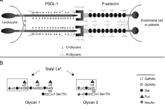
Figure 1 - A, Scheme of the in-teractions betw een human P-selectin and the amino-terminal domain of human PSGL-1. The many N-glycans on PSGL-1 and P-selectin are indicated, as are the O-glycans on PSGL-1. B, The major fucose-containing O-gly-cans of hum an HL60-derived PSGL-1. Glycan 1 is a trifucosyl-ated, monosialyltrifucosyl-ated, polylac-tosamine core-2 O-glycan and glycan 2 is a monofucosylated, disialylated core 2 O-glycan.
PSGL-1 P-selectin
Endothelial cell or platelet
Leukocyte S
S
O-Glycans
N-Glycans
A
B
Sialyl Lex
a3
a3 a
3 a3
a1-Ser/Thr
ß4 ß3 ß4 ß3 ß4
ß6
ß3 a3 ß3 a1-Ser/Thr
a3 ß4a3
GalNAc
GlcNAc
Gal
Fuc
NeuAc
S
S
Glycan 1 Glycan 2
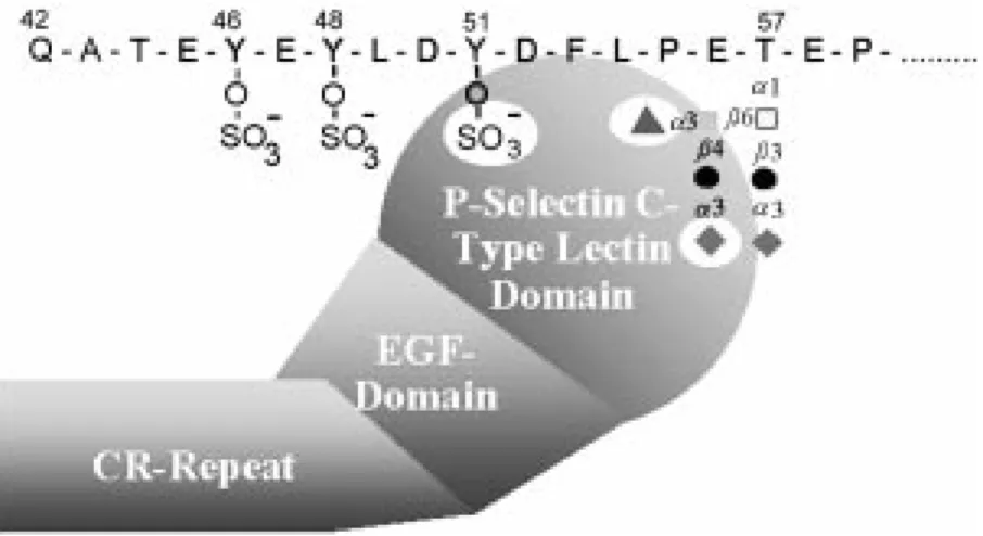
with an 18-amino acid signal sequence and a tetrapeptide consensus cleavage site for paired basic amino acid converting enzyme in leukocytes at residues 38-41 (-R-D-R-R-) (Figure 1A). Thus, the N-terminus of the mature protein begins at residue 42. There are 16 decapeptide repeating units with the consensus sequence -A-T/M-E-A-Q-T-T-X-P/L-A/T- spanning residues 118-277 in the long form of the protein and the short form is missing the residues 132-141 (11,12). Within the extracellular domain is a single Cys resi-due at position 320 that precedes the pre-dicted single transmembrane domain span-ning residues 321-341 and the cytoplasmic domain of residues 342-412. The coding region for human PSGL-1 is contained en-tirely in exon 2 of the gene, which maps to chromosome 12q24. The cDNA for the mu-rine PSGL-1 encodes a predicted 397-amino acid protein with recognizable homology to the human sequence (13). The murine pro-tein contains a predicted signal sequence and propeptide identical in size to human PSGL-1; the mature murine PSGL-1 is also predicted to begin at residue 42. However, the mouse homologue has only 10 decameric repeats with the consensus sequence -E-T-S-Q/K-P-A-P-T/M-E-A- that are obviously dif-ferent in sequence from the human PSGL-1. The highest homology between the human and murine PSGL-1 occurs in the transmem-brane (83%) and cytoplasmic domains (76%). Each subunit of human PSGL-1 contains 70 Ser and Thr residues in the extracellular domain that are potential sites for O-glyco-sylation and 3 potential sites for N-glycosy-lation. The murine PSGL-1 also contains numerous extracellular Ser and Thr residues and two potential sites for N-glycosylation. The murine PSGL-1 also contains a single unpaired extracellular Cys at residue 307 that precedes the predicted transmembrane domain. Interestingly, the human PSGL-1 contains three predicted tyrosine sulfation sites at residues 46, 48 and 51 that fall in the consensus sequence in which Tyr residues
are flanked by acidic residues. The murine PSGL-1 contains two predicted tyrosine sulfation sites at residues 54 and 56.
Structural fe ature s o f PSGL-1 re quire d fo r binding to P-se le ctin
Glyco sylatio n o f native PSGL-1
The large size and extensive glycosyla-tion of PSGL-1 present a daunting challenge to understanding how it is specifically rec-ognized by P-selectin. It was anticipated at first that PSGL-1 might be a high affinity and unique ligand for P-selectin by virtue of its mucin-like nature and the presumption that the ligand contained large amounts of the sialyl Lex
antigen (14), thereby enhancing its avidity for P-selectin. However, as discussed below, this prediction was not correct.
Treatment of purified PSGL-1 with sialidase abolishes its binding to P-selectin, confirming the cell studies that indicated a role for sialic acid in P-selectin recognition (9,14). Interestingly, treatment of neutro-phil-derived PSGL-1 with peptide N-gly-cosidase F, which removes most, if not all, the N-glycans of the molecule, does not af-fect its recognition by P-selectin, suggesting that O-glycans, but not N-glycans, are im-portant determinants (9). This conclusion is supported by the observation that treatment of either neutrophils or purified PSGL-1 with the O-sialoglycoprotease from Pasteurella
hemolytica, an enzyme that degrades
sialyl-ated mucins, blocks all interactions with P-selectin (14). Furthermore, treatment of HL60 cells with benzyl-alpha-GalNAc, which in-hibits extension of O-glycans, also reduces binding of cells to P-selectin (15). Other studies demonstrated that treatment of isolated PSGL-1 with endo-ß-galactosi-dase, a bacterial endoglycosidase capable of degrading type-2 polylactosamine repeats [-3Galß1®4GlcNAcß1-]n, significantly
pre-sumably on O-glycans, may also be impor-tant for binding.
The O-glycans of native PSGL-1 purified from human HL60 cells were determined and the results were unexpected in many ways (17). Most of the O-glycans contained a simple core-2 structure with one or two sialic acid residues and lacked fucose. Fu-cose was found in only two relatively minor O-glycans, termed glycan 1 and 2, as shown in Figure 1B. Both glycan 1 and 2 contain the sialyl Lex
antigen; however, glycan 1, but not glycan 2, contains a polylactosamine back-bone on the core-2 structure with multiple fucosyl residues. These results demonstrated that the O-glycans of PSGL-1 generally lacked fucose and that only a few O-glycans displayed the sialyl Lex
antigen, presumed to be important in P-selectin binding. The fact that glycan 2 was predicted to occur in sub-stoichiometric quantities led to the expecta-tion that glycan 1 may be the more important O-glycan for P-selectin recognition. This possibility is discussed below.
Glyco sylatio n o f re co mbinant PSGL-1
re quire d fo r binding to P-se le ctin
To explore the functional importance of core-2 O-glycans and fucose residues in PSGL-1 activity, a recombinant form of PSGL-1 was expressed in CHO cells. These cells do not express the sialyl Lex
antigen and lack a1,3-fucosyltransferases required for either Lex
or sialyl Lex
synthesis; in addi-tion, they lack the ß1,6-N-acetylglucosami-nyltransferase (C2GnT) required for core-2 O-glycan synthesis. PSGL-1 expressed in these wild type CHO cells was not able to bind P-selectin (18). However, PSGL-1 syn-thesized in CHO cells co-transfected with cDNA encoding human a 1,3-fucosyltrans-ferase III (Fuc-TIII) and the human C2GnT was a high affinity ligand for P-selectin (18,19). Such results suggested that expres-sion of both the sialyl Lex
antigen and C2GnT is required for PSGL-1 binding to P-selectin.
Tyro sine sulfatio n and the ro le o f the PSGL-1
N-te rm inus
Although glycosylation of PSGL-1 is clearly important for its binding to P-selectin, other biochemical studies of the molecule have provided several clues suggesting that sulfation was also important. The first of these clues was that PSGL-1 contained ty-rosine sulfate and that removal of tyty-rosine sulfate by bacterial aryl sulfatases abrogated binding of the molecule to P-selectin (20). Consistent with this finding, recombinant forms of PSGL-1 in which the three tyrosine residues had been mutagenized to phenylal-anine also failed to bind P-selectin (21,22). In addition, treatment of cells with sodium chlorate, an inhibitor of the ATP sulfurylase that is required for synthesis of the sulfate donor phosphoadenosine phosphosulfate (PAPS), also blocked synthesis of a func-tional PSGL-1 molecule (21,22).
was fused to the heavy chain CH2-CH3
re-gion of IgG1, and the recombinant glycopro-tein expressed in COS cells co-transfected with human Fuc-TIII, was bound by P-selectin (21). 4) Treatment of neutrophils with the cobra venom metalloproteinase mocarhagin removed the extreme N-termi-nal 10 amino acid residues from PSGL-1 and abrogated its binding to P-selectin (26). All of these experiments point to a model in which the combination of tyrosine sulfate residues and oligosaccharides on the protein are required for high affinity binding to P-selectin.
An additional approach to explore the fine structure of the PSGL-1 N-terminal do-main required for P-selectin recognition is to selectively mutate amino acids in that do-main and test the binding of recombinant PSGL-1 to P-selectin. Such site-directed mutagenesis to replace all three N-terminal Tyr residues with Phe abolishes binding of the recombinant PSGL-1 to P-selectin, but not E-selectin (18,21,22). A more detailed number of mutations reveal that any one of the three Tyr residues can support binding of the recombinant PSGL-1 to P-selectin, indi-cating that only one of the three potential tyrosine sulfate residues is necessary for bind-ing to P-selectin (27). Furthermore, muta-tion of the Thr residue at posimuta-tion 57 to Ala in the extreme N-terminus human PSGL-1 blocks binding of the recombinant molecule to P-selectin (21,22,27). Taken together, the studies on the native and recombinant PSGL-1 support the types of models shown in Figure 2, where one or more tyrosine sulfate residues acting in conjunction with nearby Thr-attached O-glycans and with a sialyl Lex
antigen on the core-2 motif are recognized by P-selectin. However, it must be stressed that this model is a mere prediction. Despite the elegant experiments described above, there is still no chemical proof that such O-linked glycans, as depicted in Figure 2, occur at Thr-57 in PSGL-1; nor is there any direct proof that there is coordinate binding of such
an O-glycan and one or more tyrosine sul-fates.
Affinity o f PSGL-1 fo r P-se le ctin
Previous studies have established that P-selectin binds to free glycans containing sialyl Lex
with relatively low affinity in the milli-molar range (0.1 to 1.0 mM). However, re-cent studies using a BIAcore apparatus and surface plasmon resonance measurements demonstrated that neutrophil-derived PSGL-1 is a high affinity ligand for P-selectin and exhibits a Kd in the range of 0.3 µM (28). The
binding is characterized by a high off-rate counterbalanced by an even higher on-rate. This high affinity between PSGL-1 and P-selectin contrasts with that observed for L-selectin binding to GlyCAM-1, where the Kd
is in the range of 0.1 mM, which was also determined by surface plasmon resonance measurements (29). The binding of recom-binant forms of PSGL-1 to P-selectin has also recently been measured and found to have a very high affinity. The Kd of soluble
P-selectin with a soluble recombinant form of PSGL-1, which was prepared using hu-man Fuc-TIII, was determined by measuring changes in intrinsic fluorescence upon ligand
binding and found to be in the range of 3 nM (30). Interestingly, when the recombinant PSGL-1 was prepared with human Fuc-TVII, the Kd was large and in the range of 80
nM (30). Although many questions remain about the relative glycosylation of native versus recombinant PSGL-1 in such studies as those cited above, these results demon-strate that PSGL-1 is a high affinity ligand for P-selectin.
D ime rizatio n o f PSGL-1
To explore the role of dimerization of PSGL-1 on its ability to mediate cell adhe-sion and recognition by P-selectin, two labo-ratories prepared a recombinant form of the molecule in which the single extracellular Cys residue in PSGL-1 was changed to Ala (31,32). Essentially both groups found that the mutated protein behaved as a monomeric protein under reducing and non-reducing conditions and that the mutated protein dis-played no affinity for P-selectin. These re-sults were taken to indicate that PSGL-1 dimerization is essential for its high affinity binding to P-selectin. However, more recent studies in our laboratory have now provided data which question the results of these stud-ies. Similar substitution of the single extra-cellular Cys with either Ala or Ser appear to have no discernible effect on binding of cells expressing the mutated protein to P-selectin (Epperson TK, Ramachandran V, Patel KD, McEver RP and Cummings RD, unpublished data). Furthermore, in the latter study it was found that the mutated PSGL-1 behaved as a non-covalent dimeric protein that was readily cross-linked in the membrane form to a dimer. In addition, a small tryptic fragment of ~9 kDa, prepared from the recombinant mole-cule and containing the extreme N-terminal domain of the molecule, bound quantita-tively to a column of immobilized P-selectin. These results suggest that covalent dimeriza-tion of PSGL-1 is not required for its func-tional association with P-selectin. However,
because PSGL-1 can non-covalently dimer-ize in the membrane, more studies will be needed in the future to define the signifi-cance of dimerization on activity of the mem-brane-bound ligand for binding to selectins.
Signaling functio ns o f PSGL-1
PSGL-1 may have much more than a passive role in mediating adhesion. It may also be an important signaling molecule to neutrophils. Upon activation of polymor-phonuclear leukocytes there is a redistribu-tion of PSGL-1 resulting in a lowering of affinity of activated cells for P-selectin (33). Incubation of neutrophils with either P-selectin or the monoclonal antibody PL1 to PSGL-1 stimulates tyrosine phosphorylation of several proteins and production of IL-8 (34). This pathway of activation appears to activate GTPase Ras and the MAP kinase cascade (34). Other studies indicate that li-gation of neutrophils with anti-PSGL-1 mon-oclonal antibodies and/or P-selectin triggers ß2-integrin-dependent and
genestein-sensi-tive cell aggregation and tyrosine phospho-rylation (35). The precise molecular mech-anism(s) by which PSGL-1 is triggering these biochemical changes are presently unclear. It is intriguing to consider that dimerization of the ligand may be more important to cytoskeletal interactions and cell signaling than to cell adhesion.
Studie s o f P-se le ctin ligands in vivo
In vivo approaches to understanding
struc-ture/function relationships of glycans in selectin ligand function have recently pro-vided exciting new insights and have partly confirmed predictions about the importance of sialyl Lex
Similarly, the binding of T lymphoblasts from Fuc-TVII-null mice to P-selectin is re-duced (37). Interestingly, it has recently been shown that both human TIV and Fuc-TVII are required to synthesize polyfucosyl-ated polylactosamine in vitro, such as found on glycan-1 (Figure 1B) (38). Whether these enzymes act cooperatively in vivo is not known. More recent studies show that the core-2 O-glycans are also critically impor-tant for neutrophil interactions with all three selectins. Neutrophils from null mice lack-ing the C2GnT bind poorly to P-selectin in fluid-phase studies, although the cells still demonstrate adhesion under shear stress to P-selectin that is much greater than that seen for Fuc-TVII-null mice (39). Although the structures of O- and N-glycans on murine leukocytes are not known, these studies strongly support the observations on human PSGL-1 that core-2 O-glycans containing the sialyl Lex
antigen are important in P-selectin recognition.
The specific in vivo functions of PSGL-1 has been explored using both blocking anti-bodies to the protein and recombinant forms. A blocking mAb to PSGL-1 (PL1) and its F(ab) fragments dramatically reduced roll-ing of human polymorphonuclear neutro-phils and HL60 cells in venules of acutely exteriorized rat mesentery, indicating that PSGL-1 is important in vivo for rolling of myeloid cells in mesenteric venules at physi-ologic shear stress (40). Another approach to study PSGL-1 function in vivo during in-flammation is to explore its role in ischemia/ reperfusion injury models, in which blood flow is blocked, thereby stimulating P-selectin expression by endothelial cells. In a rat model of ischemia/reperfusion injury, using hepatic in vivo warm ischemia and ex vivo cold ischemia in a liver transplant ex-periment (41), animals were treated with 100 µg of recombinant PSGL-1 injected through the portal vein at the time of total hepatic inflow occlusion or into the isolated organ. Treatment with the soluble
recombi-nant PSGL-1 significantly enhanced rat sur-vival and liver function and recovery. PSGL-1 may also be important for lymphocyte recruitment to sites of inflammation in vivo, since intravenous administration of antibod-ies to the extreme N-terminus of mouse PSGL-1 block migration of Th1 T-lympho-cytes into skin undergoing cutaneous de-layed-type hypersensitivity reactions (42) and block rolling of leukocytes in venules of acutely exposed mouse cremaster muscle (43). Although these studies strongly sup-port a role for PSGL-1 in leukocyte function
in vivo, many more studies are needed to
more precisely define the involvement of PSGL-1, as opposed to or in concert with other selectin ligands, in the overall response to inflammation.
Accumulation of circulating leukocytes, especially monocytes, is a recognized early event in development of atherosclerosis. Some exciting new studies are suggesting that P- and E-selectin may contribute to de-velopment of early and advanced stages of atherosclerotic lesions. Mice deficient for both P- and E-selectin (P/E-/-), combined with a deficiency in the LDL receptor (LDLR -/-) as a model system, developed fatty le-sions that were smaller than those in mice with normal P- and E-selectin (LDLR-/-, P/ E+/+) and the development of lesions was delayed (44). However, whether PSGL-1 is involved in the development of these lesions is not yet known.
Ro le o f PSGL-1 as a ligand fo r L- and E-se le ctin
All three selectins can bind weakly to simple glycans containing the sialyl Lex
still not clear (3). Interestingly, gathering evidence is indicating that PSGL-1 may be a physiological ligand for L-selectin (45-47) and may participate in some E-selectin-de-pendent adhesion. For example, neutrophil tethering to P- and E-selectin is inhibitable by blocking monoclonal antibody PL1 to PSGL-1, although the inhibition is much more efficient toward P-selectin and E-selectin (45). The results suggest that PSGL-1 is a high affinity ligand for P-selectin and perhaps a low affinity ligand for E-selectin. Furthermore, this interaction of PSGL-1 with E-selectin is not dependent on tyrosine sulfation of PSGL-1 (48). More interest-ingly, leukocyte tethering to L-selectin un-der shear stress is highly inhibitable by PL1, indicating a potential role for PSGL-1/L-selectin interactions in neutrophil-neutrophil interactions as a way of amplifying the initial leukocyte accumulation that is dependent on P-selectin (49). However, there may be other mucin-like receptors for L-selectin on leu-kocytes, as recently suggested by studies of Ramos et al. (50). All of these results are beginning to suggest that PSGL-1 is prob-ably a physiological ligand for P- and L-selectin and may contribute to some E-selectin-dependent interactions.
Future dire ctio ns
The past few years have seen an explo-sive growth in our knowledge of the struc-tures and functions of selectins, but our un-derstanding of selectin ligands involved in cell adhesion is still limited. PSGL-1 repre-sents the best characterized adhesion ligand to date, but many questions still remain. Do both P- and L-selectin dually recognize ty-rosine sulfate and sialyl Lex
residues? Are there specific binding sites for both determi-nants on these selectins? If only the extreme N-terminal domain of PSGL-1 is responsible for its interactions with P-selectin (and per-haps L-selectin), what is the function of the proximal region of the mucin? What is the role of dimerization of PSGL-1? How does ligation of PSGL-1, an extended mucin, func-tion as a signaling molecule in leukocytes? What is the function of PSGL-1 in most lymphocytes, where it is expressed in a glycoform that appears incapable of binding to P-selectin? These and many more ques-tions are being eagerly explored in many laboratories around the world, and it is an-ticipated that the coming years will yield exciting new insights into the function of PSGL-1 and related mucin selectin ligands.
Re fe re nce s
1. Yang J, Furie BC & Furie B (1999). The biology of P-selectin glycoprotein ligand-1: its role as a selectin counterreceptor in leukocyte-endothelial and leukocyte-plate-let interaction. Thrombosis and Haemo-stasis, 81: 1-7.
2. M oore KL (1998). Structure and function of P-selectin glycoprotein ligand-1. Leuke-mia and Lymphoma, 29: 1-15.
3. M cEver RP (1997). Selectin-carbohydrate interactions during inflammation and me-tastasis. Glycoconjugate Journal, 14: 585-591.
4. M cEver RP & Cummings RD (1997). Role of PSGL-1 binding to selectins in leuko-cyte recruitment. Journal of Clinical In-vestigation, 100: 485-491.
5. Low e JB (1997). Selectin ligands,
leuko-cyte trafficking, and fucosyltransferase genes. Kidney International, 51: 1418-1426.
6. M oore KL, Varki A & M cEver RP (1991). GM P-140 binds to a glycoprotein receptor on human neutrophils: evidence for a lec-tin-like interaction. Journal of Cell Biol-ogy, 112: 491-499.
7. Zhou Q, M oore KL, Smith DF, Varki A, M cEver RP & Cummings RD (1991). The selectin GM P-140 binds to sialylated, fucosylated lactosaminoglycans on both myeloid and nonmyeloid cells. Journal of Cell Biology, 115: 557-564.
8. Polley M J, Phillips M L, W ayner E, Nudelman E, Singhal AK, Hakomori S & Paulson JC (1991). CD62 and endothelial cell-leukocyt e adhesion m olecule 1
(ELAM -1) recognize the same carbohy-drate ligand, sialyl-Lew is x. Proceedings of the National Academy of Sciences, USA, 88: 6224-6228.
9. M oore KL, Stults NL, Diaz S, Smith DF, Cummings RD, Varki A & M cEver RP (1992). Identification of a specific glyco-protein ligand for P-selectin (CD62) on myeloid cells. Journal of Cell Biology, 118: 445-456.
10. Sako D, Chang XJ, Barone KM , Vachino G, White HM , Shaw G, Veldman GM , Bean KM , Ahern TJ, Furie B, Cumming DA & Larsen GR (1993). Expression clon-ing of a functional glycoprotein ligand for P-selectin. Cell, 75: 1179-1186.
Genomic organization and chromosomal localization of the gene encoding human P-selectin glycoprotein ligand. Journal of Biological Chemistry, 270: 16470-16475. 12. M oore KL, Patel KD, Bruehl RE, Li F, Johnson DA, Lichenstein HS, Cummings RD, Bainton DF & M cEver RP (1995). P-selectin glycoprotein ligand-1 mediates rolling of human neutrophils on P-selectin. Journal of Cell Biology, 128: 661-671. 13. Yang J, Galipeau J, Kozak CA, Furie BC &
Furie B (1996). M ouse P-selectin glyco-protein ligand-1: molecular cloning, chro-mosomal localization, and expression of a functional P-selectin receptor. Blood, 87: 4176-4186.
14. Norgard KE, M oore KL, Diaz S, Stults NL, Ushiyama S, M cEver RP, Cummings RD & Varki A (1993). Characterization of a specific ligand for P-selection on myeloid cells. A minor glycoprotein w ith sialylated O-linked oligosaccharides. Journal of Bio-logical Chemistry, 268: 12764-12774. 15 . Kojim a N, Handa K, New m an W &
Hakomori S (1992). Inhibition of selectin-dependent tumor cell adhesion to endo-thelial cells and platelets by blocking O-glycosylation of these cells. Biochemical and Biophysical Research Communica-tions, 182: 1288-1295.
16. M oore KL, Eaton SF, Lyons DE, Lichen-stein HS, Cummings RD & M cEver RP (1994). The P-selectin glycoprotein ligand from human neutrophils displays sialyl-ated, fucosylsialyl-ated, O-linked poly-N-acetyl-lactosamine. Journal of Biological Chem-istry, 269: 23318-23327.
17. Wilkins PP, M cEver RP & Cummings RD (1996). Structures of the O-glycans on P-selectin glycoprotein ligand-1 from HL-60 cells. Journal of Biological Chemistry, 271: 18732-18742.
18. Li F, Wilkins PP, Craw ley S, Weinstein J, Cummings RD & M cEver RP (1996). Post-translational modifications of recombinant P-selectin glycoprotein ligand-1 required for binding to P- and E-selectin. Journal of Biological Chemistry, 271: 3255-3264. 19. Kumar R, Camphausen RT, Sullivan FX &
Cumming DA (1996). Core2 beta-1,6-N-acetylglucosam inyltransferase enzym e activity is critical for P-selectin glycopro-tein ligand-1 binding to P-selectin. Blood, 88: 3872-3879.
20. Wilkins PP, M oore KL, Li F, M cEver RP & Cummings RD (1995). Tyrosine sulfation of P-selectin glycoprotein ligand-1 is re-quired for high affinity binding to P-selectin. Journal of Biological Chemistry, 270: 22677-22680.
21. Sako D, Com ess KM , Barone KM ,
Camphausen RT, Cummings DA & Shaw GD (1995). A sulfated peptide segment at the amino terminus of PSGL-1 is critical for P-selectin binding. Cell, 83: 323-331. 22. Pouyani T & Seed B (1995). PSGL-1
rec-ognition of P-selectin is controlled by a tyrosine sulfation consensus at the PSGL-1 amino terminus. Cell, 83: 333-343. 23. Li F, Erickson HP, James JA, M oore KL,
Cummings RD & M cEver RP (1996). Visu-alization of P-selectin glycoprotein ligand-1 as a highly extended molecule and map-ping of protein epitopes for monoclonal antibodies. Journal of Biological Chemis-try, 271: 6342-6348.
24. Patel KD & M cEver RP (1997). Compari-son of tethering and rolling of eosinophils and neutrophils through selectins and P-selectin glycoprotein ligand-1. Journal of Immunology, 159: 4555-4565.
25. Lim YC, Snapp K, Kansas GS, Camphausen R, Ding H & Luscinskas FW (1998). Im portant contributions of P-selectin glycoprotein ligand-1-mediated secondary capture to human monocyte adhesion to P-selectin, E-selectin, and TNF-alpha-activated endothelium under flow in vitro. Immunology, 161: 2501-2508.
26. De Luca M , Dunlop LC, Andrew s RK, Flannery Jr JV, Ettling R, Cumming DA, Veldman GM & Berndt M C (1995). A novel cobra venom metalloproteinase, mocar-hagin, cleaves a 10-amino acid peptide from the mature N terminus of P-selectin glycoprotein ligand receptor, PSGL-1, and abolishes P-selectin binding. Journal of Biological Chemistry, 270: 26734-26737. 27. Liu W , Ram achandran V, Kang J, Kishimoto TK, Cummings RD & M cEver RP (1998). Identification of N-terminal resi-dues on P-selectin glycoprotein ligand-1 required for binding to P-selectin. Journal of Biological Chemistry, 273: 7078-7087. 28. M ehta P, Cummings RD & M cEver RP
(1998). Affinity and kinetic analysis of P-selectin binding to P-P-selectin glycoprotein ligand-1. Journal of Biological Chemistry, 273: 32506-32513.
29. Nicholson M W, Barclay AN, Singer M S, Rosen SD & van der M erw e PA (1998). Affinity and kinetic analysis of L-selectin (CD62L) binding to glycosylation-depend-ent cell-adhesion molecule-1. Journal of Biological Chemistry, 273: 763-770. 30. Croce K, Freedman SJ, Furie BC & Furie B
(1998). Interaction betw een soluble P-selectin and soluble P-P-selectin glycopro-tein ligand 1: equilibrium binding analysis. Biochemistry, 37: 16472-16480. 31. Fujim ot o TT, Noda M , Takaf ut a T,
Shimomura T, Fujimura K & Kuramoto A (1996). Expression and functional charac-terization of the P-selectin glycoprotein ligand-1 in various cells. International Jour-nal of Hematology, 64: 231-239. 32. Snapp KR, Craig R, Herron M , Nelson RD,
Stoolman LM & Kansas GS (1998). Dimer-ization of P-selectin glycoprotein ligand-1 (PSGL-1) required for optimal recognition of P-selectin. Journal of Cell Biology, 142: 263-270.
33. Lorant DE, M cEver RP, M cIntyre TM , M oore KL, Prescott SM & Zimmerman GA (1995). Activation of polymorpho-nuclear leukocytes reduces their adhesion to P-selectin and causes redistribution of ligands for P-selectin on their surfaces. Journal of Clinical Investigation, 96: 171-182.
34. Hidari KI, Weyrich AS, Zimmerman GA & M cEver RP (1997). Engagement of P-selectin glycoprotein ligand-1 enhances tyrosine phosphorylation and activates mitogen-activated protein kinases in hu-man neutrophils. Journal of Biological Chemistry, 272: 28750-28756.
35. Evangelist a V, M anarini S, Sideri R, Rotondo S, M artelli N, Piccoli A, Totani L, Piccardoni P, Vestw eber D, de Gaetano G & Cerletti C (1999). Platelet/polymorpho-nuclear leukocyte interaction: P-selectin triggers protein-tyrosine phosphorylation-dependent CD11b/CD18 adhesion: role of PSGL-1 as a signaling molecule. Blood, 93: 876-885.
36. M aly P, Thall A, Petryniak B, Rogers CE, Smith PL, M arks RM , Kelly RJ, Gersten KM , Cheng G, Saunders TL, Camper SA, Camphausen RT, Sullivan FX, Isogai Y, Hindsgaul O, von Andrian UH & Low e JB (1996). The a(1,3)fucosyltransferase Fuc-TVII controls leukocyte trafficking through an essential role in L-, E-, and P-selectin ligand biosynthesis. Cell, 86: 643-653. 37. Knibbs RN, Craig RA, M aly P, Smith PL,
Wolber FM , Faulkner NE, Low e JB & Stoolman LM (1998). a (1,3)-fucosyltrans-ferase VII-dependent synthesis of P- and E-selectin ligands on cultured T lympho-blasts. Journal of Immunology, 161: 6305-6315.
38. Niem ela R, Nat unen J, M ajuri M L, M aaheimo H, Helin J, Low e JB, Renkonen O & Renkonen R (1998). Complementary acceptor and site specificities of Fuc-TIV and Fuc-TVII allow effective biosynthesis of sialyl-TriLex and related
polylactosa-mines present on glycoprotein counterre-ceptors of selectins. Journal of Biological Chemistry, 273: 4021-4026.
Fukuda M & M arth JD (1998). Core 2 oligosaccharide biosynt hesis dist in-guishes betw een selectin ligands essen-tial for leukocyte homing and inflamma-tion. Immunity, 9: 881-890.
40. Norman KE, M oore KL, M cEver RP & Ley K (1995). Leukocyte rolling in vivo is medi-ated by P-selectin glycoprotein ligand-1. Blood, 86: 4417-4421.
41. Dulkanchainun TS, Goss JA, Imagaw a DK, Shaw GD, Anselmo DM , Kaldas F, Wang T, Zhao D, Busuttil AA, Kato H, M urray NG, Kupiec-Weglinski JW & Busuttil RW (1998). Reduction of hepatic ischemia/re-perfusion injury by a soluble P-selectin glycoprotein ligand-1. Annals of Surgery, 227: 832-840.
42. Borges E, Tietz W, Steegmaier M , M oll T, Hallmann R, Hamann A & Vestw eber D (1997). P-selectin glycoprotein ligand-1 (PSGL-1) on T helper 1 but not on T helper 2 cells binds to P-selectin and supports migration into inflamed skin. Journal of Experimental M edicine, 185: 573-578. 43. Borges E, Eytner R, M oll T, Steegmaier
M , Campbell M A, Ley K, M ossmann H & Vestw eber D (1997). The P-selectin glyco-protein ligand-1 is important for recruit-ment of neutrophils into inflamed mouse peritoneum. Blood, 90: 1934-1942. 44. Dong ZM , Chapm an SM , Brow n AA,
Frenette PS, Hynes RO & Wagner DD (1998). The combined role of P- and E-selectins in atherosclerosis. Journal of Clinical Investigation, 102: 145-152. 45. Patel KD, M oore KL, Nollert M U & M cEver
RP (1995). Neutrophils use both shared and distinct mechanisms to adhere to selectins under static and flow conditions. Journal of Clinical Investigation, 96: 1887-1896.
46. Spertini O, Cordey AS, M onai N, Giuffre L & Schapira M (1996). P-selectin glycopro-tein ligand 1 is a ligand for L-selectin on neutrophils, monocytes, and CD34+ he-matopoietic progenitor cells. Journal of Cell Biology, 135: 523-531.
47. Guyer DA, M oore KL, Lynam EB, Schammel CM , Rogelj S, M cEver RP & Sklar LA (1996). P-selectin glycoprotein
ligand-1 (PSGL-1) is a ligand for L-selectin in neutrophil aggregation. Blood, 88: 2415-2421.
48. Goetz DJ, Greif DM , Ding H, Camphausen RT, How es S, Comess KM , Snapp KR, Kansas GS & Luscinskas FW (1997). Iso-lated P-selectin glycoprotein ligand-1 dy-namic adhesion to P- and E-selectin. Jour-nal of Cell Biology, 137: 509-519. 49. Walcheck B, M oore KL, M cEver RP &
Kishimoto TK (1996). Neutrophil-neutro-phil int eract ions under hydrodynam ic shear stress involve L-selectin and PSGL-1. A mechanism that amplifies initial leu-kocyte accumulation of P-selectin in vitro. Journal of Clinical Investigation, 98: 1081-1087.