The importance of the Thr
17
residue of
phospholamban as a phosphorylation
site under physiological and
pathological conditions
1Centro de Investigaciones Cardiovasculares, Facultad de Medicina, La Plata, Argentina
2Department of Pharmacology and Cell Biophysics,
University of Cincinnati College of Medicine, Cincinnati, OH, USA A. Mattiazzi1,
C. Mundiña-Weilenmann1,
L. Vittone1, M. Said1
and E.G. Kranias2
Abstract
The sarcoplasmic reticulum (SR) Ca2+-ATPase (SERCA2a) is under
the control of an SR protein named phospholamban (PLN). Dephos-phorylated PLN inhibits SERCA2a, whereas phosphorylation of PLN at either the Ser16 site by PKA or the Thr17 site by CaMKII reverses this
inhibition, thus increasing SERCA2a activity and the rate of Ca2+
uptake by the SR. This leads to an increase in the velocity of relax-ation, SR Ca2+ load and myocardial contractility. In the intact heart,
ß-adrenoceptor stimulation results in phosphorylation of PLN at both Ser16 and Thr17 residues. Phosphorylation of the Thr17 residue requires
both stimulation of the CaMKII signaling pathways and inhibition of PP1, the major phosphatase that dephosphorylates PLN. These two prerequisites appear to be fulfilled by ß-adrenoceptor stimulation, which as a result of PKA activation, triggers the activation of CaMKII by increasing intracellular Ca2+, and inhibits PP1. Several
pathologi-cal situations such as ischemia-reperfusion injury or hypercapnic acidosis provide the required conditions for the phosphorylation of the Thr17 residue of PLN, independently of the increase in PKA activity,
i.e., increased intracellular Ca2+ and acidosis-induced phosphatase
inhibition. Our results indicated that PLN was phosphorylated at Thr17
at the onset of reflow and immediately after hypercapnia was estab-lished, and that this phosphorylation contributes to the mechanical recovery after both the ischemic and acidic insults. Studies on trans-genic mice with Thr17 mutated to Ala (PLN-T17A) are consistent with
these results. Thus, phosphorylation of the Thr17 residue of PLN
probably participates in a protective mechanism that favors Ca2+
handling and limits intracellular Ca2+ overload in pathological
situa-tions. Correspondence
A. Mattiazzi
Centro de Investigaciones Cardiovasculares
Facultad de Ciencias Médicas 60 y 120, (1900) La Plata Argentina
Fax: +54-221-483-4833 E-mail:
Presented at the XX Annual Meeting of the Federação de Sociedades de Biologia Experimental,
Águas de Lindóia, SP, Brazil, August 24-27, 2004.
Research supported by PICT #08592 (FONCYT), PIP #02256 and 02257 (CONICET) and Fogarty International Research Award Grant #1-R03-TW-06294 (NIH).
Received August 9, 2005 Accepted January 2, 2006
Key words
•Phospholamban
•Thr17 site phosphorylation •ß-adrenergic stimulation
•Acidosis
Introduction
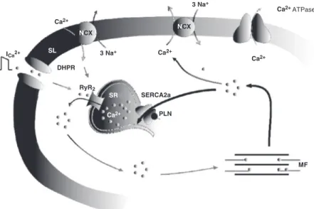
Figure 1 is a schematic illustration of the central players in the excitation-contraction coupling mechanism. Upon depolarization, Ca2+ enters the cell through L-type Ca2+
channels and triggers the release of more Ca2+ from the sarcoplasmic reticulum (SR)
through the activation of the ryanodine re-ceptors. This process, known as Ca2+
-in-duced-Ca2+ release (1), amplifies and
coor-dinates the Ca2+ signal to produce
contrac-tion by interacting with myofilament
pro-teins. The decrease in cytosolic Ca2+ to
pro-duce relaxation is mainly inpro-duced by SR Ca2+-ATPase (SERCA2a), which mediates
Ca2+ uptake into the SR, and, to a lesser
extent, by the Na+/Ca2+ exchanger, which
transfers Ca2+ to the extracellular space (2).
The activity of SERCA2a is under the control of a closely associated SR protein named phospholamban (PLN) (3). In the dephosphorylated form, PLN decreases the apparent affinity of SERCA2a for Ca2+ (4,5).
Phosphorylation of PLN relieves this inhibi-tion, thus increasing SR Ca2+ uptake.
Ex-periments on the intact heart have shown that ß-adrenoceptor stimulation phosphory-lates PLN at the Ser16 residue by
cAMP-dependent protein kinase (PKA) and at the Thr17 site by Ca2+-calmodulin-dependent
protein kinase II (CaMKII). Phosphoryla-tion of these sites reverses the inhibiPhosphoryla-tion of SERCA2a by PLN, thus increasing the af-finity of the enzyme for Ca2+ and the rate of
Ca2+ uptake by the SR. This in turn leads to
an increase in the velocity of relaxation, SR Ca2+ load and, as a consequence, increased
SR Ca2+ release and myocardial contractility
(6-9) (Figure 2). The status of phosphoryla-tion of PLN also depends on the activity of type 1 phosphatase (PP1), the major SR-phosphatase, which specifically dephospho-rylates PLN (10). Although, as stated above, ß-adrenergic stimulation phosphorylates Ser16 and Thr17 of PLN, the role of the
phos-phorylation of Thr17 has been generally
dis-regarded. The present article focuses on the role of the Thr17 site of PLN during
ß-adre-nergic stimulation and under different patho-logical conditions such as acidosis and is-chemia/reperfusion injury.
Role of the phosphorylation of Thr17
of PLN during ß-adrenergic stimulation
Although in vitro studies indicate that PKA and CaMKII phosphorylations are in-dependent of each other (11), earlier
at-Figure 1. Central players of excitation-contraction coupling (ECC) in cardiac muscle. Scheme of the ECC mechanism in cardiac muscle. Upon depolarization, Ca2+ influx through L-type Ca2+ channels or dihydropyridine receptor (DHPR) triggers the release of more Ca2+ from the sarcoplasmic reticulum (SR). This Ca2+ binds to the myofilaments (MF) to produce contraction. Part of the Ca2+ is then extruded outside of the cell through the Na+/ Ca2+ exchange mechanism (NCX), working in the forward mode, but most of it is re-taken up by SR Ca2+-ATPase (SERCA2a). Phospholamban (PLN) is a small SR protein associ-ated with SERCA2a. The state of PLN phosphorylation regulates the activity of this Ca2+ pump. RyR2 = ryanodine receptor; SL = sarcolemma.
Figure 2. Phospholamban phos-phorylation leads to an increase in SR Ca2+ uptake which accel-erates relaxation and enhances SR Ca2+ load. More Ca2+ is then available for release from the SR, resulting in increased con-tractility. Thus, phospholamban is a major regulator of myocar-dial relaxation and contractility. SR = sarcoplasmic reticulum.
ICa2+
Ca2+
Ca2+ 3 Na+
Ca2+
Ca2+ATPase 3 Na+
SERCA2a
PLN RyR2
DHPR
MF
NCX
NCX
SL
SR
tempts to phosphorylate PLN by CaMKII in the intact heart usually failed, unless cellular cAMP levels increased (6,7,12). These find-ings suggested an interaction between the PKA and CaMKII pathways for PLN phos-phorylation. The availability of transgenic models expressing wild-type PLN (PLN-WT), the Ser16→Ala mutant PLN
(PLN-S16A) or the Thr17→Ala mutant PLN
(PLN-T17A) in the cardiac compartment of PLN knock-out mice, and of phosphorylation site-specific antibodies to PLN, which precisely discriminate between the Ser16 and the Thr17
phosphorylation sites, in combination with the quantification of 32P incorporation into
PLN, helped to clarify the relative role of Ser16 and Thr17 phosphorylation (9,13,14).
Experiments in transgenic mice expressing either PLN-WT or the PLN-S16A, indicated that the phosphorylation of Ser16 of PLN is a
prerequisite for the phosphorylation of Thr17
(13). Other experiments conducted with PLN-T17A hearts further indicated that the Ser16 site can be phosphorylated
independ-ently of Thr17in vivo and that
phosphoryla-tion of Ser16 was sufficient to evoke the
maximal effect of ß-adrenergic stimulation (14).
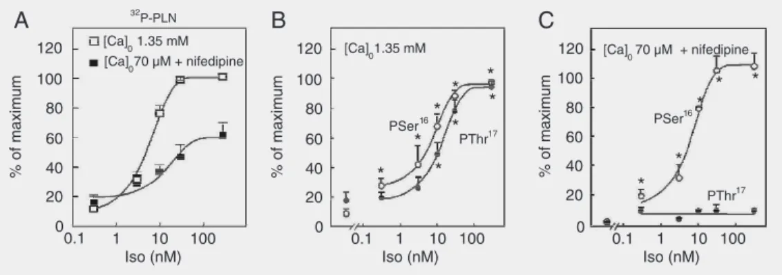
Figure 3A, shows the increase in total PLN phosphorylation produced by increas-ing isoproterenol concentration in the car-diac perfusate. When Ca2+ influx to cardiac
cells was virtually abolished by the presence of low extracellular Ca2+ (Ca2+)
o and the
Ca2+ channel blocker nifedipine to prevent
CaMKII activation, total PLN phosphoryla-tion decreased at the highest isoproterenol concentrations (10, 30, and 300 nM), but did not significantly change at the lowest levels of ß-adrenoceptor stimulation (0.3 and 3 nM). These results suggested that there was no contribution of the CaMKII pathways to the total phosphorylation of PLN at the low-est isoproterenol concentrations. Immuno-detection of site-specific phosphorylated PLN revealed an isoproterenol concentra-tion-dependent increase in both Ser16 and
Thr17 residues of PLN at normal (Ca2+) o
Figure 3. Isoproterenol (Iso) concentration-dependent increase in 32P incorporation into phospholamban (PLN) and in the phosphorylation of Ser16 and Thr17 residues at two different levels of extracellular Ca2+. A, Rat hearts were perfused with 32P and then with different isoproterenol concentrations at two different levels of Ca2+. The virtual suppression of Ca2+ supply to the heart (70 µM (Ca)o + 0.4 µM nifedipine) significantly decreased 32P incorporation into PLN (32P-PLN), at 10, 30 and 300 nM Iso, but failed to affect it at 0.3 and 3 nM Iso. Data are reported as percent maximal 32P incorporation into PLN. B and C, Densitometric analysis of the signal of immunoblots of sarcoplasmic reticulum membrane vesicles isolated from rat hearts perfused with different Iso concentrations at two (Ca2+)
o, and probed with anti-PSer16-PLN and anti-PThr17-PLN. At 1.35 mM (Ca)o, Iso increased the phosphorylation of both Ser16 and Thr17 (B). Phosphorylation of Thr17 was shifted to the right relative to the increase in the phosphorylation of Ser16. The decrease in (Ca2+)o, as shown in panel A, did not significantly affect the phosphorylation of Ser16 but inhibited the increase in the phosphorylation of Thr17 at all Iso concentra-tions (C). Data are reported as percent of the maximal signal achieved in each experimental series. *P < 0.05 compared to no drug values at normal (Ca2+)o (B) and at low (Ca2+)o (C) (Student t-test for unpaired samples). Modified from Ref. 15, with permission.
% of maximum
120
100
80
60
40
20
0
120
100
80
60
40
20
0
120
100
80
60
40
20
0 [Ca] 1.35 mM
0 [Ca]
070 µM + nifedipine
[Ca]
01.35 mM [Ca] 700 µM + nifedipine
PSer16 PThr17
PThr17
* *
* *
*
* *
*
* *
* * *
0.1 1 10 100
Iso (nM)
0.1 1 10 100
Iso (nM)
0.1 1 10 100
Iso (nM)
A 32 B C
P-PLN
% of maximum % of maximum
(Figure 3B). However, when isoproterenol was perfused in the presence of low (Ca2+)
o,
Ser16 was the only site to show a
concentra-tion-dependent increase in phosphorylation (Figure 3C). At each isoproterenol concen-tration, Ser16 was phosphorylated to a
simi-lar extent, independently of the phosphory-lation of Thr17. In order to correlate the
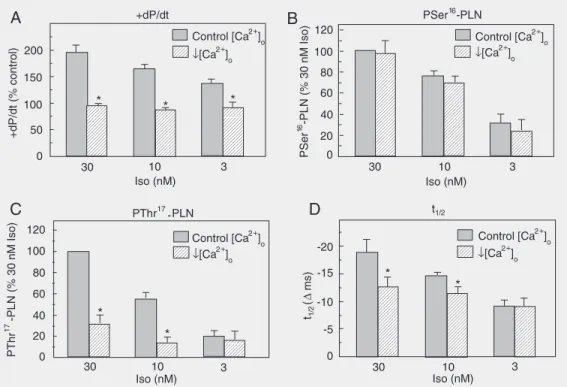
changes observed at the specific phosphory-lation sites of PLN with the mechanical ac-tivity of the heart during ß-adrenergic stim-ulation, Ca2+ supply to the cell was decreased
so as to cancel the positive inotropic effect of each isoproterenol concentration. Figure 4 shows that this maneuver decreased the phosphorylation of Thr17 in association with
a significant reduction of the relaxant effect
of the ß-agonist at 10 and 30 nM renol (9,15). However, at 3 nM isoprote-renol, decreasing (Ca2+)
o had no significant
effect on Thr17 or Ser16 phosphorylation, nor
did it modify the relaxant effect of isoprote-renol, supporting earlier experiments in which total PLN phosphorylation was evalu-ated with 32P (15,16). The lack of a
contribu-tion by Thr17 to the total PLN
phosphoryla-tion at the lowest isoproterenol concentra-tions might be attributed to a modest PKA activity, unable to cause an increase in intra-cellular Ca2+, sufficient to activate CaMKII,
as well as a significant phosphatase inhibi-tion necessary to detect the phosphorylainhibi-tion of the Thr17 residue (15), as it will be
dis-cussed below.
Figure 4. Effects of decreasing (Ca2+)o on mechanical parameters and phospholamban (PLN) phosphorylation sites at different isoproterenol (Iso) concentrations. A, Ca2+ supply to the heart was decreased so as to abolish the Iso-induced positive inotropic effect at the different concentrations. (Ca)o was 0.25 mM at 30 and 10 nM Iso and 0.50 mM at 3 nM Iso. Contractility was evaluated by the maximal rate of rise in pressure (+dP/dt). Phosphorylation of Ser16 of PLN (PSer16-PLN), induced by the different concentrations of Iso, was not affected by the decrease in (Ca2+)o (B); however, this decrease, reduced the phosphorylation of Thr17 (C) as well as the isoproterenol-induced relaxant effect (decrease in half relaxation time, t1/2) (D), at 30 and 10 nM Iso. At 3 nM Iso the decrease in (Ca2+)o did not affect either the Iso-induced phosphorylation of Thr17 or the relaxant effect. +dP/dt is expressed as percentage of control values. t1/2 is expressed as difference from control values (∆ ms). Site-specific phosphoryla-tion of PLN is expressed as percent of the signal obtained with 30 nM Iso at 1.35 mM (Ca)o. *P < 0.05 compared to the corresponding Iso concentration at control (Ca2+)o (Student t-test for unpaired observations). Modified from Ref. 15, with permission.
+dP/dt (% control)
200
150
100
50
0
Control [Ca ]2+o
30 10 3
* * *
+dP/dt
Control [Ca ]2+o PSer -PLN16
120
100
80
40
0 60
20
PSer
-16
PLN (% 30 nM Iso)
t1/2
120
100
40
20
0 80
60
PThr
-PLN (% 30 nM Iso)
17
PThr17-PLN
Control [Ca ]2+o ↓[Ca ]2+
o
↓[Ca ]2+o ↓[Ca ]2+o
30 10 3 30 10 3
-20
-15
-10
-5
0
t
(
1
/2
∆
ms)
Control [Ca ]2+o ↓[Ca ]2+
o
A B
C D
*
*
*
*
Iso (nM) Iso (nM)
Iso (nM) Iso (nM)
Taken together, these findings demon-strate: a) the additive nature of the PKA and CaMKII pathways of PLN phosphorylation, in agreement with in vitro results (11); b)
that both sites equally contribute to the total PLN phosphorylation at the highest levels of ß-adrenergic stimulation; c) that at low iso-proterenol concentrations (≤3 nM), the in-crease in PLN phosphorylation and the re-laxant effect of isoproterenol appear to be exclusively determined by the increase in the phosphorylation of the Ser16 residue.
Phosphorylation of Thr17 of PLN
in the absence of ß-adrenergic stimulation
The conclusion that CaMKII-dependent PLN phosphorylation can only occur in the intact heart in the presence of ß-adrenergic stimulation, i.e., when the cAMP levels within the cell and PKA activity increase (7,12), was in sharp contrast with the inde-pendence of both pathways of PLN phos-phorylation described in in vitro systems
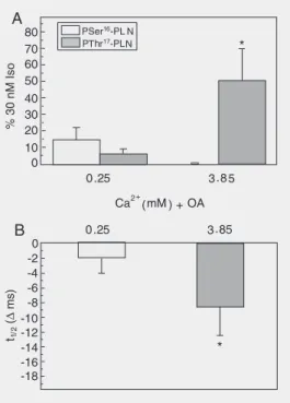
(11). Figure 5 shows that the increase in contractility (intracellular Ca2+) produced by
increasing extracellular Ca2+, in the
pres-ence of the phosphatase inhibitor, okadaic acid, evoked a significant increase in Thr17
phosphorylation associated with a relaxant effect, in the absence of any significant in-crease in cAMP levels or in the phosphory-lation of Ser16 of PLN (9). These results
indicate that the Thr17 residue can be
phos-phorylated independently of Ser16
phospho-rylation, as was described in vitro and
fur-ther emphasize that phosphatases are as im-portant as kinases in determining the level of phosphorylation of any protein. Accord-ingly, phosphorylation of the Thr17 residue
in the intact heart could be detected in two situations: a) in the presence of high extra-cellular Ca2+ (to activate CaMKII) and of
okadaic acid (to inhibit PP1), and b) at high intracellular cAMP, which, by activation of PKA, accounts for both effects, i.e., the
in-crease in intracellular Ca2+ and the inhibition
of PP1.
In the context of the experiments decribed above, it is important to discuss the apparent contradiction between some of the results of the experiments on transgenic mice (13,14) and of phosphorylation experiments con-ducted on animals with an intact PLN (7-9,15). The failure to find PLN phosphoryla-tion (i.e., Thr17 phosphorylation) in
trans-genic mice in which the Ser16 site was
mu-tated to Ala (13) - which would apparently contradict the results of the phosphorylation experiments showing that Thr17 can be
phos-phorylated independently of Ser16
phospho-rylation (9,22) - must be attributed to the fact that the lack of phosphorylation of the Ser16
site under conditions of ß-stimulation pre-cludes the increase in intracellular Ca2+
nec-essary to phosphorylate the Thr17 residue.
Moreover, the experiments showing that in transgenic mice in which the Thr17 site was
mutated to Ala, Ser16 phosphorylation was
sufficient to mediate the maximal response to isoproterenol (14), do not necessarily con-tradict the results of the phosphorylation experiments which indicate that, when both
Figure 5. Effects of increasing (Ca2+)o and inhibition of phos-phatases on the phosphoryla-tion of Thr17 of phospholamban (PLN) and mechanical relax-ation. A, The increase in (Ca2+)o in the presence of okadaic acid (OA) increased the phosphory-lation of Thr17 site of PLN. B, The increased phosphorylation of the Thr17 site was associated with a decrease in half relax-ation time (t1/2). *P < 0.05 com-pared to 0.25 mM Ca2+ + OA. (Student t-test for paired (me-chanical) or unpaired (biochemi-cal) data). Modified from Ref. 9, with permission.
% 30 nM Iso
80 70 60 50 40 30 20 10 0
PSer -PL N16
PThr -PLN17
3.8 5 0.25
3.85 0.25
* *
t
(
ms)
1/
2
∆
0 -2 -4 -6 -8 -10 -12 -14 -16 -18 A
B
ate tension (17). Acidosis produces a rapid decrease in the contraction of cardiac muscle mainly due to a decrease in myofilament Ca2+ responsiveness (18) and an impairment
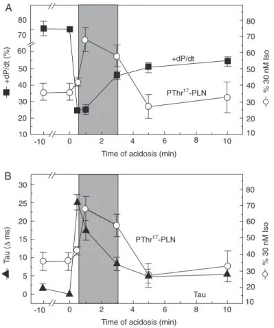
of relaxation, which occurs in spite of the decrease in the responsiveness of the con-tractile proteins and appears to be mainly produced by a direct inhibition of SERCA2a (19). This initial impairment of contractility and relaxation is followed by a spontaneous mechanical recovery. The mechanisms un-derlying this recovery are poorly understood. Figure 6 shows that phosphorylation of the Thr17 site of PLN transiently increases at the
onset of acidosis and is associated with a large part of the contractile and relaxation recoveries, most of which occurred within the first 3 min of acidosis. This phosphoryla-tion would provide a mechanism to over-come the direct depressant effect of acidosis on SERCA2a (19). Whereas Ser16 was not
phosphorylated during the period of acido-sis, the Thr17 site became dephosphorylated
after 5 min of acidosis (20), which would indicate that the phosphorylation of these sites does not contribute to the contractile and relaxation recoveries observed after 5 min of acidosis or that the phosphorylation of the Thr17 residue has “memory”,
trigger-ing a mechanism that persists throughout the period of acidosis. The increase in the phos-phorylation of the Thr17 site at the beginning
of acidosis might be attributed to the in-crease in intracellular Ca2+ that has been
shown to occur during acidosis (19), and to the acidosis-induced phosphatase inhibi-tion (21). These two phenomena provide the necessary conditions to increase the phos-phorylation of the Thr17 site (22).
Dephos-phorylation of the Thr17 residue would occur
after the recovery of phosphatase activity due to the return of intracellular pH to values close to control (23). Consistent with the important role of Thr17 phosphorylation
in the mechanical recovery from acidosis, experiments by DeSantiago et al. (24) showed absence of mechanical recovery in
myo-Figure 6. Effect of acidosis on left ventricular function and on the phosphorylation of Thr17 of phospholamban (PLN). Hypercapnic acidosis produced an initial (30 s) decrease in con-tractility (maximal rate of rise in pressure, +dP/dt) (A) and an antirelaxant effect (prolonga-tion of the time constant of left ventricular developed pressure decay, Tau) (B), followed by mechanical recovery, most of which occurred during the first 3 min of acidosis. Phosphory-lation of the Thr17 site of PLN increased simultaneously with the early recovery of contractil-ity and relaxation (A and B) and then returned to basal levels. Phosphorylation of Ser16 remained at basal levels throughout the acidosis period (not shown). Phosphorylation of Thr17 is reported as indicated in Figure 4C. +dP/dt is reported as percent of basal values. Tau is reported as ∆ ms of basal values. *P < 0.05 compared to basal values (Student t-test for paired (mechanical) or unpaired (biochemical) data). Modified from Ref. 20, with permis-sion.
sites are present, they both contribute to the total phosphorylation of PLN and to the mechanical effect of ß-adrenoceptor stimu-lation (7,9,15).
Role of the phosphorylation of Thr17
of PLN in pathological conditions
Acidosis
Intracellular acidosis is associated with a decrease in the ability of the heart to
gener--10 0 2 4 6 8 10
Time of acidosis (min) +dP/dt
PThr -PLN17
80
70
60
50
40
30
20
10
+dP/dt (%)
80
70
60
50
40
30
20
10
PThr -PLN17
80
70
60
50
40
30
20
10 30
25
20
15
10
5
0
T
au (
ms)
∆
-10 0 2 4 6 8 10
Time of acidosis (min)
% 30 nM Iso
A
B
Tau
cytes lacking PLN. Finally, a more promi-nent role of the phosphorylation of PLN residues might be expected in vivo. Sys-temic acidosis is known to increase sympa-thetic nerve activity (25). This increased sympathetic tone may produce a more persistent phosphorylation of both the Thr17
and Ser16 residues of PLN, with a further
contribution to mechanical recovery.
Stunning
Ischemic heart diseases still constitute the leading cause of mortality and morbidity in industrialized countries. Myocardial stun-ning is recognized as the reversible decrease in myocardial contractility that follows a brief ischemic episode, clinically manifested as sluggish recovery of the pump function
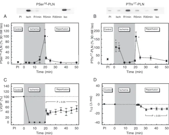
Figure 7. Time course of phospholamban (PLN) Ser16 and Thr17 phosphorylation and of left ventricular function during ischemia and reperfusion. Representative immunoblots (A and B, above) and overall results of densitomet-ric analysis of the signals of inmunoblots (A and B, below) of sarcoplasmic reticulum membrane vesicles isolated from hearts freeze-clamped after 20 min of global ischemia (Isch) and at 1, 5, and 30 min during reperfusion (R1-R30 min). The blots were probed with anti PSer16-PLN (A) and anti PThr17-PLN (B). Phosphorylation of PLN sites induced by 30 nM isoproterenol (Iso) in non-ischemic hearts is shown in the blots for reference. Ser16 phosphory-lation significantly increased after 20 min of ischemia and Thr17 phosphorylation transiently increased at 2-5 min of ischemia and at 1 min of reperfusion. Left ventricular developed pressure (LVDP) (C) decreased to virtually non-detectable levels after cessation of flow and reperfusion resulted in a partial recovery of contractility. In spite of the impairment in contractility, t1/2 (D) recovered immediately after ischemia and then significantly decreased below pre-ischemic values (PI), indicating an acceleration of ventricular relaxation. Points represent the mean ± SEM of data from 51 hearts freeze-clamped at 20 min of ischemia and 1, 2, 3, 5, 15, and 30 min of reperfusion. LVDP is reported as percent PI and t1/2 as ∆ ms of PI. Site-specific PLN phosphorylation is reported as shown in previous figures. *P < 0.05 compared to PI (Student t-test for paired (mechanical) or unpaired (biochemical) data). Modified from Ref. 35, with permission.
PSer
16-PLN (% 30 nM Iso) 140 120 100 80 60 40 20
140 120 100 80 60 40
20 0
LVDP (%)
Control Ischemia Reperfusion
P < 0.05
Control Ischemia Reperfusion Control Ischemia Reperfusion
Control Ischemia Reperfusion
PI Isch R1min R5min R30min Iso
0 PThr
17-PLN (% 30 nM Iso) 200
150
100
50
0
40
20
0
-20
-40 t1/2
(
∆
ms)
PI 0 10 20 30 40 50
Time (min)
PI 0 10 20 30 40 50
Time (min)
PI 0 10 20 30 40 50
Time (min)
PI 0 10 20 30 40 50
Time (min)
PI Isch R1min R5min R30min Iso
*
* *
*
P < 0.05
PSer16-PLN PThr17-PLN
A
C
B
after revascularization (26,27). Although the phenomenon of myocardial stunning was de-scribed over 25 years ago (28), the mechan-isms responsible for the delayed recovery of contractile function are not completely clear (29). Different types of evidence obtained in rodents and humans indicate that the ultimate cause of the alteration of myocardial contrac-tility is a decrease in myofilament Ca2+
re-sponsiveness, since the Ca2+ transient is not
altered, although contractility is decreased (29). On the other hand, experimental evidence indicates that the function of the SR is altered both in reversible and irreversible ischemia-reperfusion injury (30-34). In particular, in the case of myocardial stunning, a decrease in the activity of SERCA2a and/or in the rate of Ca2+
reuptake by the SR has been described in several species (31-34). Thus, an obvious ques-tion is: why does the intracellular Ca2+
tran-sient remain unaltered in species in which SR function is depressed? A possible explanation for this apparent contradiction is that compen-satory mechanisms can overcome the de-pressed SERCA2a activity. One possible com-pensatory mechanism is the phosphorylation of PLN. To test this hypothesis, we studied the time course of phosphorylation of PLN resi-dues at different times during ischemia/reper-fusion (35). Figure 7 shows the time course of left ventricular developed pressure (LVDP), half relaxation time (t1/2) and the
phosphoryla-tion of Ser16 and Thr17 residues of PLN during
ischemia and reperfusion. LVDP decreased to virtually undetectable levels after cessation of
flow. Reperfusion resulted in a partial recov-ery of LVDP. In spite of the reduction of contractility, t1/2 recovered immediately after
ischemia and then significantly decreased be-low pre-ischemic values, indicating an accel-eration of ventricular relaxation. Phosphory-lation of Ser16 was significantly increased at
the end of ischemia, but returned to basal values during the reperfusion period. Phos-phorylation of the Thr17 residue of PLN
tran-siently increased both at the beginning of ischemia (2-5 min) and at the beginning of reperfusion (1 min), remaining at pre-ischemic values for the rest of the reperfusion period. The decrease in Thr17 phosphorylation by
CaMKII inhibition, prolonged t1/2, which
indi-cates that the phosphorylation of Thr17, when
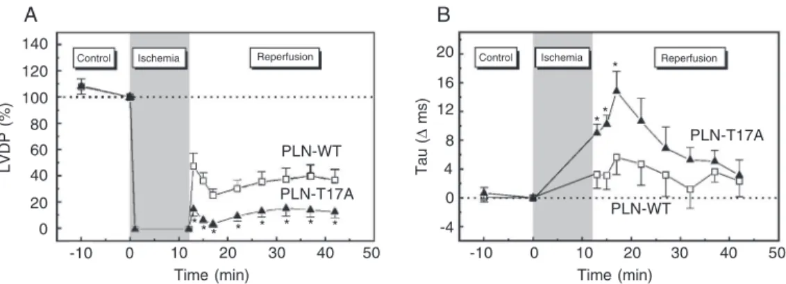
present, attenuates the impaired relaxation that occurs at the beginning of reperfusion (36). Figure 8 shows the time course of contractile and relaxation parameters during ischemia and reperfusion in PLN-WT and PLN-T17A mutant hearts. The recovery of LVDP was significantly lower in PLN-T17A mutant hearts than in PLN-WT hearts throughout the reper-fusion period, whereas the time constant of pressure decay, Tau, was significantly pro-longed at the beginning of reperfusion and then recovered to values close to control. Ad-ditional experiments also showed that the con-tractile recovery was depressed in PLN-S16A mutant hearts compared to the heart of PLN-WT mice. However, this depression only oc-curred at the beginning of reperfusion (36). These results indicate that phosphorylation of
Figure 8. Functional role of Thr17 phosphorylation in the ischemia-reperfused mouse heart. Time course of left ventricular devel-oped pressure (LVDP; A) and Tau (B) during ischemia and re-perfusion in wild-type phospho-lamban (PLN-WT) and PLN with a Thr17 to Ala mutation (PLN-T17A) mouse hearts. Mutation of Thr17 to Ala is associated with diminished contraction and re-laxation recoveries after the is-chemic insult compared to PLN-WT mouse hearts. *P < 0.05 compared to PLN-WT (Student
t-test for unpaired observations). Modified from Ref. 36, with per-mission.
LVDP (%)
140
120
100
80
60 40
20
Control Ischemia Reperfusion Control Ischemia Reperfusion
0
16
12
8
4
0
0 20 30 40 50
Time (min)
-10 0 20 30 40 50
Time (min) *
** *
10 10
-4 20
Tau (
∆
ms)
A B
* * * * * * * PLN-T17A
PLN-T17A
PLN-WT PLN-WT
Thr17, although transient, may play a
signifi-cant role in the critical phase of initial reper-fusion, favoring Ca2+ re-uptake by the SR.
This would tend to compensate for the de-pression of SERCA2a and would ameliorate Ca2+ overload, typical of the beginning of
reflow (29). Moreover, these experiments indicate that activation of SERCA2a by any other means, at this crucial moment of reper-fusion, may contribute significantly to Ca2+
handling. In agreement with this view, re-cent experiments suggested that the cardio-protection produced by cGMP-mediated stimuli in reoxygenated cells is associated with an increase in the phosphorylation of Ser16 of PLN (37).
Conclusions
Taken together, these findings suggest that the Thr17 site of PLN has an important
role, not only under physiological condi-tions, such as ß-adrenoceptor stimulation, but also in the mechanical recovery under some pathological conditions such as acido-sis and stunning. An interesting point is that, although the phosphorylation experiments reveal that the phosphorylation of Thr17 is
transient and occurs only at the beginning of both acidosis and reperfusion, the presence of this residue and/or of an intact PLN seems to be necessary for mechanical recovery dur-ing the reperfusion or acidosis period.
References
1. Fabiato A & Fabiato F (1997). Calcium release from the sarcoplas-mic reticulum. Circulation Research, 40: 119-129.
2. Bers DM (2001). Excitation-Contraction Coupling and Cardiac Con-tractile Force. 2nd edn. Kluwer Academic Publishers, Dordrecht, Netherlands.
3. Tada M, Kirchberger MA & Katz AM (1975). Phosphorylation of a 22,000-dalton component of the cardiac sarcoplasmic reticulum by adenosine 3',5'-monophosphate-dependent protein kinase. Jour-nal of Biological Chemistry, 250: 2640-2647.
4. James P, Inui M, Tada M et al. (1989). Nature and site of phosphola-mban regulation of the Ca²+ pump of sarcoplasmic reticulum. Na-ture, 342: 90-92.
5. Kim HW, Steenaart NA, Ferguson DG et al. (1990). Functional reconstitution of the cardiac sarcoplasmic reticulum Ca2+-ATPase with phospholamban in phospholipid vesicles. Journal of Biological Chemistry, 265: 1702-1709.
6. Lindemann JP, Jones LR, Hathaway DR et al. (1983). ß-Adrenergic stimulation of phospholamban phosphorylation and Ca2+-ATPase activity in guinea pig ventricles. Journal of Biological Chemistry, 258: 464-471.
7. Vittone L, Mundiña C, Chiappe de Cingolani G et al. (1990). cAMP and calcium dependent mechanisms of phospholamban phosphory-lation in intact hearts. American Journal of Physiology, 258: H318-H325.
8. Napolitano R, Vittone L, Mundiña-Weilenmann C et al. (1992). Phos-phorylation of phospholamban in the intact heart. A study on the physiological role of the Ca-calmodulin-dependent protein kinase system. Journal of Molecular and Cellular Cardiology, 24: 387-396. 9. Mundiña-Weilenmann C, Vittone L, Ortale M et al. (1996). Immuno-detection of phosphorylation sites gives new insights into the mechanisms underlying phospholamban phosphorylation in the in-tact heart. Journal of Biological Chemistry, 271: 33561-33567. 10. MacDougall LK, Jones LR & Cohen P (1991). Identification of the
major protein phosphatases in mammalian cardiac muscle which dephosphorylate phospholamban. European Journal of Biochemis-try, 196: 725-734.
11. Bilezikjian LM, Kranias EG, Potter JD et al. (1981). Studies on phosphorylation of canine cardiac sarcoplasmic reticulum by cal-modulin-dependent protein kinase. Circulation Research, 49: 1356-1362.
12. Lindemann JP & Watanabe AM (1985). Phosphorylation of phos-pholamban in intact myocardium. Role of Ca2+ -calmodulin-depend-ent mechanisms. Journal of Biological Chemistry,260: 4516-4525. 13. Luo W, Chu G, Sato Y et al. (1998). Transgenic approaches to
define the functional role of dual site phospholamban phosphoryla-tion. Journal of Biological Chemistry, 273: 4734-4739.
14. Chu G, Lester JW, Young KB et al. (2000). A single site (Ser16) phosphorylation in phospholamban is sufficient in mediating its maxi-mal cardiac responses to ß-agonists. Journal of Biological Chemis-try, 275: 38938-38943.
15. Said M, Mundiña-Weilenmann C, Vittone L et al. (2002). The relative relevance of phosphorylation of the Thr17 residue of phospholamban is different at different levels of ß-adrenergic stimulation. Pflügers Archiv, 444: 801-809.
16. Mundiña de Weilenmann C, Vittone L, de Cingolani G et al. (1987). Dissociation between contraction and relaxation: the possible role of phospholamban phosphorylation. Basic Research in Cardiology, 82: 507-516.
17. Gaskell WH (1880). On the tonicity of the heart and blood vessels.
Journal of Physiology,3: 48-75.
18. Fabiato A & Fabiato F (1978). Myofilament-generated tension oscil-lations during partial calcium activation and activation dependence of the sarcomere length-tension relation of skinned cardiac cells.
Journal of General Physiology, 72: 667-699.
myo-cytes. American Journal of Physiology, 275: H977-H987.
20. Mundiña-Weilenmann C, Ferrero P, Said M et al. (2005). Role of phosphorylation of Thr17 residue of phospholamban in mechanical recovery during hypercapnic acidosis. Cardiovascular Research, 66: 114-122.
21. Mundiña-Weilenmann C, Vittone L, Cingolani HE et al. (1996). Ef-fects of acidosis on phosphorylation of phospholamban and tropo-nin I in rat cardiac muscle. American Journal of Physiology, 270: C107-C114.
22. Vittone L, Mundiña-Weilenmann C, Said M et al. (1998). Mechan-isms involved in the acidosis enhancement of the isoproterenol-induced phosphorylation of phospholamban in the intact heart. Jour-nal of Biological Chemistry,273: 9804-9811.
23. Cingolani HE, Koretsune Y & Marban E (1990). Recovery of con-tractility and pHi during respiratory acidosis in ferret hearts: role of Na+-H+ exchange. American Journal of Physiology, 259: H843-H848.
24. DeSantiago J, Maier LS & Bers DM (2004). Phospholamban is required for CaMKII-dependent recovery of Ca transients and SR Ca reuptake during acidosis in cardiac myocytes. Journal of Molec-ular and CellMolec-ular Cardiology, 36: 67-74.
25. Brofman JD, Leff AR, Munoz NM et al. (1990). Sympathetic secre-tory response to hypercapnic acidosis in swine. Journal of Applied Physiology, 69: 710-717.
26. Braunwald E & Kloner RA (1982). The stunned myocardium: Pro-longed postischemic ventricular dysfunction. Circulation, 66: 1146-1149.
27. Kusuoka H, Porterfield JK, Weisman HF et al. (1987). Pathophysiol-ogy and pathogenesis of stunned myocardium. Depressed Ca2+ activation of contraction as a consequence of reperfusion-induced cellular calcium overload in ferret hearts. Journal of Clinical Investi-gation,79: 950-961.
28. Heyndrickx GR, Millard RW, McRitchie RJ et al. (1975). Regional
myocardial functional and electrophysiological alterations after brief coronary artery occlusion in conscious dogs. Journal of Clinical Investigation, 56: 978-985.
29. Bolli R & Marbán E (1999). Molecular and cellular mechanism of myocardial stunning. Physiological Reviews, 79: 609-634. 30. Rapundalo ST, Briggs FN & Feher JJ (1986). Effects of ischemia on
the isolation and function of canine cardiac sarcoplasmic reticulum.
Journal of Molecular and Cellular Cardiology,18: 837-851. 31. Krause SM, Jacobus WE & Becker LC (1989). Alterations in cardiac
sarcoplasmic reticulum calcium transport in the postischemic “stunned” myocardium. Circulation Research, 65: 526-530. 32. Limbruno U, Zucchi R, Ronca-Testoni S et al. (1989). Sarcoplasmic
reticulum function in the “stunned” myocardium. Journal of Molecu-lar and CelluMolecu-lar Cardiology, 21: 1063-1072.
33. Zucchi R, Ronca-Testoni S, Di Napoli P et al. (1996). Sarcoplasmic reticulum calcium uptake in human myocardium subjected to ische-mia and reperfusion during cardiac surgery. Journal of Molecular and Cellular Cardiology,28: 1693-1701.
34. Zucchi R, Cerniway RJ, Ronca-Testoni S et al. (2002). Effect of cardiac A1 adenosine receptor overexpression on sarcoplasmic reticulum function. Cardiovascular Research, 53: 326-333. 35. Vittone L, Mundiña-Weilenmann C, Said M et al. (2002). Time course
and mechanisms of phosphorylation of phospholamban residues in ischemia-reperfused rat hearts. Dissociation of phospholamban phosphorylation pathways. Journal of Molecular and Cellular Cardi-ology,34: 39-50.
36. Said M, Vittone L, Mundiña-Weilenmann C et al. (2003). Role of dual-site phospholamban phosphorylation in the stunned heart: in-sights from phospholamban site-specific mutants. American Journal of Physiology, 285: H1198-H1205.