From the Disciplines of Endocrinology, Physiology and Biostatistics, Faculty of Medicine of the Triângulo Mineiro and Disciplines of General Pathology and Physiology, University of Uberaba.
Received for publication on March 18, 2002.
C CELLS IN THE THYROID OF PINEALECTOMIZED
RATS
Marcus Aurelho de Lima, Lilian Margareth Biagioni de Lima, Leandro Luís Lopes Freitas, Patrícia Fernanda Toledo Barbosa, Maria de Fátima Borges, Luiz Reis and Gilberto de Araujo Pereira
LIMA MA de et al. - C cells in the thyroid of pinealectomized rats. Rev. Hosp. Clín. Fac. Med. S. Paulo 58(1):21-26, 2003.
PURPOSE: To study quantitatively C cells in the thyroids of non-isogenic rats to determine the possible effects of pinealectomy on the number of these cells, and consequently on the synthesis and secretion of calcitonin.
METHODS: Twenty male rats of an outbred strain (200-300 g) were used in the present study. One group of 10 animals was pinealectomized 50 days prior to sacrifice. Thyroid tissue was stained for calcitonin (Dako Corporation) at a 1:1500 dilution. The number of C cells observed was expressed as number of cells/cm2. Data were analyzed statistically by
Mann-Whitney test.
RESULTS: The number of C cells in pinealectomized and normal animals ranged from 489 to 2084 per cm2 and 227 to
1584 per cm2, respectively, a difference that was statistically significant (P <0.05).
CONCLUSIONS: These results showed consistent differences in the number of C cells after pinealectomy when compared to controls. We believe that pinealectomy increases the number of C cells in the rat thyroid.
DESCRIPTORS: Thyroid. C Cells. Pineal Gland. Rat. Calcitonin.
INTRODUCTION
There is some evidence that the pineal gland is involved in various en-docrine functions, and its effect on the thyroid has been investigated in par-ticular, by several authors who suggest that melatonin inhibits thyroid func-tion by suppressing the producfunc-tion of pituitary TSH1-3, while others state that
melatonin has a stimulatory effect4,5, or
that its effects on the thyroid are neg-ligible6,7. Thus, this is a highly
contro-versial topic.
Few studies are available concern-ing the effect of the pineal gland on the metabolism and number of C cells8. Although some studies have
shown an increase in the number of these cells9-11, of their
cytoplasm/nu-cleus ratio and of the number of secre-tary granules12 after pinealectomy (PX),
McMillan et al.13 detected no
signifi-cant differences in any of these param-eters following PX.
In view of these considerations, the objective of the present investigation was to study C cells quantitatively in non-isogenic rats to determine the pos-sible effects of PX on the number of these cells and consequently on the synthesis and secretion of calcitonin.
MATERIALS AND METHODS
Sample section
The study was conducted on 20 non-isogenic male Wistar rats (Rattus norvegicus) weighing 200 to 300 g. The animals were divided into 2 groups: control (normal rats receiving sham operations, N = 10) and pinealectomized (N = 10).
Pinealectomy
Pinealectomy was performed using the method of Haldar-Misra14. The
region selected for craniotomy was shaved and disinfected, the skin was re-tracted with a separator, and the skull was exposed. Using a circular serrate burr, craniotomy was performed at the level of the lambdoid suture. The bone fragment was removed and placed in an appropriate vessel containing physi-ological saline. The sagittal sinus was separated, the pineal gland was fully re-moved by pinching, and the bone frag-ment was returned to its place. The vari-ous anatomical planes were then su-tured. Both groups (control and pinealectomized) underwent prophy-lactic antibiotic treatment and were re-turned to their cages for recovery.
Sham surgery was performed fol-lowing the same procedure as de-scribed above, except that the pineal gland was not removed.
Thyroid collection
Under inhalation ether anesthesia, the chest of the animals was opened and the heart exposed. The left ventri-cle was perfused first with physiologi-cal saline (0.9% NaCl) until blanching of the organs was achieved (requiring about 300 mL) and then with 4% buff-ered formalin, pH 7.4, until the animal stiffened (requiring about 100 mL). The pineal region was investigated, and the gland was completely re-moved. After perfusion, the thyroid was collected together with the trachea and placed in a glass vessel containg buffered formalin, pH 7.4. The in-terval between pinealectomy and re-moval of the thyroid was 50 days.
Thyroid analysis
After dissection and fixation in 4% formalin, the thyroid-trachea specimen was cut crosswise at 1 to 2 mm inter-vals, and all fragments were dehy-drated in an increasingly concentrated ethyl alcohol series (70%, 80%, 90%, and absolute alcohol), cleared with
xylene, embedded in paraffin, and cut on a microtome into 4 mm-thick sec-tions. The sections were then dried in an oven at 37°C; some of the slides were stained with hematoxylin-eosin, and some by the immunohistochemi-cal technique for later histologiimmunohistochemi-cal analysis. The whole thyroid was stud-ied with semi-serial sections.
Immunohistochemical technique
Anticalcitonin antibody at a 1:1500 dilution was used for immuno-histochemical analysis. Sections were deparaffinized, hydrated, and pretreated with 0.01 M sodium citrate buffer in a microwave oven for 5 cy-cles of 3 minutes each at maximum power (900 W). The volume was com-pleted with sodium citrate buffer in each cycle to prevent section drying. The slides were left to rest for 20 min-utes and were then immersed in phos-phate-buffered saline (PBS), pH 7.4, for 10 minutes. The sections were then incubated with normal horse serum for 20 minutes and covered with the anti-body at the same dilution for 22 hours at 4°C. The avidin-biotin system was
used, and the material was processed using 3-amino-9-ethyl-carbazole (AEC A-5754, Sigma) as the chromogen. The sections were counterstained with Mayer hematoxylin, and a fragment of normal thyroid from an adult rat was used as positive control. The negative control was a sample of the case to be evaluated that had not been treated with the primary antibody.
Cell counts
The slides containing the thyroid fragments were submitted to micro-scopic analysis for C cell counts using a light microscope. The cells were counted in a 1 cm2 area defined with a
grid considering color (red, due to the action of AEC), morphology, and to-pography. The mean number of cells/ field was determined for each case by 2 observers.
Statistical analysis
The Mann-Whitney test was used to compare the results between normal and pinealectomized animals, with the level of significance set at 5%.
Ethics
This study conformed with the guiding principles of the Declaration of Helsinki.
RESULTS
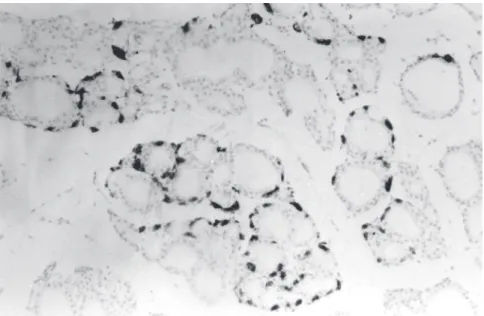
The histological sections stained with hematoxylin-eosin did not show any alterations in thyroid morphology. When the immunohistochemical method was used, C cells, which were stained red-brownish, were concen-trated in the lobes of the gland and were absent in the isthmus.
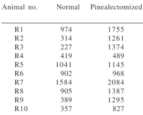
The number of C cells ranged from 227 to 1584 cells/cm2 in the normal
group and from 489 to 2084 cells/cm2
in pinealectomized animals (Table 1). The data showed a statistically signifi-cant (P <0.05) difference in C cell number in pinealectomized rats.
DISCUSSION
The results obtained in the present study did demonstrate a statistically significant increase in C cells in the thyroids of the pinealectomized group. These data disagree with those reported by Brammer et al.7 and McMillan et
al.13 in studies on rats in which they
found no evidence of a significant in-teraction between the pineal gland and the hypothalamus-pituitary-thyroid axis. However, these results agree with those reported in other studies, which have suggested the influence of the pineal gland on the number of the C cells of the thyroid11,13.
With respect to the arrangement of C cells, the observation that they were concentrated in the lobes of the gland and were practically absent in the isth-mus has also been reported by McMillan et al.13, and our data
con-firm his report. The same author stated that there were no significant differ-ences in C cell numbers between the right and left thyroid lobe.
Several studies have been con-ducted to define the relationship be-tween the pineal and the thyroid. In 1966, Ishibashi et al. proposed that PX stimulates the release of thyroxin. In 1972, Relkin13 suggested that the
pin-eal gland inhibits the secretion of thy-roid hormones, and in 1976 he ob-served a reduction in plasma TSH lev-els after intraventricular injection of melatonin in the rat14. Malendowicz
and Woznicki15 in 1986 supported this
finding of an inhibitory effect of the pineal gland on thyroid function in a
study of the role of pinealectomy and melatonin in Wistar rats, as did Haldar and Pandey16 in 1988 in a study of
pinealectomy in squirrels.
However, there is not a consensus among investigators regarding this in-hibitory effect of the pineal gland on the thyroid. In 1970, Rowe et al.6
ob-served that PX did not affect plasma TSH levels. In 1979, Brammer et al.7
observed that the TRH content of the hypothalamus, the TSH content of the pituitary and plasma, and the serum levels of free T3 and T4 were not af-fected by PX. Additionally, Peschke et al.17 showed that PX may induce a
state of prolonged hypothyroidism. Several studies18,19 have shown that
physical stress such as anesthesia, sur-gery, and electric shock inhibits the thyroid-pituitary axis. Although tran-sitory, the effect of PX on the response to TSH may be specific and may not be due to surgical stress. Barbot et al.20
suggested that TSH regulates calci-tonin synthesis in a manner similar to the regulation of thyroid hormones, and experimental studies21,22 have
sug-gested that TSH hypersecretion may be responsible for C cell hyperplasia. Cur-rently, we know that C cells, although inserted in the thyroid parenchyma, have an action that is independent of TSH and of the thyroid hormones.
The pineal gland could influence two important components of the cal-cium-regulating system in the body— that is, parathyroid and C cells—with an action that is at times stimulating and at times inhibitory9,10. Animal data
indicate that pineal melatonin is in-volved in the regulation of calcium and phosphorus metabolism by stimu-lating the activity of the parathyroid glands and inhibiting calcitonin re-lease and prostaglandin synthesis21-23.
Hence, the pineal gland may function as a “fine tuner” of calcium homeostasis.
With respect to the number and ac-tivity of thyroid C cells, Krstic10
de-Table 2 - Analyses of variance (Mann-Whitney test) of the number of C cells/cm2
between pinealectomized and normal animals.
Animals Median Minimum And Sum Ranks U Statistic Mann-Whitney
C cells/cm2 Maximum P value
Normal 661 227 1584 7 3 1 8 0,015
Pinealectomized 1278 489 2084 1 3 7
Table 1 - Number of C cells/cm2 of
thyroid in normal and pinealectomized rats.
Animal no. Normal Pinealectomized
R1 974 1755
R2 314 1261
R3 227 1374
R4 419 489
R5 1041 1145
R6 902 968
R7 1584 2084
R8 905 1387
R9 389 1295
tected hyperplasia and hypertrophy of these cells after PX. More recently, McMillan et al.13 compared the effects
of PX and sham surgery in Wistar rats studied during different seasons of the year and observed no significant dif-ferences in the number or volume of C cells of rats undergoing PX or sham surgery, nor did they detect any impor-tant seasonal variation in these param-eters.
Thus, we believe that the effect of the pineal gland on C cells may be discrete, with little influence on the number or size of these cells. However, the data obtained in the present study
regarding rat C cells will be valuable for future comparative and experimen-tal studies.
On the other hand, the reduction in melatonin cannot be considered the only explanation for the effect of pinealectomy, since this hormone is also synthesized in the retina and in enterochromaffin cells26,27. After PX,
these tissues may gradually assume the role of the pineal gland.
Consequently, future studies should be performed to observe the ef-fect of pinealectomy on C cell prolif-eration and function over time, con-tributing to the elucidation of the
re-lationship between C cells and the pin-eal gland. In summary, this study pro-vides preliminary evidence for the po-tential efficacy of melatonin for con-trolling C cell proliferation, and pos-sibly for co-treatment of medullary carcinoma. Therefore, it should be ex-tended by other experiments related to both C cell proliferation and secretion.
ACKNOWLEDGMENTS
The research was supported by FAPEMIG (5000599) and CNPq (800908/92-9).
RESUMO
LIMA MA de e col. - Células C em tireóides de ratos pinealecto-misados. Rev. Hosp. Clín. Fac. Med. S. Paulo 58(1): 21-26, 2003.
OBJETIVO: Estudar quantitativa-mente células C de tireóides em ratos
não isogênicos para determinar os pos-síveis efeitos da pinealectomia no nú-mero destas células e conseqüentemen-te na sínconseqüentemen-tese e secreção de calcitonina.
MÉTODO: Vinte ratos machos com peso de 200 a 300g foram utili-zados no presente estudo. Um grupo de
pela análise de variância (ANOVA) e teste Mann-Whitney.
RESULTADOS: O número de cé-lulas C em animais pinealectomizados e normais variou de 489 a 2084 célu-las/cm2 e 227 a 1584 células/cm2,
res-pectivamente.
CONCLUSÕES: Estes resultados mostraram diferenças consistentes (p<0,05) no número de células C após a pinealectomia quando comparada aos controles. Nós acreditamos que a
pinealectomia aumenta o número de células C nas tireóides dos ratos.
DESCRITORES: Tiróide. Células C. Glândula Pineal. Rato. Calcito-nina.
REFERENCES
1 . RELKIN R - Effects of pinealectomy and constant light and darkness on thyrotropin levels in the pituitary and plasma of the rat. Neuroendocrinology 1972; 10:46-52.
2 . MANNISTO PT, TOIVONEN M, TUOMINEN R - Time relationships and reversal by two indoleamines of the pinealectomy-inhibited thyrotropin secretion in male rats. Med Biol 1987; 65:267-272.
3 . MESS B, PETER L - Effect of intracerebral serotonin administration on pituitary-thyroid function. Endocrinol Exp 1975; 9 :105-113.
4 . ISHIBASHI T, HAHN DW, SRIVASTAVA L et al. - Effect of pinealectomy and melatonin on feed consumption and thyroid hormone secretion rate. Proc Soc Exp Biol Med 1966; 122 :644-647.
5 . SOUTTO M, GUERRERO JM, OSUNA C et al. - Nocturnal increases in the triiodothyronine/ thyroxine ratio in the rat thymus and pineal gland follow increases of type II 5’ – deiodinase actvity. Int J Biochem Cell Biol 1998; 30: 235-241.
6 . ROWE JW, RICHERT JR, KLEIN DC, REICHLIN S - Relation of the pineal gland and environmental lighting to thyroid function in the rat. Neuroendocrinology 1970; 6:247-254.
7 . BRAMMER GL, MORLEY JE, GELLER E et al. - Hypothalamus-pituitary-thyroid axis interactions with pineal gland in the rat. Am J Physiol 1979; 236:416-420.
8 . KHAVINSON VKH, KVETNOI IM, POPUCHIEV UV et al. -Effect of pineal peptides on neuroendocrine system after pinealectomy. Arkr Patol 2001; 63: 18-21.
9 . MILINE R, KRSTIC R - Sur l’histophysiologie correlative de la glande pineale et des glandes parathyroides. Z. Zellforsch Mikrosk Ana 1966; 69:428-437.
10. KRSTIC R - Die treffermethode in der Messung der Parathyreozytenaktivitat nach der Epiphysektomie. Esperientia 1966; 22:336-337.
11. CSABA G, BARATH P - The effect of pinealectomy on the parafollicular cells of the rat thyroid gland. Acta Anat 1974; 88:137-146.
12. CSABA G, BARATH P - Morphological changes of the thymus and the thyroid gland after postnatal extirpation of pineal body. EndocrinolExp 1975; 9:59-67.
13. MCMILLAN PJ, HEIDBUCHEL U, VOLLRATH L - Number and size of rat thyroid C cells: no effect of pinealectomy. Anat Rec 1985;212:167-171.
14. WAYNFORTH HB, FLECKNELL PA - Specific surgical operations. In: WAYNFORTH HB, FLECKNELL PA -Experimental and surgical technique in the rat. 2ed. San Diego, CA, Academic Press, 1992. p.279-284.
16. HALDAR C, PANDEY R - Effect of pinealectomy and photoperiod on testis and thyroid gland of the Indian tropical palm-squirrel Funambulus pennanti. Indian J Exp Biol 1988; 26:516-519.
17. PESCHKE D, PESCHKE E, PEIL J et al. - Circannual serum creatine kinase patterns after ganglionectomy and pinealectomy of the Wistar rat. J Pineal Res 1989; 6:375-383.
18. FORTIER C, DELGADO A, DUCOMMUN P et al. - Functional interrelationships between adenohypophysis, thyroid, adrenal cortex and gonads. Can Med Assoc J 1970; 103:864-874. 19. SIMPKINS JW, HODSON CA, MEITES J - Differential effects of
stress on release of thyroid-stimulating hormone in young and old male rats. Proc Soc Exp Biol Med 1978; 157 :144-147.
20. BARBOT N, GUYETANT S, BELDENT V et al. - Thyroidite chronique auto-immune et hiperplasie des cellules C. Ann Endocrinol 1991; 52:109-112.
21. BIDDINGER PW, BRENNAN MF, ROSEN PP - Symptomatic C-cell hyperplasia associated with chronic lymphocytic
thyroiditis. Am J Surg Pathol 1991; 15:599-604.
22. KATOH R - Experimental thyroid tumorigenesis induced by 3-amino-1,2,4-triazole (AT) and diidropropanolnitrosamine (DIPN). J Iwate Med Ass 1983; 13:379-405.
23. HAKANSON DO, BERGSTROM WH - Pineal and adrenal effects on calcium homeostasis in the rat. Pediatr Res 1990; 27: 571-3 .
24. HAKANSON DO, PENNY R, BERGSTROM WH - Calcemic responses to photic and pharmacologic manipulation of serum melatonin. Pediatr Res 1987; 22: 414-6.
25. SANDYK R, ANASTASIADIS PG, ANNINOS PA et al. - Is post menopausal osteoporosis related to pineal gland functions? Intern J Neuroscience 1992; 62: 215-25.
26. PANG SF, BROWN GM - Regional concentrations of melatonin in the rat brain in the light and dark period. Life Sci 1983; 33:1199-1204.