DOI: http://dx.doi.org/10.18363/rbo.v76.2019.e1750 Original Article / Collective Health - Stomatology
Analysis of Dental Students’ Perspective on the Use
of Laser Therapy in the Treatment of Oral Lesions
Associated with Herpes Type 1
Luana Kelle Batista Moura,1 Daniela Bezerra da Silva,1 Bárbara Soares Sousa,1 Eduardo Souza de Lobão Veras,1 Marta Rosado de Oliveira Campos1
1Dental School, University Center UNINOVAFAPI, Teresina, PI, Brazil • Conflicts of interest: none declared.
AbstrAct
Objective: to analyze the dentistry students’ perspective about the use of laser therapy in the treatment of herpes type 1. Material and Methods: this is a descriptive
exploratory methodological study with a quantitative approach conducted from May to June 2018, with 146 undergraduate students of dentistry from a private higher education institution in the city of Teresina – Piauí – Brazil, through a structured questionnaire. Students who did not go through the subjects of semiology, periodontics, and oral pathology and who failed in the mentioned subjects were excluded. Those who presented approval in these subjects and who are performing clinical care were included. Data were inserted and analyzed in SPSS version 13.0 with a margin of error of 5%, using descriptive, inferential and comparative analysis techniques. This study was approved by the Ethics and Research Committee under protocol number 2.861.891. Results: regarding the knowledge about the lesion color, 48.63% stated that red color was related to herpes. 40.41% did not know the size of the lesion. About the duration of the lesion, the study revealed that 39.4% knew the frequency of lesion duration and, when associated with the texture, 45.20% did not know about the Herpes simplex type lesion. The study verified that 75% did not know which type of laser should be chosen for treatment, as well as 56.16% did not know the proper time to reuse the therapy, and 65.75% did not know the recurrence characteristic after using laser, with 80.13% being unaware of the period of relapse after the use of laser therapy. Conclusion: it was concluded that the knowledge of herpes simplex and laser therapy represented relevance, since the students are not aware of herpes simplex pathology and the laser therapy in the management of herpes. Laser therapy was considered an effective technology, which will allow rapid, effective and painless treatment, and will contribute to the reduction of drug interventions and improve the benefit-cost ratio.
Keywords: Herpes virus; Laser therapy; Pathology; Dentistry
Introduction
T
he search for innovations in the treatment of oral herpes is seen as a challenge in current Dentistry, since the pathology is worldwide spread, with the presence of signs and symptoms that compromise aesthetics, also interfering in social life, well-being and patient’s quality of life.Herpes simplex is a viral infection, derived from the herpes simplex virus (HSV). These oral viral infections have two distinct types, called: HSV-1, responsible for lesions on the face and trunk, and HSV-2, which is also related to the lesion in the genital area.1,2 In both types, the infection is
usually acquired through direct contact with an injury or with infected body fluids, such as saliva or exudate from active lesions and is influenced by factors such as: low immunity, local trauma, stress, fatigue and periods of hormonal changes.3,1
Infections caused by HSV represent one of the most common Sexually Transmitted Infections (STIs), as they have a seroprevalence of 60 to 80% in the world population, which can mean a vast viral reservoir.2 They present as the
main clinical manifestations gingivostomatitis with regional adenopathy, keratoconjunctivitis,2 accompanied by small,
intra and perioral vesicles, preceded by pain, fever, tingling, burning, discomfort, malaise and dysphagia, accompanied or not by symptoms as headache and gingival inflammation.2,4-7
In the literature, there are several treatments proposed to herpes, such as the use of drugs based on acyclovir. 2,4-7
However, the seek for an alternative has been constant to achieve fast, painless and efficient treatment, as proposed by use of the low-level laser therapy that has been shown to be feasible by producing an analgesic effect in the irradiated area and capable of acting as an anti-inflammatory and analgesic.9,10
The laser is a non-ionizing electromagnetic radiation, with characteristics of monochromaticity, coherence, directionality with the possibility of focusing on small areas. It is characterized by being a safe technique, if used correctly and with a high possibility of becoming the therapy of choice in these cases, as it promotes a decrease in associated pain, rapid healing, improving the patient’s prognosis.11 Studies
prove that low-level laser provides rapid improvement in tissue repair, anti-inflammatory and analgesic action, in addition to decreasing the number of recurrences and delaying new manifestations of HSV.12
Even with substantiated data that show the success of the treatment of oral herpetic lesions, there are still few educational institutions that offer dental services to the community, which invest in this technology, aiming the reduction of injuries and rapid effectiveness in the treatment performed. Such technological innovation can directly influence the aggregation of new knowledge for academics
2
Rev. Bras. Odontol. 2019;76:e1750and future dental professionals about low-level lasers for the treatment of herpes and, consequently, results in a limitation in the excellence of patient care.
Given the above, the aim of this study was to analyze the dentistry students’ perspective about laser therapy in the treatment of oral lesions associated with HSV-1.
Material and Methods
This is an exploratory, descriptive study with a quantitative approach that was carried out at the Integrated Health Center of the Dentistry Graduation Course at UNINOVAFAPI, Teresina/Piauí - Brazil. The health center has a wide structure for providing services in the health care areas. With these services, patients can be treated with integrity, care and reception. The center has an agreement with the Unified Health System (SUS) and the Family Health Program (PSF), serving people of seven neighborhoods around the institution, providing around 60,000 services in the various areas offered.
Research participants
The participants of the study were students from the 4th to the 10th semester of the Dentistry course, who were asked questions about their knowledge related to herpes simplex type 1 and laser therapy in its treatment. The sample had a total of 146 students, of which 43 were from the 4th semester, 63 from the 5th semester, 59 from the 6th semester, 46 from the 7th semester, 41 from the 8th semester, 49 from the 9th semester, and 56 from the 10th semester. The sample was a simple random type. In this type of sample, the premise is that each component of the population studied has the same chance of being chosen to compose it. The technique that guarantees this equal probability is the random selection of individuals, for example, by drawing lots using the sample size calculation (Figure 1).
Data collection and analyses
Data collection was carried out using a structured questionnaire with socio-demographic data and questions about the clinical characteristics of herpes simplex type 1, using laser therapy as a form of treatment, in addition to collecting studies about laser therapy and its management.
The data were analyzed using descriptive, inferential statistical techniques and comparative analyzes. Wilcoxon and T test for paired samples were used. The data were analyzed using SPSS (Statistical Package for the Social Sciences) in version 13.0. The error margin of the statistical tests was 5% of significance.
Ethical aspects
The project was sent to the Research Ethics Committee of the University Center UNINOVAFAPI, as it involves research involving human beings, and achieved the ethical consent under protocol number 2,861,891. Participants received the Free and Informed Consent Term to sign up, according to the guiding principles set out in Resolution No. CNS 466/12.
Results
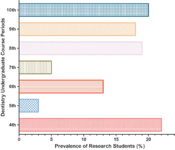
In the interviewed population of 146 individuals, it was found that 108 were female and 38 males, which shows the prevalence of women in this private Dentistry course. In this study, it can also be observed that the prevalence of students who participated in the study was 22% from the 4th semester, 19% from the 8th semester, 18% from the 9th semester and 20% from the 10th semester, which demonstrates that the majority of the interviewees were in the beginning of the Dental School Graduation Course with insipient clinical care and pathology and periodontics knowledge, as well as in partial and final semesters the students have continuous clinical practice in various specialties of Dentistry, without focusing on the basic diagnostic of oral cavity (Figure 2).
This result corroborates with the findings about the knowledge of the lesion’s color, since 48.63% of the interviewees pointed out the red color as a characteristic of the lesion and 21.23% reported not knowing when it is related to herpes (Figure 3). Regarding the description of the type of elementary lesion, most of the students, totaling 23%, did not know the correct description of the type of lesion (vesicle, crust, ulcer, pseudomembrane) of clinical manifestation of HSV-1. It was observed that 40.41% are unaware of the size of the lesion and only 28.08% described it as rounded. In concern to the duration of the injury, the study revealed that 39.4% knew the frequency of injury duration, 33 (56%) were unaware of the time and 27.04 (4%) presented other types of frequency, demonstrating the prevalence of lack of knowledge.
Another relevant finding in the study was the knowledge about clinical signs, with 34.93% of the students pointing the vesicles on the lips as a prevalent sign of the disease and
Figure 1. Sample size calculation formula. Legend: n = Sample; Z =
Confidence level (95%); P = Amount of expected success (%) (50%); Q = Amount of expected error (%) (50%); N = Population Size; e = Level of accuracy 5%.
The excluded participants were students from the 1st to the 3rd semester, who have not yet taken courses in Semiology, Periodontics and Oral Pathology, which are prerequisites for identifying lesions in the oral cavity. In addition, students from the 4th to the 10th semester who failed in the respective subjects were also excluded. The participants included in the study were undergraduate students from the 4th to the 10th semester of the Dentistry course at UNINOVAFAPI who passed the prerequisite subjects and who are undergoing clinical care to identify possible injuries.
Figure 2. Graph on prevalence of undergraduate students of dentistry by each semester of course.
4
Rev. Bras. Odontol. 2019;76:e1750Healing occurs within 1 to 10 days of the onset of initial symptoms. And regardless of the treatment, these lesions evolve to cure in a period of 7 to 14 days, without leaving scars.1
Infection caused by HSV-1 can be initial or recurrent and has morphological characteristics and a lytic replicative cycle, with a short replicative cycle similar to those of other members of the Herpesviridae family, causing latent and/ or persistent infection in the host’s sensory neuronal cells.18
Prodromal signs and symptoms such as burning, itching, pain, twinge, localized heat or erythema in the involved epithelium, appear six to 24 hours before the development of the lesions, in which multiple small erythematous papules develop, forming groups of vesicles filled with liquids .19
Clinical symptoms of recurrent infection occur in phases, following the sequence: prodromal, vesicle, ulcer and crust. Some of these steps may have a short duration and seem to be imperceptible to the individual. Symptoms such as a burning sensation or itching appear in 46 to 60% of individuals, lasting about six hours. Approximately 25% of facial recurrences do not progress beyond the prodromal or vesicular stage. Recurrent infection lesions are almost always red macules that quickly become very infectious vesicular and later form ulcers. Healing occurs within 1 to 10 days of the onset of initial symptoms.20
These lesions progress to healing in a period of 7 to 14 days without scarring, regardless of treatment. In immunocompromised patients, the time of infection becomes prolonged and the clinical manifestations are more extensive and severe. Among the greatest difficulties during the clinical course of the infection are: aesthetic damage, functional discomfort that occurs through burning, itching and pain, in addition to contact trauma.20
For the proposed treatments, the therapeutic use of low-level lasers has been constant in the health sciences as a choice of innovative method due to its anti-inflammatory, analgesic, anti-edematous effects and its contribution to tissue repair. The clinical application for the treatment of acute and chronic pain is currently a well-defined procedure. The literature also highlights the speed of wound repair. Its application, in cases of herpes simplex, provides great relief to the patient, contributing to the interruption of the virus cycle and fast repair of the clinical aspect.1
Lasers are classified into high and low level. The high-level ones are used for the removal, cutting and coagulation of tissues, while the low-level lasers are used in tissue repair processes, such as muscle, joint, nerve, bone and skin injuries. A wide variety of lasers can be found in the literature in order to promote the tissue healing process, including: Helium-Cadmium, Argon, Helium-Neon, Krypton, Gallium and Aluminum arsenide and carbon dioxide. However, the success 26.71% were unaware of any existing sign related to viral
disease.
The study also assessed the level of knowledge about the use of lasers in the treatment of herpes simplex. It was found that 75% of students are unaware of which type of laser should be chosen for treatment, as well as 56.16% are unaware of the time for reusing the therapy, 65.75% did not know the recurrence characteristic after use of laser therapy and 80.13% are unaware of the recurrence time after using this approach. When related to the association of drug therapies and the laser therapy, 57.53% present low knowledge, which demonstrates relevance in the implementation of clinical protocols for the use of lasers in dentistry considering type, relapse of disease and drug interactions.
Discussion
Herpes simplex, or herpes type 1, is characterized as an ulcerative mucocutaneous viral infection, with virus of the family Herpesviridae [herpes simplex type 1 (HSV-1) and type 2 (HSV-2)] as etiologic agent and it is directly related to several orofacial pathologies presenting a seroprevalence of 60 to 90% in the world population, which can mean a vast viral reservoir, in addition to a public health problem.
Oral herpetic lesions affect about 20% - 40% of the general population and are more prevalent among low socioeconomic status groups. It is estimated that 90% of the world population is infected with HSV-1 and that women of reproductive age are greatly affected. Currently, it is still a topic of great relevance in the scientific environment.13,14,15
The infection usually affects children from 6 months to 5 years of age, adolescents and adults around 20 years of age, with adults having periods of greater occurrence. The prevalence of infection increases gradually since childhood, reaching 70% or 80% in adults. The prevalence in adults and adolescents varies from 10 to 15%.1 Herpetic infection in
neonates is less frequent.16
Upon clinical examination, it is observed that the lesions, due to recurrent herpes simplex infection, have red macules1
that progress to small yellowish vesicles,12,17 normally grouped
in the mucosa and skin and tend to coalesce, breaking and releasing white-yellowish liquid, forming a serosanguineous crust that coagulates, becoming adherent and facilitating tissue regeneration.2,3,7 They form ulcers covered by a grayish
membrane and an erythematous halo surrounding it.1,12
The pathology is self-limiting and the duration of manifestations of primary infection is 10 to 14 days after an incubation period of up to 26 days, with signs and symptoms regressing spontaneously. It is stated in the literature that spontaneous involution and complete healing without sequelae occur in 8 to 10 days.2 After this period, the virus
stay latent in the trigeminal nerve ganglion, located in the petrous portion of the temporal bone.7
of low-level therapy and its respective effects is dependent on the wavelength, power, dose and time applied.21
Due to its wide use in lesions, the low-level laser can be controlled by the operator, through applications in appropriate doses or fluences, with wavelengths (color) addressed to the specific cell site.1
Low-level laser therapy (LLLT) is a form of phototherapy that uses red or near infrared photons. These wavelengths are generally chosen because they present a good transmission through the tissues and are able to reach efficiently in more internal layers.22
The effects of LLLT have been described as: (1) primary (act as modulators of cellular function); 2) secondary (lead to pain relief effects or induce tissue healing). These effects depend on the adequacy of the irradiation parameters. To provide therapeutic doses, it is necessary to observe optical characteristics of the tissue involved, in addition to the depth and size of the lesion.22
A more clinically useful approach is to apply high amounts of energy to the vesicles (6 J to 10 J per point). Usually, individuals report a significant decrease in the symptom of pain, and a feeling of mild paresthesia can also be found. Three applications with intervals of two to three days between sessions may be necessary. In the phase of ulcer, the dentist must observe the clinical characteristic of the lesion, which is often covered by a crust. In this case, an energy of 2-3 J per point can be applied to the edges of the lesion. The crust can significantly compromise the passage of light and interfere with the results of LLLT. In case of non-crusted ulcerations, the professional should check if there are still small vesicles. If this occurs, the higher energies described above must be adopted.22
Internal or external stimuli such as stress, immunosuppression, high fever, trauma and ultraviolet light can trigger the virus and lesions appear once more.23
Recurrences occur two to three times a year. With the LLLT in place, the pruritus phase is when the best results are obtained in relation to treatment. According to a recent systematic review,24 among the three characteristic phases of HSV-1
(pruritus, vesicles and crust), the ideal phase to start treatment is the first one. The use of LLLT, at this stage, can inhibit the outbreak of wounds by stimulating the microcirculation25
and the local immune system.26 The LLLT proposes to reduce
the number of recurrences and delay new manifestations.27
The recurrence of the manifestation of HSV-1, in the studied patient, demonstrated effectiveness in the control for a period of 12 months.28
There are reports of several therapies for the treatment of herpes.29 Among the drug therapies, acyclovir and its prodrug
valacyclovir, penciclovir and its prodrug fanciclovir are the most commonly used antiviral agents for the treatment of herpes simplex, which can be carried out systemically or topically. Studies indicate greater efficacy of treatment when it is systemically administrated and initiated in the prodromal phase.30 In addition to systemic treatment, topical
acyclovir, penciclovir and docosanol are therapeutic options for recurrent herpes labialis but are less effective than oral antivirals.31
Honarmand et al. showed that treatment with diode laser reduced recovery time and pain intensity faster than treatment with acyclovir cream.14 LLLT provided, in the
present clinical case, a rapid improvement in tissue repair, anti-inflammatory and analgesic action , in addition to decreasing the number of recurrences and delaying new manifestations.28 Early recognition, careful monitoring and
careful care for wounds will prevent long-term sequelae when complications occur.32 It is important to be aware of
small vesicles in the gums, which later form characteristic ulcerations of acute herpetic gingivostomatitis (AHGS).33 The
virus incubation period lasts an average of 7 days after the appearance of signs and symptoms, which include diffuse vesicles and gingivitis, progressing to superficial ulcerations throughout the oral mucosa, fever, malaise, headache, cervical lymphadenopathy and severe pain, making feeding difficult.34,35
Conclusion
It was concluded that undergraduate students of dentistry are unaware of the herpes simplex pathology and the treatment with laser therapy for it, as well as the advantages of this approach. In view of the study, it is suggested that the use of this technology must be implemented through protocols in educational institutions, in order to promote knowledge for undergraduate students and dentists. There is a need to build a protocol for the care of injuries with the use of laser therapy to improve care and teaching in the Dental School.
4. Gama CRB, Passos MD, Varella R, Lasmar R, Gama GF, Oliveira L, et al. Avaliação clínica da unicaria tomentosa no tratamento e controle de lesões decorrentes de Infecção pelo vírus herpes simplex. DST J Bras Doenças Sex Transm. 2010;22(4):215-21.
5. Tagliari NAB, Kelman RG, Diefenthaler H. Therapeutic aspects of infections caused by Herpes Simplex Virus Type-1. Rev. Perspect. 2012;36(133):191-201. 6. Passos MRL, Junior JE, Cavalcanti SMB, Salles RS. Genital herpes on the penisand topic use of Uncaria Tomentosa: case report. DST. J. Bras. Doenças References
1. Vazzoller RMS, Fernandes RD, Sena RMM, Desenna AM. Tratamento do herpes simples por meio da laserterapia – relato de casos. RcITPAC. 2016;9(3):01-10.
2. Geller M, Neto MS, Ribeiro MG, Oliveira L, Naliato EC, Abreu C, et al. Revisão Bibliográfica, Herpes Simplex: Clinical Update, Epidemiology and Therapeutics. DST J Bras Doenças Sex Transm. 2012;24(4):260-66.
3. Al-Maweri SA, Kalakonda B, AlAizari NA, Al-Soneidar WA, Ashraf S, Abdulrab S, et al. Efficacy of low-level laser therapy in management of recurrent
6
Rev. Bras. Odontol. 2019;76:e1750Submitted: 12/15/2019 / Accepted for publication: 12/30/2019
Corresponding author Luana Kelle Batista Moura
E-mail: luanamoura@uninovafapi.edu.br
1. Luana Kelle Batista Moura – DDS; PhD. Contribution: Writing of the manuscript: effective scientific and intellectual participation for the study, data acquisition, preparation of the manuscript, draft of the manuscript, critical review and final approval. ORCID: 0000-0003-4917-7598
2. Daniela Bezerra da Silva – Undergraduate student of Dentistry. Contribution: Intellectual participation for the study; data acquisition; critical review and final approval. ORCID: 0000-0003-0397-6036
3. Bárbara Soares Sousa – Undergraduate student of Dentistry. Contribution: Intellectual participation for the study, data acquisition, critical review and final approval. ORCID: 0000-0002-9467-9565
4. Eduardo Souza de Lobão Veras – DDS; PhD. Contribution: Intellectual participation for the study, data acquisition, critical review and final approval. ORCID: 0000-0003-4629-8909
5. Marta Rosado de Oliveira Campos – Undergraduate student of Dentistry. Contribution: Intellectual participation for the study, data acquisition, critical review and final approval. ORCID: 0000-0001-8550-4688
Avaliação do efeito de pomada de própolis para tratamento de herpes labial recorrente – um estudo piloto. Arq. cienc. saúde Unipar. 2017;21(1):13-8. 8. Serna-Ojeda JC, Ramirez-Miranda A, Navas A, Jimenez-Corona A, Graue-Hernandez EO. Herpes simplex virus disease of the anterior segment in children. Cornea. 2015; 34: S68-S71.
9. Pedrazini MC, Cury PR, Araujo VC, Wassal T. Efeito da lisina na incidência e duração das lesões de herpes labial recorrente. Rev Gaúch. Odontol. 2007; 55(1):7-10.
10. Carvalho RR, Eduardo FP, Ramalho KM, Antunes JLF, Bezinelli LM, Magalhães MHCG, et al. Effect of laser phototherapy on recurring herpes labialis prevention: an in vivo study. Lasers Med. Sci. 2010; 25(3), 397-402. 11. Lizarelli RFZ. Protocolos Clínicos Odontológicos. Uso do Laser de Baixa Intensidade. 4nd ed. São Paulo: Edição; 2010.
12. Martins MLS, Arantes ACS, Nicolau RA. Tratamento de herpes simples tipo 1 com laser de baixa intensidade (660 nm) – relato de caso clínico. Revista Univap. 2017;22(41):61-67.
13. Wilson SS, Fakioglu E, Herold BC. Novel approaches in fighting herpes simplex virus infections. Expert Rev. Anti-infect. Ther. 2009;7(5):559–68. 14. Honarmand M, Farhadmollashahi L, Vosoughirahbar E. Comparing the effect of diode laser against acyclovir cream for the treatment of herpes labialis. J Clin Exp Dent. 2017;9(6):729-32.
15. Zverev VV, Makarov OV, Khashukoeva AZ, Svitich OA, Dobrokhotova YE, Markova EA, et al. In vitro studies of the antiherpetic effect of photodynamic therapy. Lasers Med. Sci. 2016;31(5):849–55.
16. [The Spanish Society of Paediatric Infectious Diseases guidelines on the prevention, diagnosis and treatment of neonatal herpes simplex infections]. An Pediatr (Barc). 2018 Jul;89(1):64 e1-e10.
17. Fatahzadeh M, Schwartz RA. Human Herpes Simplex Virus Infections: Epidemiology, pathogenesis, symptomtology, diagnosis, and management. J Am Acad Dermatol. 2007;57(5):737-56.
18. Ferreira DC, Martins FO, Romanos MTV. Impacto do laser de baixa intensidade na supressão de infecções pelos vírus Herpes simplex 1 e 2: estudo in vitro. Rev. Soc. Bras. Med. Trop. 2009;42(1):82-5.
19. Neville B. Patologia Oral e Maxilofacial. Rio de Janeiro: Elsevier Brasil; 2016.
20. Núñez SC, Ribeiro MS, Garcez AS. PDT – Terapia Fotodinâmica Antimicrobiana na Odontologia. Terapia Fotodinâmica Antimicrobiana/ Aplicação Clínica em Herpes Labial. Rio de Janeiro: Elsevier Brasil; 2013. P. 249-58.
Mini Curriculum and Author’s Contribution
potência na cicatrização de feridas cutâneas. Rev. Col. Bras. 2014;41(2):129-33. 22. Garcez AS, Ribeiro MS, Núñez SC. Laser de Baixa Potência: Princípios Básicos e Aplicações Clínicas na Odontologia. Terapia Laser de Baixa Potência em Lesões Orais/Herpes. 1nd ed. Rio de Janeiro; 2012.
23. Honarmand M, Farhadmollashahi l, Vosoughirahbari E. Comparing the effect of diode laser against acyclovir cream for the treatment of herpes labialis. J Clin Exp Dent. 2017;6:729-32.
24. Eduardo CP, Aranha ACC, Simões A, Bello-Silva MS, Ramalho KM, Esteves-Oliveira, M, et al. Laser treatment of recurrent herpes labialis: a literature review. Lasers Med. Sci. 2014;29(4):1517–29.
25. Queiroz LS, Wollmann DER, Nicolau RA, Pacheco MTT. Effect of LED irradiation on microcirculation of auricular mouse. Rev. Assoc. Paul. Cir. Dent. 2008; 62(2):138-42.
26. Piva JADAC, Abreu EMDC, Silva VDS, Nicolau RA. Ação da terapia com laser de baixa potência nas fases iniciais do reparo tecidual: princípios básicos. Arq Bras Dermatol. 2011;86(5):947-54.
27. Gonzalez BM, Hernandez A, Estevez A. Tratamiento del herpes simple labial com láser de baja potencia. Colom. Med. 2008;39(2):175-81.
28. Martins MLS, Arantes ACS, Nicolau RA. herpes simplex type1 treatmentwithlow-level laser (lambda 660 nm)-clinical case report. Revista Univap. 2016;22(41):61-7.
29. Consolaro A, Consolaro MF. Diagnóstico e tratamento do herpes simples recorrente peribucal e intrabucal na prática ortodôntica. Rev. Dent. Press Ortodon. Ortop. Facial. 2009;14(3):16-24.
30. Cunningham A, Griffiths P, Leone P, Mindel A, Patel R, Stanberry L, et
al. Current management and recommendations for access to antiviral therapy
of herpes labialis. J. Clin. Virol. 2012, 53(1):6 -11.
31. Usatine RP, Tinitigan R. Nongenital herpes simplex virus. Am Fam Physician. 2010;82(9):1075-82.
32. Zaouak A, Benmously R, Hammami H, Fenniche S. A case of herpes simplex virus reactivation after fractional ablative carbon dioxide laser to treat a burn scar. J Cosmet Laser Ther. 2019;21(3):145-6.
33. Marcucci G. Fundamentos de odontologia - Estomatologia. 1nd ed. Rio de Janeiro: Guanabara Koogan; 2005.
34. 34. Amir J, Harel L, Smetana Z, Varsano I. The natural history of primary herpes simplex type 1 gingivostomatitis in children. Pediatr Dermatol. 1999;16(14): 259-63.
35. George AK, Anil S. Acute Herpetic Gingivostomatitis Associated with Herpes Simplex Virus 2: Report of a Case. J Int Oral Health. 2014;6(3):99-102.