Luis Korrodi Mineiro
Marques Gregório
Characterization of PPP1 interacting proteins in male
reproduction
Caracterização de proteínas que interagem com a
PPP1 na reprodução masculina
Luis Korrodi Mineiro
Marques Gregório
Characterization of PPP1 interacting proteins in
male reproduction
Caracterização de proteínas que interagem com a
PPP1 na reprodução masculina
Dissertação apresentada à Universidade de Aveiro para cumprimento dos requisitos necessários à obtenção do grau de Doutor em Bioquímica, realizada sob a orientação científica da Doutora Margarida Sâncio da Cruz Fardilha, Professora Auxiliar Convidada da Secção Autónoma das Ciências da Saúde da Universidade de Aveiro e co-orientação científica da Doutora Odete A.B. da Cruz e Silva, Professora Auxiliar da Secção Autónoma das Ciências da Saúde da Universidade de Aveiro.
Esta dissertação teve o financiamento da FCT através da bolsa SFRH/BD/42334/2007.
Dedico este trabalho ao meu avô, João Alves Mineiro, que é um exemplo para mim a nível pessoal e professional. Estás sempre presente e nunca serás esquecido.
o júri
presidente Prof. Dr. Amadeu Soares
Professor Catedrático, Departamento de Biologia, Universidade de Aveiro
Prof. Dr. Georg H. Lüers
Professor Associado da Universidade de Hamburg-Eppendorf
Prof. Dr. Srinivasan Vijayaraghavan
Professor Associado da Universidade de Kent
Prof. Dr. Odete Abreu Beirão da Cruz e Silva
Professora Auxiliar com Agregação da Universidade de Aveiro
Prof. Dr. Pedro José Esteves
Professor Coordenador do Instituto Politécnico de Saúde do Norte da Cooperativa de Ensino Superior Politécnico e Universitário
Prof. Dr. Margarida Sâncio da Cruz Fardilha
agradecimentos Este trabalho não teria sido possível sem a ajuda de muitas pessoas às quais agradeço o apoio dado:
Ao meu orientador, Prof. Edgar da Cruz e Silva, por ter sido durante a primeira parte do meu doutoramento um mentor para mim.
À minha co-orientadora e mais tarde orientadora, Margarida Fardilha que me acompanhou neste percurso, aguentou as minhas teimas e teve a paciência para me animar sempre que alguma coisa não corria pelo melhor. A ela agradeço o meu crescimento científico.
À minha co-orientadora, Profa. Odete da Cruz e Silva pela disponibiliadade, força e incentivo prestados, em especial na recta final.
A todo o grupo do Laboratório de Transdução de Sinais em especial à Sara Esteves por todo o seu inestimável apoio desde o inicio deste trabalho, pela camaradagem e por ter razão. Às novas estupendas adições, Mónica, João e Joana pela ajuda na recta final. Sem vocês este trabalho não seria tão condimentado.
Ao grupo de Neurociências em especial à Sandra Vieira pelo seu grande apoio em parte do trabalho.
To Dr. Vijay for the collaboration in the transgenic mice work, for all the fruitful scientific discussions and all the support during the one year stay in Kent, USA. Also to all lab members, Nilam, Ramdas, Teja, Sromona, David and Meenakshi for the support and friendship. Also to Cheryl, for being my borrowed mother and for helping me in the american journey.
Ao Dr. Pedro Esteves pelas diversas discussões futebolísticas mas sempre científicas e também à Joana e à Ana pela ajuda na parte final do trabalho. Aos meus amigos por animarem esta etapa e tornarem-na mais fácil, em especial ao Gonçalo pelo companheirismo e aventura por diferentes terras e à Sofia por me acompanhar nos momentos laboratoriais mais hilariantes. Aos colegas de casa passados e presentes, em especial ao Tiago, o eterno comparsa nesta academia.
À minha namorada, Rita, por todo apoio prestado a todos os níveis e por ser uma pessoal especial. Obrigado por me aguentares.
À minha família, em especial aos meus pais e às minhas avós por me terem sempre apoiado em todas as escolhas que fiz e por serem quem são. Sem vocês isto não teria sido possível.
palavras-chave PPP1, fosfatase, Tctex1d4, citoesqueleto, microtúbulos, tubulina, fosforilação, dineína, inibidor 2, PPP1R2, PPP1R2P3, espermatozoide, testículo
resumo A fosforilação reversível de proteínas é um importante mecanismo de controlo em eucariotas. A fosfoproteína fosfatase 1 (PPP1) é uma fosfatase de serina/treonina envolvida em vários processos celulares. Existem três isoformas da subunidade catalítica (α/CA, δ/β/CB e γ/CC) com pequenas diferenças nos terminais amino e carboxílico. O gene PPP1CC sofre ainda
splicing alternativo para produzir duas isoformas, a PPP1CC1 ubíqua e a
PPP1CC2 enriquecida em testículo e específica de esperma. A localização e especificidade de substratos da PPP1 está dependente da formação de complexos oligoméricos com proteínas que interagem com a PPP1 (PIPs). O objetivo principal desta tese foi estudar novas PIPs, específicas de testículo e esperma, a fim de melhor caracterizar o papel desta fosfatase e dos respetivos complexos na reprodução em mamíferos. Com este fim, estudou-se a presença, localização e possíveis funções de uma PIP previamente conhecida, PPP1R2, e de duas novas PIPs, PPP1R2P3 e Tctex1d4.
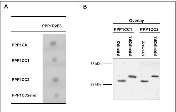
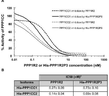
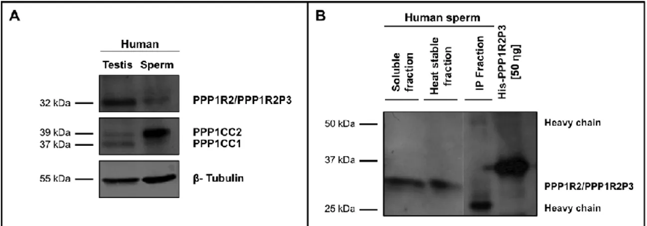
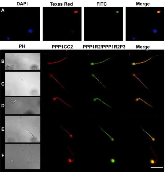
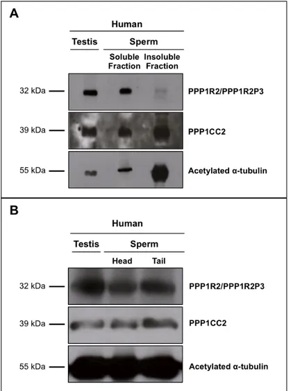
PPP1R2 e PPP1R2P3 estão presentes em esperma humano colocalizando com a PPP1CC2, na cabeça e na cauda. A hipótese é que as holoenzimas localizadas na cabeça terão um papel na reação acrossómica, enquanto que as holoenzimas presentes no axonema são relevantes para o controlo da motilidade flagelar. De seguida foram estudados os pseudogenes da PPP1R2, em termos de história evolutiva e de possíveis funções. Na espécie humana, a PPP1R2 tem 10 pseudogenes, 7 deles específicos de primatas. Estudos de bioinformática e dados de expressão mostram que os PPP1R2P1/P3/P9 são os pseudogenes com maior probabilidade de serem transcritos e traduzidos. Também identificámos o PPP1R2P9 em esperma humano e mostrámos que alguns pseudogenes poderão estar associados a estados fisiopatológicos. Isto indica que o processo de evolução poderá estar ligado á formação de novos genes ou ao controlo do mRNA da PPP1R2. A sobre-expressão da PPP1R2 ou PPP1R2P3 em testículo de ratinho também foi realizada, para caracterizar os mecanismos envolvidas na função dos complexos PPP1R2/PPP1R2P3-PPP1CC2 na espermatogénese e fisiologia dos espermatozoides.
A dineína de cadeia leve, Tctex1d4, foi encontrada como interagindo com a PPP1C e como estando presente em testículo de ratinho e em esperma humano. Demonstrámos que a Tctex1d4 e a PPP1 colocalizam no centro organizador de microtúbulos e nos microtúbulos e que o motivo de ligação à PPP1 presente na Tctex1d4 parece ser importante para manter a PPP1 no centro organizador de microtúbulos e/ou para disromper ou atrasar o seu movimento ao longo dos microtúbulos emergentes. Estes resultados abrem novos caminhos para os possíveis papéis do complexo Tctex1d4-PPP1 na dinâmica dos microtúbulos, motilidade do esperma, reação acrossómica e na regulação da barreira hemato-testicular, provavelmente, através da via de sinalização do TGFß. A análise do motivo de ligação à PPP1 mostra que este é altamente conservado entre os mamíferos, com exceção das Pikas, sugerindo que esta perda aconteceu antes da radiação das Pikas, há 6-20 milhões de anos atrás. Através de um rastreio por mutações demonstrámos que a capacidade da Tctex1d4 se ligar à PPP1 é mantida nas Pikas, embora o motivo de ligação à PPP1 esteja disrompido.
keywords PPP1, phosphatase, Tctex1d4, cytoskeleton, microtubules, tubulin,
phosphorylation, dynein, inhibitor 2, PPP1R2, PPP1R2P3, spermatozoa, testis
abstract Reversible phosphorylation of proteins is an important intracellular control mechanism in eukaryotes. Phosphoprotein Phosphatase 1 (PPP1) is a major serine/threonine protein phosphatase involved in a wide range of cellular processes. Three closely related catalytic subunit isoforms (/CA, δ//CB and
/CC) exist with only minor differences at their N- and C-terminus. PPP1CC gene can also undergo tissue-specific processing to yield a ubiquitously expressed PPP1CC1 and the testis-enriched and sperm-specific PPP1CC2 isoforms. PPP1C exists in the cell as an oligomeric complex binding to a spectrum of PPP1 interacting proteins (PIPs), which modulate both its intracellular localization and substrate specificity.
The main goal of this thesis was to study novel PIPs in testis and sperm, in order to further characterize the role of PPP1CC2 and the respective complexes in mammalian reproduction. To this end we addressed the presence, localization and putative roles of a previously known PIP, PPP1R2, in testis and sperm, and two novel PPP1CC2 testis/sperm specific PIPs, PPP1R2P3 and Tctex1d4. PPP1R2/PPP1R2P3 were shown to be present in human sperm co-localizing with PPP1CC2, in the head and tail. It was shown that PPP1R2P3 is a heat stable inhibitor of PPP1CC that cannot be phosphorylated by GSK3. We hypothesize that the holoenzymes localized in the head may have a role in the acrosome reaction while the axoneme bound holoenzymes are relevant for the control of flagellar motility. To further address the PPP1R2 significance, its pseudogenes were described in terms of evolutionary history and putative functions. In human specie, PPP1R2 has ten pseudogenes most of them primate-specific. Besides PPP1R2P3, bioinformatic studies and expression data show that PPP1R2P1, PPP1R2P2 and PPP1R2P9 are the pseudogenes with more probability of being transcribed and eventually translated. Moreover, we identified PPP1R2P9 in human sperm and showed that several pseudogenes appear to be associated with physiological and pathological states. This indicates that evolution processes might be in part related with the formation of new genes or in the control of the parental PPP1R2 message. Overexpression of human PPP1R2 or PPP1R2P3 in mouse testis was also pursued to provide the molecular tools to initiate the characterization of the mechanisms behind PPP1R2/PPP1CC2 and PPP1R2P3/PPP1CC2 role in spermatogenesis and sperm physiology.
Dynein light chain, Tctex1d4, was found to bind to PPP1C and to be present in mouse testis and human sperm. Tctex1d4-PPP1CC complex was shown to co-localize in the microtubule organizing centre and in microtubules. Moreover, the Tctex1d4 PPP1 binding motif seems to be important to retain PPP1CC in the microtubule organizing centre, and also to disrupt or delay its movement along microtubules. These results open new avenues to the possible roles of Tctex1d4-PPP1 complex in microtubule dynamics, sperm motility, acrosome reaction and in regulation of the blood testis barrier possibly via TGFß signaling. Moreover, PPP1 binding motif is highly conserved among mammals, except in Pikas, suggesting that this event happened before the Pikas radiation, 6-20 Million years ago. Mutational screening shows that the ability of Tctex1d4 to bind to PPP1 is maintained in Pikas, although the PPP1 binding motif is disrupted.
Index
Abbreviations ... i
Introduction ... 1
Protein phosphorylation - kinases and phosphatases ... 1
Phosphoprotein Phosphatase 1 (PPP1) ... 3
Phosphoprotein phosphatase 1 – Phosphatase Interacting Proteins (PIPs) ... 5
Phosphoprotein phosphatase 1 binding motif ... 8
Protein phosphatase 1 role in testis and sperm ...10
Testis and sperm PPP1CC/PIP complexes ...13
Objectives ...20 References...21 Chapter II ...33 Introduction ...33 References...34 Chapter II.A ...35
Discovery and characterization of two forms of PPP1R2 in human sperm ...35
Abstract ...36
Introduction ...37
Material and Methods ...39
Results and Discussion ...45
Conclusion ...58
Acknowledgements ...61
References...62
Chapter II.B ...67
The evolution of PPP1R2 related pseudogenes ...67
Abstract ...68
Background ...69
Methods ...71
Results and Discussion ...76
Conclusions ... 100
Acknowledgements and Funding ... 101
References... 102
Chapter II.C ... 107
Overexpression of PPP1R2 and PPP1R2P3 in mouse – a transgenic approach . 107 Introduction ... 107
Objective ... 113
Material and Methods ... 114
Results and Discussion ... 122
Conclusion ... 134
Chapter III ... 141
Introduction ... 141
References... 142
Chapter III.A ... 143
TCTEX1D4, a novel Protein Phosphatase 1 interacting protein involved in tubulin dynamics in testis and sperm ... 143
Abstract ... 144
Introduction ... 145
Materials and Methods ... 147
Results ... 152
Discussion... 166
References... 171
Chapter III.B ... 175
An intriguing switch in the novel PPP1C binding partner Tctex1d4, between the binding motif and a glycosylation site, in Pikas (Ochotona sp.) ... 175
Abstract ... 176
Introduction ... 177
Materials and Methods ... 179
Results ... 183 Discussion... 187 Acknowledgements ... 191 References... 192 Chapter IV. ... 197 Conclusion ... 197 References... 201 Appendix ... 205
Abbreviations
ACN acetonitrile
AKAP A-kinase-anchoring protein APC/C anaphase-promoting complex
Arg arginine
ASPP1/2 apoptosis-stimulating of p53 protein 1 and 2, also known as PPP1R13B and PPP1R13A respectively
ATP adenosine triphosphate
Bad Bcl2-associated agonist of cell death BCA bicinchoninic acid assay
Bcl-2 B-cell CLL/lymphoma 2
BEB bayes empirical bayes
BLAST basic local alignment search tool
bp base pair
BRCA1 breast cancer 1
BSA bovine serum albumin
BTB blood testis barrier
Ca2+ calcium
cAMP cyclic adenosine monophosphate CASA computer assisted sperm analyser
CASK calcium/calmodulin-dependent serine protein kinase CDC25C cell division cycle 25 homolog C
CDH cadherin
CDK cyclin-dependent kinase
cDNA complementary DNA
CHAPS 3[(3-Cholamidopropyl)dimethylammonio]-propanesulfonic acid CID collision-induced dissociation
CKII casein kinase II
CMV-IE cytomegalovirus immediate early COS-7 monkey kidney fibroblast cell line
CPI-17 protein kinase C potentiated inhibitor 17, also known as PPP1R14A
CT C-terminal
CTD carboxy-terminal domain
Cre causes recombination
CReP constitutive repressor of eIF2alpha phosphorylation, also known as PPP1R15B
DAPI 4,6-diamidino-2-phenylindole
DARPP32 dopamine and cAMP regulated phosphoprotein, also known as PPP1R1B
ddH2O double distilled water
DIC differential interference contrast DMEM Dulbecco’s Modified Eagle medium dN non- synonymous substitutions per site
DNA deoxyribonucleic acid
dS synonymous substitutions per site
DTA diphtheria toxin A
ECL enhanced chemiluminescence
EDTA ethylenediaminetetraacetic acid EF1α elongation factor 1α
EGTA ethylene glycol tetraacetic acid ELM eucaryotic linear motif
ERK extracellular signal-regulated kinase
EST expressed sequence tags
FA formic acid
FAK focal adhesion kinase
FCP TFIIF-associating CTD phosphatase
FDR false discovery rate
FEL fixed-effect likelihood FITC fluorescein isothiocyanate
FLP yeast-derived Flip
FRT flipase recognition target
FSIP fibrous sheath interacting protein FSP95 fibrous sheath protein of 95 kDa
GADD34 growth arrest and DNA damage-inducible protein 34, also known as PPP1R15A
gDNA genomic DNA
GBPI1 gastrointestinal and brain-specific PP1-inhibitory protein 1, also known as PPP1R14D
GC guanine-cytosine
GC2 germ cells
GEO gene expression omnibus
GL hepatic glycogen-targeting protein phosphatase 1 regulatory subunit GL also known PPP1R3B
Glu glutamic acid
GM protein phosphatase 1 regulatory subunit GM, also known PPP1R3A GSK3 glycogen synthase kinase 3
hAAT human α1-antitrypsin
Hb2E protein phosphatase 1 regulatory subunit 3F, also known as PPP1R3F HEPES 4-(2-hydroxyethyl)-1-piperazineethanesulfonic acid
hsp90 heat shock protein 90
I1 inhibitor 1, also known as PPP1R1A I2 inhibitor 2, also known as PPP1R2 I2-L inhibitor 2-like, also known as PPP1R2P3 IACUC institutional animal care and use committee
ICs intermediate chains
IFT intraflagellar transport
IHC immunohistochemistry
IPP5 inhibitor-5 of protein phosphatase 1, also known as PPP1R1C IPTG isopropyl-β-D-thio-galactopyranoside
IUP intrinsically unstructured protein
JHDM1D jumonji C domain containing histone demethylase 1 homolog D KEPI kinase-enhanced PP1 inhibitor, also known as PPP1R14C KIAA1443 protein phosphatase 1, regulatory subunit 3E, also known as
PPP1R3E
LB loading buffer
LCs light chains
Leu leucine
LICs light intermediate chains
LINE long interspersed nuclear elements LRT likelihood ratio test
LTQ linear ion trap
LTR long terminal repeat
MAOA monoamine oxidase A
MAPK mitogen activated protein kinase
Mg2+ magnesium
ML maximum-likelihood
Mn2+ manganese
MoMLV moloney murine leukemia virus
mPrm mouse protamine promoter
mRNA messenger RNA
MTOC microtubule organizing center
MYPTs myosin phosphatase targeting subunit, also known as PPP1R12A/B. MYPT3 is known as PPP1R16B
NCBI National Center for Biotechnology Information Nek2 nimA-related protein kinase 2
Neurabins I/II neuronal binding proteins, also known as PPP1R9A/B
NIPP1 nuclear inhibitor of protein phosphatase 1, also known as PPP1R8
NJ neighbor-joining
NOM1 nucleolar protein with MIF4G domain 1
NT N-terminal
ORF open reading frame
p85 protein phosphatase 1 myosin-binding subunit of 85 kDa, also known as PPP1R12C
PBS phosphate buffered saline
PBS-T phosphate buffered saline with Tween
PCDH protocadherin
PCR polymerase chain reaction
PDHA2 pyruvate dehydrogenase (lipoamide) alpha 2 PGK1 phosphoglycerate kinase 1
PH phase contrast
PHACTR phosphatase and actin regulator
PHI1 phospholipase C beta-3 neighboring gene protein, also known as PPP1R14B
PIP phosphatase interacting protein
PKA cAMP-dependent protein kinase
PMSF phenylmethylsulfonyl fluoride
PNUTS protein phosphatase 1 nuclear targeting subunit, also known as PPP1R10
PP protein phosphatase
PPM protein phosphatase Mg2+ or Mn2+ dependent
PPP phosphoprotein phosphatase
PPP1BM PPP1 binding motif PPP1C PPP1 catalytic subunit
PPP1CA/B/C PPP1 catalytic subunit alpha/beta/gamma isoform PPP1R PPP1 regulatory subunit
Pro proline
PTG protein targeting to glycogen, also known as PPP1R3C RAP-1 ras related protein 1
RAP1GDS1 GTP-GDP dissociation stimulator 1 REL random effect likelihood
RIPA radio-immunoprecipitation assay RIPP1 ribosomal inhibitor of PP1
RNA ribonucleic acid
RSV-LTR rous sarcoma virus long terminal repeat
RT room temperature
RT-PCR reverse transcriptase PCR RT-qPCR real time quantitative PCR
RUNX1 Runt-related transcription factor 1 SARP several ankyrin repeat protein
SDS sodium dodecyl sulfate
sds22 protein phosphatase 1 regulatory subunit 22, also known as PPP1R7 SDS-PAGE SDS polyacrylamide gel electrophoresis
Ser serine
SETD4 SET domain containing 4
SGCD sarcoglycan delta
SI spectral index
SINE Short interspersed nuclear elements SLAC single likelihood ancestor counting
SLC5A7 solute carrier family 5 choline transporter, member 7
SLC37A3 solute carrier family 37 (glycerol-3-phosphate transporter), member 3 ST6GAL2 ST6 beta-galactosamide alpha-2,6-sialyltranferase 2
STPP serine-threonine protein phosphatase
SV-40 simian virus 40
Szp1 spermatogenic zip protein 1
TAKAP-80 testis-specific A-kinase-anchoring-protein TAP transporter , ATP-binding cassette, sub-family B TAT-1 transactivating regulatory protein 1
TAU microtubule-associated protein tau TBS tris buffered saline
TBS-T tris buffered saline with tween
Tctex5 t-complex-associated-testis-expressed 5, also known as PPP1R11 TFIIF transcription factor IIF
TGFβ transforming growth factor beta
Thr threonine
TIMAP transforming growth factor β inhibited membrane-associated protein, also known as PPP1R16B
TIMD4 T-cell immunoglobulin and mucin domain containing 4
tk thymidine kinase
TLRR testis leucine-rich repeat
TPCK tosyl phenylalanyl chloromethyl ketone Tsga2 testis specific gene A2
Tyr tyrosine
URI unconventional prefoldin RPB5 interactor
UTR untranslated region
Chapter I
Introduction
Introduction
Protein phosphorylation - kinases and phosphatases
Reversible phosphorylation of structural and regulatory proteins is an important intracellular control mechanism in eukaryotes affecting up to 30% of the proteome. It plays a central role in the control of almost all cellular functions including metabolism, signal transduction, cell division and memory [1-3]. Intracellularly, protein phosphorylation regulates a variety of important functions, including subcellular localization, protein degradation and stabilization, as well as biochemical activities [4].
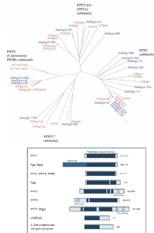
The phosphorylation state of a protein is a dynamic reversible process involving both protein kinases (add the phosphate), and protein phosphatases (PPs, remove the phosphate). Unlike protein kinases, that all belong to a single gene family, PPs are divided into several distinct and unrelated protein/gene families. The Tyr-specific PP family, as well as including the Tyr-specific PPs, also comprises the so-called dual specificity PPs (capable of dephosphorylating Ser, Thr and Tyr residues). Within the Ser/Thr-specific protein phosphatases (STPPs) three distinct gene families have been described: the PPMs (Mg2+ or Mn2+ dependent protein phosphatases), the FCPs (TFIIF-associating CTD phosphatases) and the PPPs (phosphoprotein phosphatases). The PPM family comprises the Mg2+-dependent PPs, such as pyruvate dehydrogenase, PP2C, and relatives [5]. The FCP family comprises FCP1 and SCPs 1-3 PPs [6, 7]. Sequence analysis using three well characterized genomes, allowed the construction of a phylogenetic tree and the division of PPP family in four subfamilies: PPP1 (Fig I.1A right branch), PPP2 (PPP2/PP2A, PPP4 and PPP6, Fig I.1A upper branch), PPP3/PP2B/Calcineurin (Fig I.1A, left branch) and PPP7 (PPP5 and PPP7, Fig I.1A, lower branch). These gene subfamilies share high homology in the catalytic domains (Fig I.1B, dark blue bars) but differ in the N- and C-terminal domains (Fig I.1B) [5, 8, 9]. Besides these intracellular PPs involved in signal transduction, there are also unrelated non-specific alkaline and acidic PPs that are usually found either in specialized intracellular compartments or in the extracellular milieu [10].
Figure I. 1: Evolutionary and structural relationship between the different PPP families. A. Phylogenetic tree, depicting the relationships between Homo sapiens (Hs, red), Drosophila melanogaster (Dm, blue) and budding yeast Saccharomyces cerevisiae (Sc, brown) protein serine/threonine phosphatases of the PPP family (figure taken from
[4]
). B. Domain organization of PPP family members. The size in amino acids is shown on the right (adapted from[11]
).The sequencing of entire genomes has revealed that approximately 3% of all eukaryotic genes encode protein kinases or PPs [12]. Surprisingly, there appear to be 2-5 times fewer PPs than protein kinases. This imbalance is even more pronounced when the analysis is limited to STPPs and Ser/Thr-kinases, particularly in vertebrates. The human
A
genome, for instance, encodes approximately 20 times fewer STPPs than Ser/Thr-kinases. Thus, it is often concluded that, whereas, the diversity of the Ser/Thr-protein kinases has kept pace with the increasing complexity of evolving organisms, the STPPs apparently have not. However, in the past two decades it has become evident that the diversity of STPPs is achieved not only by the evolution of new catalytic subunits, but also by the ability of a single catalytic subunit to interact with multiple regulatory subunits [1, 13].
Phosphoprotein Phosphatase 1 (PPP1)
Phosphoprotein Phosphatase 1 (PPP1) is a major STPP involved in a wide range of cellular processes such as cell cycle progression, protein synthesis, muscle contraction, glycogen metabolism, cytokinesis, and neuronal signaling [1, 3 ].
The PPP1 catalytic subunit (PPP1C) is expressed from one (Saccharomyces cerevisiae) to eight (Arabidopsis thaliana) different isoforms in the eukaryotic genomes. In mammalian genomes three separate genes encode for three closely related isoforms (/A, δ//B and /C). These isoforms are > 90% identical in amino acid sequence, with minor differences, primarily at their N- and C-terminus [1]. PPP1CC gene also undergoes tissue-specific alternative splicing to yield a ubiquitously expressed PPP1CC1 (1) isoform and the testis-enriched and sperm-specific PPP1CC2 (2) isoform. [14-18] (Fig I.2).
Figure I. 2: Schematic representation of PPP1CC gene exon-intron organization. Exons are shown in blue boxes with numbers. Alternative splicing of exon 7 originates the testis-enriched and sperm-specific PPP1CC2 isoform. Amino acids corresponding to the exon 7 specific C-terminal are shown. * denotes the termination codon.
It has already been shown that PPP1 isoforms are expressed in a variety of mammalian cells, although they localize intracellularly in a distinct and characteristic manner. PPP1CA was found to be ubiquitous in all mouse tissues except in skeletal and heart muscles. PPP1CB is also ubiquitous in all mouse tissues except in skeletal muscle and PPP1CC1 has higher levels in brain, small intestine and lung compared to other tissues and not detectable in heart and spleen. PPP1C2 is the isoform in higher quantity in testis and virtually the only one expressed in spermatozoa but with low amounts in brain, lung, spleen and thymus [19]. Specifically in brain, the different PPP1 isoforms were shown to be present in different regions and revealed also specific subcellular localization [18, 20]. While PPP1CB is the predominant isoform associated with microtubules in the neuronal cell body, PPP1CC1 and PPP1CA are preferentially concentrated in the dendritic spines [20, 21]. Tissue expression of PPP family members is shown in Table 1.
Table I. 1: Tissue expression of the main PPP family members.
Protein Phosphatase Tissues
PPP1CA Ubiquitous; more predominantly in brain [19]
PPP1CB Ubiquitous; more predominantly in liver and kidney
[19]
PPP1CC1 Ubiquitous; more predominantly in brain, small
intestine and lung [19]
PPP1CC2 Yes, low abundance [20]
PPP2 Ubiquitous; more predominantly in brain [22]
PPP3 Ubiquitous; more predominantly in brain [23]
PPP4 Ubiquitous; more predominantly lung, liver and
kidney [24]
PPP5 Ubiquitous; more predominantly in brain [25, 26]
PPP6 Ubiquitous; more predominantly in heart and
skeletal muscle [27]
PPP7 Ubiquitous; more predominantly in sensory organs
During the cell cycle, phosphorylation status, activity, and subcellular localization of PPP1 changes. Studying PPP1 localization during the cell cycle, Andreassen et al. demonstrated that the distribution of PPP1 isoforms in cells was highly dynamic. PPP1CA/B/C1 localize to distinct subcellular compartments during both interphase and mitosis [29]. PPP1 is expressed in various cellular compartments, but it is most abundant in the nucleus. Within the nucleus, PPP1CA associates with the nuclear matrix, PPP1CC1 localizes to the nucleolus, and PPP1CB is associated with whole chromatin. During mitosis, PPP1CA localizes to centrosomes, while PPP1CC1 is associated with microtubules of the mitotic spindle. In contrast, PPP1CB is strongly localized to chromosomes [30-32].
Although most biochemical studies have not directly addressed the significance of the different isoforms, it is now well established that these distinct subcellular localizations, activity and phosphorylation status are due in part to the interacting proteins [17, 29, 31]. Moreover PPP1C tissue specificity could be a determinant of which subset of different regulators are available to. Also, during evolution, other proteins present in the same tissues, and/or with the same subcellular localization, might have gained the ability to bind to a specific isoform, giving rise to new functions, and extending the repertoire of known regulators [33].
Phosphoprotein phosphatase 1 – Phosphatase Interacting Proteins (PIPs)
PPP1C exists in the cell as an oligomeric complex. The PPP1C binds a spectrum of interacting proteins, PPP1 interacting proteins (PIPs), also known as PPP1 regulatory subunits (PPP1R), which modulate both PPP1C intracellular localization and substrate specificity, and may also function as target subunits [1, 3, 34]. This implies differences in the specificity of interaction of a particular PPP1 isoform with a particular PIP, which may in turn exhibit subcellular compartmentalization or tissue specific enrichment. For example, PPP1R9A/neurabin I, targets PPP1C to the actin-rich post-synaptic density, where the complex regulates the dendritic spine morphogenesis and maturation. In contrast PPP1R9B/neurabin II preferentially binds to the PPP1CC1 isoform over the other two isoforms, PPP1CA and PPP1CB [35-38].
During the past two decades, a variety of approaches have identified more than two hundred PIPs. However, considering the number of phosphatases and phosphoprotein substrates encoded for by the human genome many more remain unknown [2, 5, 29, 31,
38, 39]. PIPs are divided in four major categories: substrates, substrate specifiers, targeting subunits or inhibitors of the catalytic activity [39].
Some PIPs, like BRCA1 [40], the protein phosphatase CDC25C [41], the apoptotic protein Bad [42], caspase 2 [43] and protein kinase Nek2 [44] are PPP1 substrates. In contrast to the others, the last two are maintained in an inactive state by PPP1. While PPP1R2 [45] and PPP1R14A [46] are both substrates and inhibitors of PPP1, Nek2 [47] and PPP1R16B [48] are dephosphorylated by PPP1 but also target other proteins to PPP1 mediating their dephosphorylation. PIPs can also target PPP1 to specific structures, such as the nucleus (PPP1R10) [49], nuclear membrane (AKAP149) [50], nucleoli (NOM1) [51], chromatin (Repo-man) [52], centrosome (Nek2) [44], plasma membrane (integrin αIIB) [53], actin cytoskeleton (PPP1R9 subfamily) [54], microtubules (TAU), myosin (PPP1R12 subfamily and PPP1R16A) [55], glycogen particles (PPP1R3 subfamily) [56], endoplasmic reticulum (PPP1R15A) [57], mitochondria (URI, Bcl2) [58, 59].
Finally, some PIPs are true PPP1 inhibitors, as they block the access to the active site and inhibit the dephosphorylation of all substrates, for instance PPP1R2 and PPP1R11 [45]. Additionally, several regulators can show preferential binding to a specific PPP1 isoform. Some examples are given in Table I.2.
Table I. 2: PPP1 regulatory subunits. Regulatory subunit genes nomenclature and the function of the proteins coded by these genes are given. Also, the specific PPP1C isoform that was found to bind to these regulatory subunits is shown (adapted from [2]).
Regulatory Subunit
PPP1C
isoform Family Holoenzyme function
References supporting function Gene Alternatives names PPP1R1A Inhibitor 1, I1 PPP1 inhibitor, glycogen metabolism, synaptic plasticity
and muscle
[60, 61]
PPP1R1B DARPP32 PPP1CA Neurotransmission [62, 63]
PPP1R1C IPP5 PPP1CA Apoptosis [64]
PPP1R2 Inhibitor 2, I2 PPP1CA PPP1CB PPP1CC PPP1 inhibition, phosphorylated by Pro-directed kinases [65, 66] PPP1R3A GM, RGL, PPP1R3, PP1G Glycogen metabolism [56, 67, 68] PPP1R3B GL,
PPP1R4 PPP1CA Glycogen metabolism [69]
PPP1R3C PTG,
PPP1R5 Glycogen metabolism [70-72]
PPP1R3D PPP1R6 PPP1CC Glycogen metabolism [68]
PPP1R3E KIAA1443,
FLJ00089 Glycogen metabolism [73]
PPP1R3F Hb2E Glycogen metabolism; Depletion
resulted in G2 M arrest [74]
Regulatory Subunit
PPP1C
isoform Family Holoenzyme function
References supporting function Gene Alternatives names PPP1R7 Sds22 PPP1CB
Mitosis, regulation of sperm function and epithelial cell
polarity and shape
[75-77] PPP1R8 NIPP1, ARD-1 PPP1CA PPP1CB PPP1CC RNA splicing [78, 79]
PPP1R9A Neurabin I PPP1CA Neurabin
family
Dendritic spine signaling, synaptic plasticity and synaptic
transmission [37, 38, 80, 81] PPP1R9B Spinophilin, Neurabin II PPP1CA PPP1CB PPP1CC Neurabin family
Dendritic spine signaling, synaptic plasticity and synaptic
transmission [37, 38, 81, 82 , 83] PPP1R10 PNUTS, p99, CAT53 PPP1CA
RNA splicing, chromosome decondensation, apoptosis, proteasomal degradation and
retinal synaptic activity
[84-89] PPP1R11 Inhibitor 3, HCG-V, TCTE5, TCTEX5, IPP3 PPP1CA PPP1CC
Inhibits PPP1, apoptosis, sperm function [90-92] PPP1R12A MYPT1, M110, MBS, M130 PPP1CB MYPT
family Myosin/actin targeting [55]
PPP1R12B MYPT2, PP1bp55, M20 splice form PPP1CB MYPT family
Myosin/actin targeting; target subunit of myosin phosphatase
in heart
[93]
PPP1R12C p85, LENG3 PPP1CB MYPT
family Myosin/actin targeting [94]
PPP1R13A TP53BP2, p53BP2, ASPP2 PPP1CA PPP1CC Apoptosis [95, 96] PPP1R13B ASPP1,
p53BP2-like PPP1CA Apoptosis [97]
PPP1R14A CPI-17 family PHI
Inhibits smooth muscle myosin phosphatase increasing muscle
contraction
[46, 98]
PPP1R14B PHI-1 family PHI endothelial and epithelial cells Modulates retraction of [46, 99]
PPP1R14C KEPI, CPI-17like PHI family PKC-dependent PPP1 inhibitor regulated by morphine; regulation of signaling pathways
important for drug reward and addiction [46, 100, 101] PPP1R14D shorter isoform, GBPI-1, CPI17like PHI family Inhibits PPP1 when phosphorylated (Brain/Stomach) - activated by PKC and inactivated by PKA [46, 102 ] PPP1R15A GADD34 PPP1CA PPP1CB PPP1CC GADD34 and related
Protein synthesis, regulation of calreticulin exposure, TGFbeta
signaling [57, 103, 104]; PPP1R15B CReP PPP1CA GADD34 and related Protein synthesis [105 , 106]
PPP1R16A MYPT3 PPP1CB MYPT
family Myosin/actin targeting, translocation of nuclear receptors [107 , 108] PPP1R16B TIMAP, ANKRD4 Regulation of pulmonary endothelial barrier [48]
Phosphoprotein phosphatase 1 binding motif
PIPs are structurally unrelated, but most of them share a short, degenerate RVXF-type docking motif that binds to a hydrophobic groove located on a surface behind the PPP1C active site [109]. Frequently, this motif is flanked N-terminally by four or five basic residues and C-terminally by four or five acidic residues. However the binding of this motif does not change the PPP1 conformation and functions only to anchor the PIPs to PPP1. This binding is essential, as it brings PPP1 into close proximity with its PIPs and promotes secondary interactions that will contribute to PPP1 isoform selection and regulates the activity and substrate specificity of the holoenzyme [39]. Presently, the Hendrickx pattern of the RVxF motif is the one in which both the specificity and the sensitivity are relatively high compared with the Wakula and Meiselback previous definitions [109-111]
Other PPP1 binding motifs have also been described. The SILK motif, present in PPP1R2 and other regulators, occurs N-terminal to the RVxF motif and binds also to a PPP1 hydrophobic groove, different from RVxF, which faces opposite to the catalytic site. This motif doesn’t change the conformation of PPP1 and serves for anchoring [65, 112]. A motif present in the MYPT family of proteins (PPP1R12 subfamily) is the myosin phosphatase terminal helicoidal element or MyPhoNE. This motif is also present N-terminally of RVxF, binds to a shallow hydrophobic cleft of PPP1 and contributes to substrate selection [55]. Other motifs already described, but not well studied, include the anti-apoptotic family members of Bcl2 motif juxtaposed the RVxF motif that confers an apoptotic signature [113, 114] and the C-terminal RARA motif, which is also required, besides RVxF, for the binding of PPP1R15A to PPP1C [57] (Table I.3).
These PPP1 signatures in the different PIPs permit the identification and characterization of new PIPs that interact with PPP1, and are a key to understanding the myriad of functions of PPP1 and its subcellular role.
Table I. 3: PPP1 binding motifs. Information about each PPP1 binding motif known to date is presented, as well as, the specific pattern. Examples of PIPs for each motif are also shown. aa, amino acid.
Motif PIP Reference
RVxF motif
[RK]-X(0,1)-[VI]-{P}-[FW] X(0,1) is any aa, present or absent
{P} represents any aa except P
PPP1R8 PPP1R10 [110] [HKR]-[ACHKMNQRSTV]-V-[CHKNQRST]-[FW] PPP1R10 [111] [K54R34L4]-[K28R26S10T9A8M3V3H4N3Q3]-[V94I6]-{FIMYDP}-[F83W17]
{FIMYDP} represents any aa except F/I/M/Y/D/P numbers show the respective percentage of each
aa calculated from all the known PIPs
PPP1R8 PPP1R10 [109] SILK motif K-[GS]-I-L-[RK] -X(7-107)-[RK]-X(0,1)-[VI]-{P}-[FW] X(7-107) means that SILK motif needs to be from 7
to 107 aa of distance from the RVxF motif; X(0,1) is any aa, present or absent;
{P} represents any aa except P
NOM-1 WBP11 [51] [115, 116] MyPhoNE motif R-X-X-Q-[VIL]-[KR]-X-[YW] X is any aa PPP1R12A PPP1R12B [55] PPP1R2 degenerate motif R-[KR]-X-H-Y X is any aa PPP1R2 [112] K-S-Q-K-W PPP1R2 [45] Other motifs [RK]-X(0,1)-[VI]-X-F-X-X-[RK]-X-[RK] X(0,1) is any aa, present or absent
X is any aa Bcl-2 Bad Bcl-X Bcl-W [114] [42] R-A-R-A PPP1R15A [57] R-N-Y-F iASPP [117]
Protein phosphatase 1 role in testis and sperm
Several PPP family members have shown to be expressed in cells from testis and/or spermatozoa, suggesting an important function in spermatozoa formation (Table I.3). All PPP1C isoforms (A/B/C1/C2), are expressed in mammalian testis [118], whereas to date only PPP1CA and PPP1CC2 were shown to be present in spermatozoa, with the latter being more abundant. Ppp1CC gene null male mice were shown to be infertile due to impaired spermatogenesis, leading to the absence of epididymal spermatozoa [119]. Although PPP1CA expression was increased and its localization altered, it could not substitute for PPP1CC, further suggesting a specific role for the sperm-specific PPP1CC2 in sperm differentiation and morphogenesis [118].
Table I. 4: Testis and sperm expression of the main PPP family members. Protein
Phosphatase Testis Sperm
PPP1CA Yes [120] Yes [121] Fardilha, unpublished data)
PPP1CB Yes [120] No
PPP1CC1 Yes [120] No
PPP1CC2 Yes, highly abundant
[15, 16, 120, 122] Yes [14, 15, 123]
PPP2 Yes [124] Yes [123, 125]
PPP3 Yes [126, 127] Yes [128]
PPP4 Yes, highly abundant
[24, 124] ND
PPP5 Yes [26] ND
PPP6 Yes, highly abundant
[27, 124] ND
PPP7 Yes, highly abundant
[28] ND
Testes contain hundreds of tightly packed seminiferous tubules, each one composed of several layers of peritubular myoid cells. The peritubular myoid cells are responsible for the irregular contractions of the seminiferous tubules which propel fluid secreted by the supporting Sertoli cells, together with testicular spermatozoa into the lumen and through the tubular network [129].
Spermatogenesis takes place in the seminiferous tubules and is the process by which spermatozoa are produced. It can be divided into three major phases: (1) proliferation and differentiation of spermatogonia, (2) meiosis and (3) spermiogenesis (transformation of round spermatids into spermatozoa) and spermiation (release of spermatozoa from the supporting Sertoli cells) [130, 131]. Interstitial Leydig cells are responsible for testosterone production, which is essential for maintenance of spermatogenesis.
In testis, PPP1CC2 is localized in the cytoplasm of secondary spermatocytes and round spermatids, as well as elongating spermatids and testicular and epididymal spermatozoa, while PPP1CC1 expression is observed mainly in Leydig cells but also weakly in all stages of spermatogenesis in both the cytoplasm and nuclei and PPP1CA in spermatogonia, peritubular cells, pachytene spermatocytes and interstitial Leydig cells [118].
After spermiation, spermatozoa exit the seminiferous tubules through a system of genital ducts and enter the first part of the epididymis. The epididymides are divided, morphologically and functionally into caput, corpus and cauda. The sperm mature during their passage through the caput and corpus, whereas the cauda functions predominantly for storage. Epididymal sperm maturation involves a series of modifications: (1) remodeling of the sperm plasma membrane, (2) changes in composition and cellular localization of the proteins, (3) alteration of the glycoproteins content and (4) changes in pH and in the levels of Ca2+ and cAMP [125, 132, 133]. Sperm maturation involves the interaction with proteins that are synthesized and secreted in a region-dependent manner from the epididymal epithelium. These maturation steps allow spermatozoa to acquire the progressive motility.
Given that spermatozoa are terminally differentiated cells, essentially devoid of transcriptional and translational activity, they are an ideal model system to study the regulation of PPP1 in relation to motility and metabolism. Of note, several lines of evidence have demonstrated that PPs are direct players in the acquisition of sperm movement. Previous results show that PPP1CC2 activity is correlated with motility since phosphatase inhibitors were able to induce motility in completely immotile bovine caput
epididymal sperm and to stimulate the kinetic activity of mature caudal sperm. Intriguingly, these effects were completely independent of calcium and cAMP meaning that PPP1R1 is not involved [14, 123].
Several studies have demonstrated that PPP1CC2 is important in regulating sperm motility [14, 76, 134, 135]. In sperm, PPP1CC2 is present along the entire flagellum including the midpiece, consistent with a role in sperm motility, but it is also found in the posterior and equatorial regions of the head, suggesting a role in the acrosome reaction [135]. In Chlamydomonas PPP1C is primarily, but not exclusively, anchored in the central pair apparatus, associated with the C1 microtubule and at less extent to the outer doublet microtubules, suggesting that PPP1 can control both dynein arms and thereby flagellar motility [136].
The PPP1 driven endogenous regulation of protein phosphorylation and sperm motility, could represent an important mechanism for physiological regulation of a cell that encounters dramatically different environments, as it journeys through the seminiferous tubules and the female reproductive tract. Previous results [14, 123] provide strong support for a novel unifying hypothesis, based on the observation that PPP1 is present in sperm and that pharmacological modulation of its activity profoundly affects sperm motility. In other cell types, PPP1 has been implicated in the control of diverse processes such as cellular metabolism, muscle contraction, mitosis, neurotransmitter release, etc. [1, 3, 137]. Regulation of these processes involves complex intracellular pathways, initiated by activation of distinct receptors and second messenger systems [138]. However, the precise role played by STPPs and their regulation, have only recently started to be elucidated. The available data demonstrate their highly regulatable nature, contrary to previous opinions. One particularly interesting mechanism for controlling PPP1 activity involves its inhibition by heat-stable PP inhibitors PPP1R1, and PPP1R2, phosphoproteins whose state of phosphorylation controls their inhibitory activity. PPP1R1 is phosphorylated by cAMP-dependent protein kinase and dephosphorylated by calcium/calmodulin-dependent PPP3 [139]. Thus, PPP1 is also involved in the cross-talk between the intracellular messengers, calcium and cAMP [138].
Furthermore, other maturation steps are acquired by spermatozoa in the female tract and prepare them for the fertilization process: capacitation, hyperactivation and acrosome reaction [140, 141]. It has already been shown the involvement STPPs in hyperactivated motility cAMP-dependent phosphorylation [142]. Also, Visconti and co-workers have recently shown that inhibition of STPPs induces capacitation-associated signaling by tyrosine phosphorylation [143].
Testis and sperm PPP1CC/PIP complexes
The diversity of PPP1 function is achieved by its capacity to form functionally distinct multimeric complexes. Significantly, some testis/sperm-specific PIPs have been identified. For example, the spermatogenic zip protein 1 (Szp1), a member of the basic helix-loop-helix family of transcription factors, which binds to PPP1CC2 in mouse testis [144]. Overexpression of Szp1 and loss of PPP1CC in the testis show similar phenotypes, such as spermatogenic arrest and germ cell apoptosis [145]. Another example is endophilin B1t, this testis enriched isoform of endophilin B1a was shown to bind PPP1CC2 but did not interact with a mutant form of PPP1CC2, lacking the specific C-terminus, nor with PPP1CA [146]. Moreover, the somatic isoform did not interact with any of the PPP1C isoforms and the characteristic punctuate expression pattern of endophilin in testis, was absent in PPP1CC null mice. Also, endophilin B1t was able to inhibit a recombinant PPP1CC2 activity [146]. Other proteins have also been implicated in the regulation of PPP1CC2 in testis/sperm either by protecting (14-3-3/YWHA) or inhibiting (PPP1R2-like, PPP1R7 and PPP1R11) PPP1CC2 activity during sperm maturation correlating with increased spermatozoa motility [15, 125, 135, 144, 147, 148].
PPP1R2 is capable of inhibiting the catalytic subunit of PPP1 leading to the production of a stable PPP1C-PPP1R2 complex. GSK-3 phosphorylates PPP1R2 in the complex, relieving the inhibition and producing active PPP1 (Fig I.3). This biochemical pathway is likely to be operative in mammalian sperm since preliminary studies have identified a PPP1R2-like activity and also the presence of GSK-3 in mammalian sperm [14, 123].
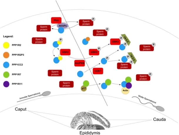
Figure I. 3: Illustrative scheme of the proteins involved in the acquisition of sperm motility based on PPP1CC2 regulation. AKAPs anchoring seem to modulate PPP1CC2 and PKA activities inducing sperm motility and phosphorylation of several proteins. PPP1CC2 and GSK3 phosphorylation by AKT and an unknown , respectively, lead to their inactivation and their possible binding to the bridging molecule 14-3-3, in epididymal cauda. PPP1CC2 is maintained active in caput due to the phosphorylation of a PPP1R2-like protein by GSK3. In cauda, GSK3 activity lowers and therefore a PPP1R2-like protein inhibits PPP1CC2 leading to sperm motility. PPP1R7 binds to PPP1CC2 in cauda sperm also inhibiting the phosphatase. In caput, PPP1R7 is prevented to inhibit PPP1CC2 due to a p17 unknown protein. Also, a multimeric complex has been identified composed by PPP1CC2, PPP1R7, actin and PPP1R11, where PPP1CC2 was inactive. (adapted from [149]).
PPP1CC/PPP1R11 complex
PPP1R11/I3 is a potent heat-stable PPP1 inhibitor [90, 150] and is a human homologue of the mouse t-complex expressed protein 5 (Tctex5), being genetically linked to the male sterility phenotypes of impaired sperm tail development and poor sperm motility in t complex mice [151-153]. Tctex5 gene synergistically with Tsga2 gene is a candidate for the “curlicue” and for the “stop” sperm phenotypes. The first phenotype is a chronic, negative bend of both the flagellar middle-piece and principal piece of spermatozoon, while the second phenotype prevents sperm from t-haplotype homozygotes from penetrating zona-free egg [154-156]. Tctex5 was shown to be present
in sperm protein lysates localizing to nuclei of pachytene spermatocytes, round spermatocytes, cytoplasm of Sertoli cells, in testis; cilia, secretion bodies and nuclei of epithelial cells and interstitium smooth muscle cells in the epididymis. In epididymal mouse spermatozoa Tctex5 is present in the head and principal piece of the tail [154] (Fig I.4). These are also the locations where PPP1CC2 is expressed [147].
Figure I. 4: Schematic representation of the subcellular localization of PPP1-PIP complexes in spermatozoon (adapted from [149]).
PPP1CC/PPP1R7 complex
A yeast sds22 homologue, PPP1R7, was identified in sperm [76], and inhibits the PPP1 catalytic subunit in rat liver nuclei [157]. Consistently, a PPP1R7 homologue was also identified in rat testis in association with PPP1CC2 [158]. The expression pattern of rat PPP1R7 matches that of PPP1CC2, suggesting that its involvement in spermatogenesis is correlated with the control of PPP1CC2 activity. Furthermore PPP1R7 was also identified in motile caudal spermatozoa as a regulator of PPP1CC2 catalytic activity [135]. Additionally, sds22 has consensus sites for phosphorylation by GSK3, PKA and CDK2 (calmodulin dependent kinase II), all present in sperm.
In male germ cells PPP1CC2, PPP1R11, PPP1R7 and actin form a multimeric complex in which PPP1CC2 is inactive [159]. The stability of the complex depended on
functional PPP1 interaction sites in PPP1R7 and PPP1R11, indicating that PPP1 mediates the interaction between these two proteins, forming a catalytically inactive complex in the germ cell [160]. The function of this complex in sperm motility, if any, still needs to be elucidated (Fig I.3).
PPP1CC/14-3-3 complex
PPP1CC2, as well as other PPP1 isoforms, have at the C-terminus a consensus TPPR amino acid sequence containing a threonine residue (T311) that can be phosphorylated by CDK2, reducing its activity [161-163]. The proportion of phosphorylated PPP1CC2 in caudal sperm is higher than in caput epididymal sperm and is localized to the posterior region of the sperm head, the equatorial region, implicated in sperm-egg binding, and in the principal piece of the sperm tail [164]. Interestingly, CDK2 knockout mice are viable but male and female are sterile [165, 166]. Vijayaraghavan et al. proposed that regulation of PPP1CC2 activity by CDK2 phosphorylation might be a mechanism for developing sperm motility. This might be achieved through binding of PPP1CC2 to the bridging molecule 14-3-3. In sperm 14-3-3 binds PPP1CC2 [125] (Fig I.3). The 14-3-3 protein is highly conserved among eukaryotic cells and acts as an adaptor protein in cellular signaling and metabolism. More than 100 binding partners have been identified, using affinity chromatography coupled with proteomic analysis [167-170]. 14-3-3 and its binding partners are regulators of protein-protein interactions during spermatogenesis [171]. It is consistent that 14-3-3 appears to regulate diverse cellular events such as cell cycle, apoptosis, protein trafficking, cytoskeleton rearrangements and metabolism [172]. The study of 14-3-3 interactome in bull sperm identified many proteins involved in different cellular events, from acrosome reaction to metabolism [173]. In particular, GSK3 was found to bind 14-3-3 [173, 174] (Fig I.3). Sperm 14-3-3 protein is present in the post-acrosomal region of the head and the principal piece, similar to PPP1CC2 [125] (Fig I.4). As already stated, changes in tyrosine and serine phosphorylation of GSK3 occur in parallel with motility stimulation in sperm [175]. The exact functions of 14-3-3 through binding to sperm phosphoproteins are still subject of extensive research.
PPP1CC/AKAPs complexes
The cyclic AMP (cAMP)-dependent protein kinase (PKA) is a ubiquitous, multifunctional enzyme involved in the regulation of several cellular events. PKA
holoenzyme consists of four subunits, two catalytic and two regulatory (RI and RII). Subcellular targeting to the vicinity of preferred substrates is a means of restricting the specificity of these enzymes [176, 177]. Compartmentalization of PKA is mediated through association of its regulatory subunits with A-kinase anchoring proteins (AKAPs) [176]. To date, over 40 AKAPs have been identified, and in testis/sperm there are three AKAPs that have been directly related to PPP1CC2 (AKAP220, AKPA3 and AKAP4) and many more show a similar localization.
AKAP220/AKAP11 binds PKA and PPP1, being a competitive inhibitor of PPP1 [178]. In testis, AKAP220 associates with PKA, where it may target the kinase to peroxisomes [179]. AKAP220 mRNA is expressed at high levels in human testis and in isolated human pachytene spermatocytes and round spermatids [180]. AKAP220 is present in human male germ cells and mature sperm and like RIIα, is located in the midpiece and is probably associated with cytoskeletal structures [180] (Fig I.4). The midpiece associated AKAP220 could serve to anchor PKA and/or PPP1CC2, directly regulating the contractile machinery in the sperm axoneme. Furthermore, it has been shown that disruption of RII interaction with AKAPs, by membrane-permeable peptides, causes the arrest of sperm motility [181].
AKAP4/AKAP82 cDNA was first isolated from a mouse testis cDNA-expression library [182, 183]. Mouse Akap4 expression was only detected in testis and it was determined that transcription is initiated at 20-22 days after birth and the mRNA is present in spermatids but not in pachytene spermatocytes [182, 184]. In RNA from testes extracts of hamster, guinea pig, rabbit, ram, and human, transcripts that hybridized to the mouse Akap4 cDNA were found [184]. AKAP4 protein was synthesized as a precursor present throughout the principal piece in testicular sperm. A small peptide corresponding to the precursor fragment and a higher molecular weight protein, possibly a phosphorylated form of the precursor, are present in epididymal sperm in low amounts. In mouse sperm, AKAP4 was detected throughout both the longitudinal columns and the semi-circumferential ribs of the fibrous sheath [185] (Fig I.4). All these findings correlate with a restricted temporal and spatial expression of the AKAP4 protein, present only in spermatogenetic cells and the predominant protein in the fibrous sheath of the sperm flagellum. Targeted disruption of the Akap4 gene causes absence of sperm motility together with a complete lack of fibrous sheath on the principal piece of mature mice sperm, causing male mice to be infertile [186]. In spermatozoa, Akap4 gene knockout mice that lack flagellar movement, exhibit a significant change in the activity and
phosphorylation of PPP1CC2 [147]. This suggests the involvement of AKAP4 in the regulation of PPP1CC2 activity in the principal piece of mouse spermatozoa.
AKAP3/AKAP110, also referred to as FSP95, undergoes tyrosine phosphorylation during in vitro capacitation of human sperm. Northern blot analysis of RNA from 50 human tissues determined that the transcripts are found only in testis, more specifically in round spermatids. Using a rat antiserum to recombinant protein, FSP95 was found only in the fibrous sheath and localized to the circumferential ribs of human sperm [187] (Fig I.4). Brown et al. reported that AKAP4 anchors AKAP3 and two novel spermatogenetic cells specific proteins, Fibrous sheath interacting proteins 1 and 2 (FSIP1 and FSIP2) [188].
Given that many AKAPs have been shown to be present in germ cells and localized to compartments related to motility where PPP1CC2 is also present they might be putatively involved in motility acquisition.
S-AKAP84 localizes to the midpiece of mouse elongating spermatids and co-localizes with mitochondria [189] (Fig I.4). Its splice variants D-AKAP-1 [116] and AKAP121 [190] were also detected in mouse testis and GC2 germ cells, respectively. TAKAP-80, was isolated by screening a rat testis expression library [191]. The corresponding protein was detected in rat testis and in purified fibrous sheath fractions from rat epididymal sperm (Fig I.4). The levels of protein were higher in mature compared to immature rat testis, correlating with the mRNA levels [191]. AKAP28, was detected in human testis, and is highly enriched in the axoneme structure (Fig I.4). It is likely to play a role in signaling mechanisms necessary for ciliar beating frequency [192]. WAVE1 localization in spermatocytes and round spermatids coincided with Golgi apparatus, whereas in elongated spermatids and testicular sperm localized to the mitochondrial sheath [193] (Fig I.4).
Recent data showed that inhibition of protein phosphatases with calyculin A resulted in an enhancement of the phosphorylated state at the activation loop of the PKA catalytic subunit in the mouse sperm principal and midpieces [194]. Also, PKA RII and PPP1CC2 are co-localized in the principal and midpieces. PPP1 and PPP3 suppress full activation of PKA, as well as enhancement of the phosphorylated states of other flagellar proteins, in order to prevent precocious changes of flagellar movement from the progressive type to hyperactivation [194].
Together, these findings suggest that the AKAP/PKA/PPP1 complex is really important for regulation of sperm motility (Fig I.3).
Of marked interest is the fact that each PPP1/PIP complex has a specific sperm subcellular location (Fig I.4). Clearly PPP1CC2/PIP complexes are essential regulatory
components in the signaling transduction cascades involved in sperm motility acquisition during epididymal transit. Defects in any component of these signaling cascades will give rise to pathological anomalies, leading to male infertility. Therefore, the study of new PPP1CC2/PIP complexes in testis and sperm are extremely important in a physiological and pathological point of view.
Objectives
The aim of this work was to study new PPP1CC2/PIP complexes in testis and sperm and to characterize their important physiological role.
Therefore, this thesis addresses a previously well known somatic PIP in testis/sperm, named PPP1R2 and two new PPP1CC2 testis/sperm specific PIPs that were found in a yeast two-hybrid technique using a human testis cDNA library.
Chapter II comprises two sections in paper format and one section in thesis format. In the first section (paper format), the objective was to study the presence of PPP1R2 and PPP1R2P3 (PPP1R2 pseudogene 3) proteins in human sperm as well as their specific sperm localization. PPP1R2P3 is a new PIP, also designated as IL (Inhibitor 2-Like/PPP1R2P3, NCBI Id: NM_206858) [149, 195]. Further, phosphorylation studies in PPP1R2P3 in vitro were also pursued. In the second section (paper format), the objective was to study the PPP1R2 pseudogenes using bioinformatics to understand their evolution and significance in mammals. In the third section (thesis format), transgenic mice expressing human PPP1R2 or PPP1R2P3 were produced to understand the role of these proteins in spermatogenesis and sperm maturation.
Chapter III comprises two sections. The first section (paper format) addresses the identification and characterization of a novel dynein light chain, Tctex1d4, in testis and sperm as a new binding partner of PPP1CC. In the second section (paper format) bioinformatics and molecular biology were integrated to understand Tctex1d4 evolution and the RVxF modification that occurred in Pika.
By unraveling these three new proteins that interact with PPP1CC and are present in testis and sperm the thesis aim to provide new insights about PPP1CC2 function in these tissues.
References
1. Cohen, P.T., Protein phosphatase 1--targeted in many directions. J Cell Sci, 2002.
115(Pt 2): p. 241-56.
2. Fardilha, M., et al., The Physiological Relevance of Protein Phosphatase 1 and its Interacting Proteins to Health and Disease. Current Medicinal Chemistry, 2010.
17(33).
3. Ceulemans, H. and M. Bollen, Functional diversity of protein phosphatase-1, a cellular economizer and reset button. Physiol Rev, 2004. 84(1): p. 1-39.
4. Philip, C., The regulation of protein function by multisite phosphorylation – a 25 year update. Trends in biochemical sciences, 2000. 25(12): p. 596-601.
5. Barford, D., A.K. Das, and M.-P. Egloff, The Structure and Mechanism of Protein Phosphatases: Insights into Catalysis and Regulation. Annual Review of Biophysics and Biomolecular Structure, 1998. 27(1): p. 133-164.
6. Yeo, M., et al., A novel RNA polymerase II C-terminal domain phosphatase that preferentially dephosphorylates serine 5. J Biol Chem, 2003. 278(28): p. 26078-85. 7. Gallego, M. and D.M. Virshup, Protein serine/threonine phosphatases: life, death,
and sleeping. Curr Opin Cell Biol, 2005. 17(2): p. 197-202.
8. Cohen, P.T., Novel protein serine/threonine phosphatases: variety is the spice of life. Trends Biochem Sci, 1997. 22(7): p. 245-51.
9. Honkanen, R.E. and T. Golden, Regulators of serine/threonine protein phosphatases at the dawn of a clinical era? Curr Med Chem, 2002. 9(22): p. 2055-75.
10. Sood, P.P. and M.A. Majid, Qualitative and quantitative changes of acid and alkaline phosphatases in the testis and epididymis of mice in relation to single high dose of alpha-chlorohydrin. Acta Eur Fertil, 1987. 18(1): p. 33-8.
11. Fardilha, M., Characterization of PP1 Interactome from human testis, 2004, Universidade de Aveiro.
12. Plowman, G.D., et al., The protein kinases of Caenorhabditis elegans: A model for signal transduction in multicellular organisms. Proceedings of the National Academy of Sciences, 1999. 96(24): p. 13603-13610.
13. David, B., Molecular mechanisms of theprotein serine/threonine phosphatases. Trends in biochemical sciences, 1996. 21(11): p. 407-412.
14. Smith, G.D., et al., Primate sperm contain protein phosphatase 1, a biochemical mediator of motility. Biol Reprod, 1996. 54(3): p. 719-727.
15. Shima, H., et al., Protein phosphatase 1 gamma 2 is associated with nuclei of meiotic cells in rat testis. Biochem Biophys Res Commun, 1993. 194(2): p. 930-7. 16. Kitagawa, Y., et al., Protein phosphatases possibly involved in rat
spermatogenesis. Biochem Biophys Res Commun, 1990. 171(1): p. 230-5.
17. Fardilha, M., Da Cruz e Silva, O.A.B., Da Cruz e Silva, E.F, A importância do mecanismo de "splicing" alternativo para a identificação de novos alvos terapêuticos. Acta Urológica, 2008. 25: p. 1: 39-47.
18. da Cruz e Silva, E.F., et al., Differential expression of protein phosphatase 1 isoforms in mammalian brain. J Neurosci, 1995. 15(5 Pt 1): p. 3375-89.
19. Takizawa, N., et al., Tissue distribution of isoforms of type-1 protein phosphatase PP1 in mouse tissues and its diabetic alterations. J Biochem, 1994. 116(2): p. 411-5.
20. Strack, S., et al., Differential cellular and subcellular localization of protein phosphatase 1 isoforms in brain. The Journal of Comparative Neurology, 1999.
413(3): p. 373-384.
21. Ouimet, C.C., E.F. da Cruz e Silva, and P. Greengard, The alpha and gamma 1 isoforms of protein phosphatase 1 are highly and specifically concentrated in

![Figure I. 4: Schematic representation of the subcellular localization of PPP1-PIP complexes in spermatozoon (adapted from [149])](https://thumb-eu.123doks.com/thumbv2/123dok_br/15913379.1092959/41.892.122.785.306.695/figure-schematic-representation-subcellular-localization-complexes-spermatozoon-adapted.webp)