REVIEW ARTICLE
Vascular Pathways of Testosterone: Clinical Implications
Margarida Lorigo1&Melissa Mariana1&Nelson Oliveira2&Manuel C. Lemos1&Elisa Cairrao1 Received: 2 August 2019 / Accepted: 15 November 2019# Springer Science+Business Media, LLC, part of Springer Nature 2019
Abstract
Cardiovascular diseases (CVD) are one of the leading causes of death worldwide. Testosterone (T) is an important sex hormone that triggers several genomic and non-genomic pathways, leading to improvements of several cardiovascular risk factors and quality of life in men. At the vascular level, the key effect of T is the vasorelaxation. This review discusses the molecular pathways and clinical implications of T in the vascular system. Firstly, the mechanisms involved in the T vasodilator effect will be presented. Then, it will be discussed the association of T with the main risks for CVD, namely metabolic syndrome, type 2 diabetes mellitus, obesity, atherosclerosis, dyslipidaemia and hypertension. Several studies have shown a correlation between low T levels and an increased prevalence of several CVD. These observations suggest that T has beneficial effects on the cardiovas-cular system and that testosterone replacement therapy may become a therapeutic reality for some of these disorders.
Keywords Androgens . Vasorelaxation . Risk factors . Cardiovascular diseases . Metabolic syndrome . Diabetes mellitus . Obesity . Atherosclerosis . Dyslipidaemia . Blood pressure
Introduction
The predominant endogenous androgen in the bloodstream is testosterone (T) [1]. Circulating T is mainly bound to serum proteins, such as sexual hormone-binding globulin (SHBG)
and albumin [2] being the T concentrations higher than its solubility. Only 2–3% of all circulating T is free [1], reaching its maximum in 30-year-old men, and continuously decreas-ing 1–2% per year afterwards [3,4]. From the molecular point of view, T can be converted to dihydrotestosterone (DHT) by the 5α-reductase enzyme or to oestradiol by aromatase [5]. DHT has more affinity for the androgen receptor (AR), being the most active androgen. Nevertheless, the plasma DHT con-centration is much lower compared to T [6] since the adult male testes produce more T than DHT. However, these levels can vary with the circadian rhythm and stress. Moreover, T is not a male-exclusive hormone. Women produce androgens in the ovaries and adrenal glands [3,7] and, during the pregnan-cy, the fetoplacental unit synthesizes androgens and oestrogens that play an important biological role in these de-velopment stages [8]. The chemical structure of T can be seen in Fig.1.
T presents genomic and non-genomic actions at the vascu-lar level, and both effects may overlap. The key T effect at the vascular level is the vasorelaxation, a non-genomic action (rapid effect) [9]. Over the years, several studies have agreed that T is an important sex hormone that triggers genomic and non-genomic pathways, leading to improvements of several cardiovascular risk factors and quality of life in men [10]. So, Associate Editor Yihua Bei oversaw the review of this article
* Elisa Cairrao [email protected] Margarida Lorigo [email protected] Melissa Mariana [email protected] Nelson Oliveira [email protected] Manuel C. Lemos [email protected] 1
CICS-UBI - Centro de Investigação em Ciências da Saúde, University of Beira Interior, 6200-506 Covilhã, Portugal
2
UDI-IPG - Research Unit for Inland Development, Polytechnic Institute of Guarda, Av. Dr. Francisco de Sá Carneiro, 6300-654 Guarda, Portugal
https://doi.org/10.1007/s12265-019-09939-5
it is very important to understand the relationship between the risk factors for cardiovascular diseases (CVD) and T, based on the knowledge of T vasodilator mechanisms. Thus, in the first topic of this paper, the mechanisms involved in the T vasodi-lator effect will be briefly presented and then, in the subse-quent sections, it will be discussed the association of T with the main risks for the CVD, namely metabolic syndrome, type 2 diabetes mellitus, obesity, atherosclerosis, dyslipidaemia, and hypertension. The final aim of this review is to clarify the clinical implications of testosterone in the vascular system.
Genomic and Non-genomic Overlapping
Vasodilatory Mechanisms of Testosterone
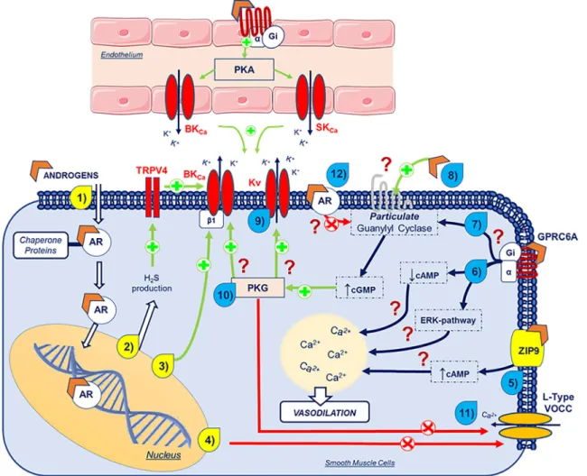
As previous described, at the vascular level, T presents geno-mic and non-genogeno-mic overlapping actions that lead to an im-provement of the risk factors for the CVD development/ progression [10].The genomic effects of androgens, including those of T, are initiated by their passage through the cellular plasma mem-brane into the cytoplasm. Then, they dissociate from the chap-erone proteins and bind to AR, leading to alterations in spe-cific genes. These alterations include an increase of the hydro-gen sulphide (H2S) production, and consequently vasodilation via transient receptor potential vanilloid 4 (TRPV4) and large-conductance Ca2+-activated K+ channels (BKCa) [10]. However, there are only a few studies regarding the androgen genomic effects at the cardiovascular level. In animal arteries, it has been suggested that androgens lead to an increase in the expression of calcium and potassium channels [11,12] as well as that of soluble guanylate cyclase [13]. Moreover, an in-crease in the expression of voltage-dependent calcium chan-nels in rat heart [14] and T-type calcium currents in neonatal rat heart [2] by androgens have also been reported. Regarding the human arteries, it has been shown that androgens cause another gene alteration that modifies the vascular tone, by downregulation of L-type voltage-operated Ca2+ channels
(L-type VOCC) and upregulation of β1 subunit of BKCa channels [15].
As previously mentioned, the main non-genomic effect of T on the vasculature is the vasorelaxation [9]. The major mechanisms involved in this effect include the nuclear or membrane receptor activation, influence of vascular endothe-lium and activation or blocking of ion channels. Concerning the activation of androgen nuclear receptors, the participation of AR in vasodilation is controversial, as some authors sug-gested the presence of this receptor on the cell membrane [16]. Moreover, some authors observed that the T vasodilatory ef-fect was inhibited by the AR antagonist [17,18]. On the other hand, several authors demonstrated that the use of the AR antagonist (flutamide) [19–21], albumin-bound androgen an-alogues, which cannot pass through the membrane [19] and protein synthesis inhibitors [22] are not involved in T vasodi-lator effect. Besides, some authors also observed that T in-duced vasodilation in arteries without the nuclear functional AR [20,23,24], thereby demonstrating that this effect was not mediated by AR [20]. Several hypotheses have been proposed to explain the rapid androgens’ vascular responses, namely a steroid nuclear receptor translocation to the cell membrane surface, nonspecific effects of steroids on plasma membrane fluidity or direct allosteric modification of ion channels de-pendent of binders and G protein-coupled receptors (GPRCs) [25]. However, concerning T, only nuclear receptor transloca-tion to the cell membrane surface has been reported. Recently, four new membrane AR have been suggested to be responsi-ble for the non-genomic effects of androgens. Two of them are GPCR (OXER1 and GPR6A) and the other ion channels/ transporters for calcium and zinc (TRPM8 and ZIP9) respec-tively [23,24].
Regarding the vascular endothelium, the key contributor to the androgen vasorelaxation effect is controversial. Some studies demonstrated that vascular endothelium has a major role in the T vasodilator mechanism [26,27], depending on species and gender. On the contrary, others demonstrated that this effect may occur through an independent-endothelium pathway [28–30], by direct action of T on the vascular smooth muscle. This last pathway seems to involve activation of the receptor coupled to Gi/oprotein, activating protein kinase A, which in turn leads to a hyperpolarization and consequently small conductance calcium-activated potassium (SKCa) and BKCachannels activation [31].
Concerning the effects on ion channels, several studies suggested that T exerts its vasodilatory effects by modulating different K+and/or Ca2+channels [15,21,32–34]. The vas-cular tone is controlled by the ion flow, and the activation of K+ channels represents the key mechanism involved in the relaxation [35,36]. This activation of K+ channels causes a membrane hyperpolarization that closes the Ca2+channels, leading to vasorelaxation [37,38], and showing the close re-lationship between the channels mentioned. Most works agree
that T activates the K+channels and/or inhibits the VOCC. In this case, the authors suggested that T can directly inhibit these channels [30,39] or that this inhibition can occur indirectly by the K+channels activation, mainly the KVand BKCachannels (due to PKG activation upon intracellular cGMP increase) [40]. Since there is no consensus regarding the exact mecha-nism of these T effects, more studies must be performed to establish if they are direct or due to the activation of a signal-ling pathway.
The overlap between genomic and non-genomic T actions makes it difficult to define the broad vascular effects of andro-gens. Increases in androgen concentrations can induce a rapid first phase effect followed by a broader, second phase; the genomic effects can modulate the effects of the first phase. To our knowledge, there are only two studies that demonstrate simultaneously both types of effects and this analysis is crucial to determine the repercussions of conceivably long-lasting therapy with androgens or a maintained increase in androgen levels. Er et al. (2007), using rat ventricular myocytes, dem-onstrated that the genomic T effects on L-type VOCC are completely antagonized by the non-genomic effects following T addition [41]. More recently, Saldanha et al. (2013) demon-strated that the genomic effects of androgens changed the expression of the BKCa β1-subunit, and the BKCa and KV channels activities change the non-genomic T action. These authors also suggested that androgens could be beneficial for the prevention of hypertension, once androgens have direct vasodilator effects and the increase of the BKCachannels ex-pression results in greater involvement of KVchannels as me-diators of vasodilation [15].
The vasodilator mechanism of testosterone, including the genomic and non-genomic actions, is illustrated in Fig.2. As can be seen in the figure, both effects may overlap.
Association of Testosterone with Risk Factors
for CVD
Testosterone was initially considered to be harmful to the car-diovascular system, as men have a high prevalence of CVD and cardiovascular morbidity and mortality are over 2-fold greater in men compared to women [42]. However, several clinical and epidemiological studies have challenged this idea. It was observed that men with CVD, type 2 diabetes mellitus (T2DM), obesity, metabolic syndrome (MeSy) and dyslipidaemia had low levels of T [43–45]. Also, the preva-lence of CVD increases in ageing men, when T production declines. These observations suggest that this hormone has beneficial effects on the cardiovascular system and that the testosterone replacement therapy (TRT) may become a thera-peutic reality for some of these pathologies. However, more recent studies have shown that TRT may be associated with an increased incidence of adverse cardiovascular events, leading
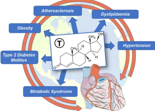
to an intensified controversy regarding the cardiovascular ben-efits of TRT [46–48]. Thus, in this section, we will address the association of T with the main risk factors for CVD develop-ment, namely metabolic syndrome, type 2 diabetes mellitus, obesity, atherosclerosis, dyslipidaemia, and hypertension. An abstract illustration of the main risk factors for CVD is repre-sented in Fig.3.
Testosterone and Metabolic Syndrome and Type 2
Diabetes Mellitus
The MeSy is a complex combination of risk factors for CVD and diabetes. These factors include dysglycemia (i.e. elevated fasting glucose), raised blood pressure, dyslipidaemia (i.e. elevated triglyceride levels and low high-density lipoprotein (HDL) cholesterol levels) and obesity [42, 49]. The molecular mechanisms behind this complex clinical condition can be explained by the bi-directional mechanism between hypogonadism and obesity—a mechanism that involves not only adipocytes but Leydig cells and hypothalamic hormones responsible for pituitary-testicular axis control. Also, excessive lep-tin production (as observed in obesity) may be an ally of this mechanism, as it disrupts testicular [50]. Patients with MeSy have twice the risk of developing CVD in the next 5–10 years than individuals without MeSy and a 5-fold increased risk for developing T2DM [49]. The most popular definitions used for MeSy are [49, 51]:
WHO (World Health Organization) 1999:
Presence of insulin resistance or glucose > 6.1 mmol/L (110 mg/dL), 2 h glucose > 7.8 mmol (140 mg/dL) (required) along with any two or more of the following: HDL cholesterol < 0.9 mmol/L (35 mg/dL) in men, < 1.0 mmol/L (40 mg/dL) in women
Triglycerides > 1.7 mmol/L (150 mg/dL)
Waist/hip ratio > 0.9 (men) or > 0.85 (women) or body mass index (BMI) > 30 kg/m2
Blood pressure > 140/90 mmHg
NCEP (National Cholesterol Education Program) ATP3 2005:
Presence of any three or more of the following:
Blood glucose greater than 5.6 mmol/L (100 mg/dL) or drug treatment for elevated blood glucose
HDL cholesterol < 1.0 mmol/L (40 mg/dL) in men, < 1.3 mmol/L (50 mg/dL) in women or drug treatment for low HDL-C
Blood triglycerides > 1.7 mmol/L (150 mg/dL) or drug treatment for elevated triglycerides
Waist > 102 cm (men) or > 88 cm (women)
Blood pressure > 130/85 mmHg or drug treatment for hypertension
IDF (International Diabetes Federation) 2006:
Waist > 94 cm (men) or > 80 cm (women) along with the presence of two or more of the following:
Blood glucose greater than 5.6 mmol/L (100 mg/dL) or diagnosed diabetes
HDL cholesterol < 1.0 mmol/L (40 mg/dL) in men, < 1.3 mmol/L (50 mg/dL) in women or drug treatment for low HDL-C
Blood triglycerides > 1.7 mmol/L (150 mg/dL) or drug treatment for elevated triglycerides
Blood pressure > 130/85 mmHg or drug treatment for hypertension
Note: NCEP and IDF definitions are very similar except in the waist parameter of 102 vs. 94 cm in men and 88 vs. 80 cm in women.
Fig. 2 Schematic representation of the overlap vasodilator genomic and non-genomic mechanisms of androgens. The yellow numbers represent the genomic actions and the blue numbers represent the non-genomic actions. (1) Androgens cross the plasma membrane and enter the cyto-plasm, where it dissociates from chaperone proteins and bind to androgen receptors (AR). (2) Androgens increase the H2S production and leads to
vasodilation via TRPV4 and BKCa. (3) Androgens upregulateβ1 subunit
BKCa. (4) Androgens downregulate L-type VOCC. (5) Androgens
acti-vate the ZIP9 and lead to a cAMP increase. (6) Through binding to GPRC6A (Giα-activation), androgens lead to a cAMP decrease and
ERK pathway activation. These two mechanisms modulate intracellular Ca2+levels and lead to vasodilation through mechanisms still unknown.
(7) Through binding to Giα, androgens are thought to activate particulate
guanil cyclase, by a mechanism that is not yet known. (8) Androgens are thought to activate protein receptors, leading to a cGMP increase by particulate guanil cyclase activation. This increase in cGMP levels leads
to PKG activation. Through PKG activation, androgens activate (9) Kv
and (10) BKCachannels and inactivate (11) L-type VOCC channels
through mechanisms still unknown. (12) Via androgen receptors (AR), it is thought that these sex hormones inhibit particulate guanil cyclase. Legend: , peptide receptor; , androgens; /green arrows, stimu-lation; /red arrows, inhibition; ?, unknown mechanism; AR, androgen receptors; BKCa, large-conductance Ca2+-activated K+channels; Ca2+,
calcium; cAMP, cyclic adenosine monophosphate; cGMP, cyclic guano-sine monophosphate; ERK, extracellular signal-regulated kinase; GPRC6A, G protein-coupled receptor family C group 6-member A; H2S, hydrogen sulphide; HSP, heat shock proteins; K+, potassium; Kv,
voltage-gated K+channels; L-Type VOCC, L-type voltage-operated Ca2+ channels; PKG, protein kinase G; TRPV4, transient receptor potential vanilloid 4; ZIP9, Zrt- and Irt-like protein 9
Although some studies have attempted to clarify the mo-lecular mechanisms underlying T2DM, these remain unclear. However, most studies agree that there is insulin resistance, β-cell dysfunction, and apoptosis, as well as oxidative stress, mitochondrial dysfunction and inflammation [52].
According to the latest systematic reviews and meta-anal-ysis, there is clear evidence that low levels of T are associated with an increased risk for MeSy [50,53–56] and T2DM [54,
57,58]. Corona et al. (2011) suggested that MeSy can be considered as an independent cause associated with male hypogonadism. Although only a few randomized controlled trials have been reported, TRT seems to be a reliable treatment to improve metabolic control, as well as central obesity [53]. It was also reported that T treatment has beneficial effects on all MeSy components [59]. The main criterion for the diagnosis of hypogonadism is the low blood T level in men, which impairs general physical and mental health status and is diag-nosed in ~ 2% of elderly men. Total and free T levels of less than 11 nmol/L and 0.22 nmol/L, respectively, are the mini-mum criteria for the hypogonadism diagnosis in Europe [42,
60]. In 2013, a review performed by Corona et al. (2013) about male hypogonadism suggested that TRT was able to improve central obesity in subjects with MeSy, and glucometabolic control in patients with MeSy and T2DM. However, the authors admitted that the number of studies was too small to draw firm conclusions [54]. Regarding the two studies performed by Brand’s group, a meta-analysis per-formed in 2011 suggested a sex-dependent association be-tween T and MeSy, as the total and free T levels were lower in men with MeSy and higher in women with MeSy, when compared to men and women without pathologies. However,
there are no indications for a sex-specific association between SHBG and MeSy, since, in both men and women, MeSy is associated with lower SHBG levels [55]. In 2014, Brand et al. suggested that low total and free T were associated with ab-dominal obesity, hypertriglyceridemia, hyperglycaemia, low HDL levels and hypertension [56]. In the same year, Cai et al. (2014) suggested that, in T2DM hypogonadal men, TRT could improve glycaemic control and decrease triglycer-ide levels. However, the authors of this systematic review concluded that the limited number of participants and the con-founding factors must be considered when assessing the met-abolic effects of TRT and its long-term influence on hypogonadal men with T2DM [61].
Another meta-analysis was performed to study the as-sociation between T and T2DM. Ding et al. (2006) showed that high T levels were associated with an elevat-ed risk of developing T2DM in women and with a lower risk in men, and higher SHBG levels induced a lower risk of T2DM in women than in men [58]. Corona et al. (2011) had suggested that T2DM can be independently associated with male hypogonadism, although TRT seems to improve glucometabolic control as well as fat mass in T2DM subjects [57]. More recently, a cohort study and meta-analysis with postmenopausal women suggested that the association of SHBG with T2DM did not change with the menopause status, whereas the associations between the endogenous sex hormones and T2DM occurred only in postmenopausal women. SHBG and total oestradiol are independent risk factors for the development of T2DM in women and no association was observed between total or free T with T2DM [62].
Fig. 3 Abstract illustration of the main risk factors for
Moreover, several studies clearly showed that TRT induced metabolic beneficial effects in MeSy and T2DM patients. The two meta-analyses performed by Corona et al. in hypogonadal MeSy patients suggested some benefits of TRT in the reduc-tion of waist circumference, fasting glucose, and insulin resis-tance, and in the increase of HDL levels [53,54]. The reduc-tion of fasting glucose, glycated haemoglobin (HbA1c), tri-glyceride and body fat were also suggested in hypogonadism and T2DM patients submitted to TRT, although total choles-terol, HDL cholescholes-terol, blood pressure, and body mass index did not seem to be affected [54,61]. In contrast, patients with MeSy and/or T2DM in the absence of classic hypogonadism did not show these TRT benefits regarding glycaemic control (assessed by HbA1c) or constitutional symptoms reported by Ageing Male Symptom score [63].
In summary, most of the mentioned studies showed that low T levels are associated with an increased risk of MeSy and T2DM. Recent systematic reviews and meta-analyses showed that this beneficial effect is more important in MeSy and T2DM hypogonadal males, and the beneficial effects are not only associated with fasting glucose, glycated haemoglobin and cholesterol levels, but also with obesity, which will be explored in the next section.
Testosterone and Obesity
According to the WHO, overweight and obesity are defined as abnormal or excessive fat accumulation that presents a risk to health, in which a person with BMI≥ 25 kg/m2is generally considered overweight, while a BMI≥ 30 kg/m2is considered obese [64]. Obesity is directly or indirectly related to metabol-ic disorders and the development and progression of T2DM. It leads to inflammation of adipose tissue with consequent dysglycemia and insulin resistance, alterations in the lipid me-tabolism and in the blood pressure, which together may lead to endothelial dysfunction and atherogenesis [65]. Because obe-sity has been implicated in the pathophysiology of CVD, it is important to understand the relationship between obesity and androgen deficiency, and how it contributes to the several pathophysiological states of the cardiovascular system [66].
As it is well known, obesity leads to a larger size of adipose cells (the so-called adipocyte hypertrophy) [67], which is a determinant factor to this metabolic disorder. These metabolic changes may lead to a reduction of T. In fact, it was demon-strated that low levels of this sex hormone are associated with increased fat mass (mainly central adiposity but also visceral adiposity [66,68,69]), and with the reduction of lean mass, in men [70]. A bidirectional relationship between T and obesity supports the hypogonadism-obesity cycle [70], but it is still unclear whether obesity induces low levels of T or vice versa. It has been suggested that androgen deficiency is a conse-quence, and not a cause, of obesity [71]. This seems to agree with other studies that suggest that obesity promotes a faster
decline of T and SHBG levels with age [66,67,72], and that weight loss increases circulating T levels in obese men [73–75]. Even if the complete mechanism is not yet defined, these studies suggest that adipocyte hypertrophy and metabol-ic changes lead to decreased T levels [67].
In adipose tissue, particularly in visceral fat, T is converted to 17β-oestradiol by aromatase [68] which is highly expressed in adipocytes [70]. After this enzymatic conversion, oestrogens act on the hypothalamic–pituitary–gonadal (HPG)-axis to inhibit the release of hypothalamic gonadotropreleasing hormone (GnRH), which in turn in-hibits the release of pituitary luteinizing hormone (LH) and ultimately contributes to a decrease of gonadal T [70]. Low T levels will lead to lower oestrogen levels from aromatization, reducing the activated ER and, therefore, increasing the vis-ceral fat deposition and insulin resistance in men. Thus, T suppresses visceral fat deposition through its aromatization to oestradiol, which seems to be inversely related to T levels and to be dependent on ERα and aromatase [69]. An increase in visceral fat will increase adipocyte size and aromatase ex-pression, leading to greater conversion of T to oestrogen, thereby lowering T levels. Obesity can lead to decreased levels of T and, in turn, low T levels also contribute to an increase in adiposity [69,70]. Furthermore, the adipose tissue is also an endocrine organ secreting several factors that will influence the pathogenesis of obesity and hypogonadism. This process is considered as the hypogonadism-obesity-adipocytokine cycle [70]. In addition to oestrogens, there are inflammatory adipocytokines (e.g. tumour necrosis factor-al-pha, TNFα, and interleuk6, IL-6) whose hypothalamic in-hibitory action is the same, leading to the reduction of T levels [70,76]. Leptin, a hormone derived from adipose tissue that regulates body weight and food intake, and that stimulates GnRH under normal conditions, is increased with increased adiposity [77]. This may render the hypothalamus resistant to stimulation (unresponsiveness) and unable to lead to a normal T production [70]. Leptin interference in androgen production (via the LH-hCG pathway) [78] is another explanation for the low T levels. Accordingly, it was recently demonstrated that a high-fat diet decreases T levels through the suppression of testicular leptin and Janus kinase/signal transducers and acti-vators of transcription (JAK-STAT) pathway [79]. The fact that Leydig cells have insulin receptors and are sensitive to hyperinsulinemia also indicates another direct point of inter-action between the production and secretion of this hormone [76,80]. It seems clear that an increase in adiposity negatively affects the T secretion through multiple pathways [81], con-tributing progressively to CVD.
Taken together, these studies show an inverse relationship between low T levels and obesity, based on a bidirectional mechanism [70], where each of the two components potenti-ates the adverse effects of the other, increasing the risk of CVD [82]. Theoretically, when multiple changes are
pathologically linked by bidirectional relationships, a thera-peutic intervention directed at any of them may lead to an improvement of all associated conditions [83]. However, whatever the causality direction, T seems to be the key to this process. TRT has been shown to be effective and to improve some obesity components, i.e. decreased visceral and body fat mass, BMI and waist circumference, and increased lean mass, thus improving the body composition [69]; TRT also resulted in cardiometabolic factors improvement [70]. However, the mechanisms underlying T effects on obesity, or vice versa, are still not fully understood. Obesity is based on metabolic homeostasis dysregulation (involving different signalling pathways) but the way it disrupts the endocrine environment [84], or the causal relationship with T, have not yet been fully unveiled and therefore require additional investigations. The relationship between low T levels, obesity, and CVD is very complex and multifactorial, remaining, so far, poorly under-stood [82].
Testosterone and Atherosclerosis
Atherosclerosis is characterized by lipid accumulation and mononuclear leukocyte infiltration in the tunica intima of ves-sels. The molecular mechanisms that may be involved in the development and progression of atherosclerosis include the low-density lipoproteins (LDL) levels increase. Several con-ditions, mainly hypertension, smoking, and diabetes, have al-so been asal-sociated to the induction of this pathology. As it affects the cardiac circulation, it can cause myocardial infarc-tion or stable angina pectoris. Atherosclerosis may also under-lie ischaemic stroke and transient cerebral ischaemic attack, and lead to the formation of aneurysms, including in the aorta [85]. The onset and progression of arteriosclerosis can be de-tected by a surrogate carotid marker called intima-media thickness (IMT). This biomarker is measured by carotid ultra-sonography and it is used worldwide since it is a simple, reproducible and non-invasive method [86].
Several cross-sectional and longitudinal studies have shown an inverse association between T levels and carotid IMT [87–92]. In a study with 403 men, aged 73–94 years,
Van den Beld et al. (2003) observed that T, estrone, and insullike growth factor I (IGF-I) concentrations were in-versely correlated with mean IMT, and this association was stronger in the group without CVD [87]. Fukui et al. (2003) enrolled 253 men with T2DM, with a mean age of 62 years, and suggested that free T concentrations were inversely corre-lated with mean IMT and plaque score. Patients with low concentrations of free T (< 10 pg/mL) had greater IMT and plaque score mean than those with higher concentrations of free T [88]. De Pergola et al. (2003), in a study with 127 glucose-tolerant and obese men, aged 18 to 45 years, showed that T levels were negatively associated with IMT, regardless of body fat and other well-known cardiovascular risk factors,
suggesting that T may have a direct anti-atherosclerotic effect. Furthermore, the authors also observed an inverse association between T levels and BMI, fat mass, and waist circumference [90]. In a study of 195 men, aged 73 to 91 years, Muller et al. (2004) suggested that low free T levels and high oestradiol levels were inversely related to the thickening of the carotid IMT and these associations were independent of cardiovascu-lar risk factors [91]. Makinen et al. (2005), in a study of 239 men (40–70 years), demonstrated an association between T levels, andropause and carotid IMT, suggesting that men with andropause symptoms, together with impaired sex hormone status and increased carotid IMT, might benefit from TRT to decelerate the progression of atherosclerosis and protect them from its clinical sequelae (i.e. coronary heart disease, ischemic stroke, and peripheral vascular disease) [92]. In 2006, Svartberg et al. (2006) suggested that total the T levels of 1482 men, aged 25–84 years, were significantly inversely related to the carotid IMT, and this association was indepen-dent of age, CVD risk factors and lifestyle factors but was not independent of BMI [89]. In a study of 354 men, aged 65 years, Soisson et al. (2012) showed that low free T com-bined with high C-reactive protein (CRP) levels were associ-ated with elevassoci-ated carotid IMT in elderly men, and this asso-ciation with carotid IMT was not observed for SHBG nor oestradiol [93]. Farias et al. (2014), in a study of 115 male patients, aged younger than 70 years, found a negative corre-lation between carotid IMT and total T concentration in middle-aged men with T2DM. Men who had low T levels also had more atherosclerotic plaques, endothelial dysfunction and higher levels of high-sensitivity CRP [94]. Moreover, Lee et al. (2014), in a study with 50-year-old men with sexual dysfunction (308) observed a significant negative correlation between total T levels and the Framingham Risk Score (FRS) [95]. This is a scoring system derived from the Framingham heart study conducted in the United States and is an instru-ment that uses a simple past medical history and gender-specific cholesterol levels in asymptomatic patients to predict the incidence rate of CVD in the next 10 years [96,97]. In this sense, Lee et al. (2014) suggested that a higher T level may decrease the 10-year risk of CVD, and the occurrence of ath-erosclerosis, coronary artery disease and coronary events [95]. In contrast to these studies, Lee et al. (2016), using a large cohort of 3164 men from 4 ethnic origins (Caucasian, African American, Hispanic American and Asian American), aged 45 to 84 years, and without known CVD, observed that lower free T was associated with a higher relative risk of coronary artery Ca2+score > 0 and lower total T was associated with higher log coronary artery Ca2+score. Lower total and free T were associated with lower IMT [98].
In summary, high levels of T seem to have an anti-atherosclerotic effect, although one study did not observe such a correlation. This discrepancy needs to be clarified, mainly considering some CVD risk factors such as diabetes or
obesity, which also appear to be involved in the progression of atherosclerosis.
Testosterone and Dyslipidaemia
Dyslipidaemia is one of the main risk factors for CVD [70,
99]. The molecular mechanisms of dyslipidaemia mainly in-volve an imbalance of the lipid profile, characterized by low levels of HDL and high levels of LDL [99,100]. Additionally, dyslipidaemia may include high levels of total cholesterol, triglycerides (TG) and very low-density lipoprotein (VLDL) [70], as well as reduced activity of lipoprotein lipase, a protein associated with increased risk of coronary artery disease (CAD) [100].
As previously mentioned, T can lead to dyslipidaemia, con-tributing to the development of CVD. Specifically, low T levels are an integral part of the MeSy, characterized not only by dyslipidaemia, but also by obesity, diabetes and hyperten-sion [101]. Haffner et al. (1993) showed that there is a corre-lation between low levels of T and high levels of LDL, total cholesterol and TG, and low levels of HDL [102]. Moreover, Agledahl et al. (2008) inversely associated the total T with TG, and positively with HDL [103]. Following these authors, Makinen et al. (2008) correlated T directly with HDL and inversely with total cholesterol and TG [104]. Other authors, in addition to the positive association of T with HDL [105], also found an inverse relationship with VLDL levels [105,
106]. TRT was associated with beneficial decreases in LDL, total cholesterol and TG [70,107]. Increases in HDL levels have also been reported after TRT, although there are contra-dictory studies where a decrease [108,109] or no change [110] occurred. An explanation for these differences has not yet been found, but it is assumed that the T stimulation of reverse cholesterol transport may lead to increased HDL-C consump-tion [70].
Overall, individuals with low T levels seem to have a pro-atherogenic lipoprotein pattern, and thus, a higher propensity to develop MeSy and CVD. Although multiple factors con-tribute to atherosclerosis, dyslipidaemia has been considered as the main cause of this disease [111], with T playing a key role. In this sense, TRT induces a more favourable lipid pro-file, contributing to a decrease in CVD occurrence. In sum-mary, androgens seem to provide a protective effect against the development and/or progression of atherosclerosis [111,
112].
Testosterone and Blood Pressure
Physiologically, blood pressure can be defined by Ohm’s law, as being proportional to cardiac output and vascular resistance to blood flow. However, several other factors, in an integrated manner, characterize the molecular mechanism of this risk factor. Specifically, the baroreceptors detect acute pressure
changes in vessels; natriuretic peptides are increased due to higher pressure on the heart; the renin-angiotensin-aldosterone system influences vascular tone homeostasis; the adrenergic system influences heart rate, contraction and vas-cular tone; and other local mediators, such as nitric oxide and endothelin, that cause relaxation or vascular contraction, re-spectively. Taken together, all these factors make blood pres-sure changes a complex mechanism [113].
Regarding the association between T and blood pressure, Zitzmann and Nieschlag, in 2007, demonstrated beneficial effects of T in the diastolic and systolic blood pressure as well as resting heart rate, which decreased significantly during treatment with T in hypogonadal men [114,115]. Later, other authors studied the association between the increase or defi-ciency of androgens with gestational hypertension and pre-eclampsia. A positive correlation between the increase of an-drogen levels and preeclampsia was observed [116–118]. It was also demonstrated that the placental aromatase enzyme, that is responsible for the conversion of androgens to oestrogen, is deficient in placental ischemia and preeclamptic pregnancy [8,119]. On the other hand, other epidemiological studies have shown that maternal serum levels of androgens, such as dehydroepiandrosterone (DHEA), DHEA sulphate, androstenedione, and T, are elevated in healthy pregnant women [120,121]. In accordance, other studies have shown that T and its 5-reduced metabolites (5α- and 5β-DHT) pro-duced a marked reduction in the blood pressure of hyperten-sive and normotenhyperten-sive rats [122]. Particularly, 5β-DHT led to a substantial acute antihypertensive effect in hypertensive rats [123], which may be mainly due to a blockade of Ca2+entry through L-type VOCC [124]. Concerning the hypertensive disorders of pregnancy, Perusquia et al. (2018) demonstrated, for the first time in a rat model of preeclampsia, that andro-gens, mainly DHEA and 5β-DHT, attenuate hypertension in vivo due to their vasorelaxant effect, and this is a non-genomic mediated response [125]. In conclusion, an excess or insufficient androgen production during pregnancy may trigger the development of preeclampsia or gestational hyper-tension. Thus, these studies point out that the administration of androgens in women with hypertensive disorders of pregnan-cy is a near possibility. Nevertheless, further clinical studies are needed.
Testosterone and Heart Diseases
As described above, low levels of T appear to be harmful to the cardiovascular system due to the observed increase of CVD risk factors [45, 126]. T not only influences vascular reactivity but affects peripheral resistance, cardiac electro-physiology and cardiac output [126]. It is not clear how T influences ventricular repolarization mechanisms, but it ap-pears to act by activating K+ channels and simultaneously reducing the activity of L-type Ca2+ channels, thereby
increasing the repolarization reserve [69]. Decreased T is also associated with heart diseases such as coronary artery disease (CAD), heart failure (HF), ischaemic stroke, and atrial fibril-lation (AF) [127].
Regarding CAD, some studies have shown an inverse as-sociation between T levels and CAD. Phillips et al. (1983) were the first authors to demonstrate that exists an inverse relationship between free T levels and CAD degree [128]. Similar results were later obtained by Zhao et al. (1998) [129]. In 2000, English et al. (2000) demonstrated that men have lower levels of T than men with normal coronary angio-grams. Interestingly, the authors also found low levels of 17ß-estradiol, which is suggestive of a direct action of T on the CAD pathogenesis [43]. Several investigations also evaluated the carotid artery by ultrasonography and demonstrated that low T levels were associated with carotid IMT [89,93,130,
131], total carotid plaque area [132], and intermittent claudi-cation [133]. Rosano et al. (2007) in their investigations also demonstrated that low T levels and 17β-estradiol are associ-ated with coronary artery disease in male patients with angina. These results also show that there is an inverse relationship between the degree of CAD and plasma T levels suggesting that low plasma T may be involved with the increased risk of CAD in men [134]. The clinical relevance of low oestradiol levels after normalization of T levels needs further investiga-tion as both sex hormones are tightly linked.
Concerning HF, this heart disease is usually associated with an increase in peripheral vasoconstriction [126]. The most common cause of HF and impaired left ventricular dysfunc-tion is coronary artery disease (CAD) with resulting cardiac ischemia, leading to decreased contractility and weakness of the heart muscle. It is common for men with HF to have low T levels and this leads to increased mortality [135] as they are associated with reduced heart function, loss of lean muscle mass and strength, chronic anaemia and insulin resistance [126]. Some studies have suggested that there also appears to be an inverse relationship between total and T-free levels and HF grade [69], in which case the prevalence of hypogonadism is greater than 40% [136]. It has been specu-lated that low T levels may be linked to an anabolic/catabolic imbalance, a typical feature of advanced HF. Some research supports this hypothesis as they report that low T levels are associated with some clinical features of advanced HF (e.g. reduction of muscle mass, decreased exercise capacity, energy handling, fatigue dyspnoea, and final cachexia) [136]. Patients with HF have decreased exercise capacity and muscle fatigue, but this is not necessarily related to the myocardial dysfunc-tion degree [136], a fact that is supported by other investiga-tions [137,138]. Studies by Jankowska et al. (2006) demon-strated that low T levels independently predisposed to im-paired exercise capacity, whereas other indices related to the heart disease progression were not [137]. Moreover, Srinivas-Shankar et al. (2010) also reported that age-related decline in
T contributes to a gradually impaired exercise capacity in older men, and T treatment for 6 months has been shown to prevent age-associated loss of lower limb muscle strength and improve body composition, quality of life, and physical func-tion [138].
Some authors also tried to understand the role of T on stroke or myocardial infarction. Total production of T and free T levels appears to be significantly inversely associated with a higher incidence or severity of a stroke or transient ischemic attack (TIA), to the 6-month mortality and the infarct size [127, 139]. Differences between patients and control subjects in se-rum 17 β-estradiol levels were not found suggesting that T may directly affect the ischemic stroke pathogen-esis in men [139]. Mäkinen et al. (2005) also observed that there is a positive correlation between testosterone deficiency and increased carotid IMT. Thus, these au-thors also suggested a possible protective role of T for the development of atherosclerosis in middle-aged men [92]. In the same sense, Yeap et al. (2009) also evi-denced that, in older men, lower T levels predict inci-dent stroke and TIA, after adjusting for conventional risk factors for CVD [140]. This can have as several consequences, such as an increase in carotid IMT, the formation of an abdominal aortic aneurysm or even the appearance of lone atrial fibrillation (AF), as demon-strated later by the same authors [141]. Older men with abdominal aortic aneurysm have reduced low free T levels, suggesting that an impaired gonadal function may be also involved in arterial dilatation as well as occlusive vascular disease in older men [141].
Recent evidence confirms that low T levels are a risk factor not only for ischemic stroke but also for AF [142,
143]. An independent association between lone AF and low levels of T was reported [144], suggesting that T may be protective for the regulation of myocardial elec-trical conduction. In the same sense, Zhang et al.(2017) demonstrated, in castrated mice, that T deficiency is re-sponsible for the late Na+ current increase, which pro-logues the action potential repolarization and consequent-ly, promote an increased susceptibility to AF [145]. Even if most studies point to an association of low T levels with an increased risk of developing AF, it has been also shown that low levels of oestradiol [142] and DHT [146] may increase the risk of incident AF. Moreover, some studies have shown that elevated T levels may also be predictive of AF [147, 148]. More studies are neces-sary to understand these contradictory data.
Taken together, these studies demonstrated that T seems to be protective for the development of heart diseases suggesting, therefore, that low levels of this sex hormone are a good marker for heart disease’s diagnosis.
Potential Therapeutic Application
of Testosterone
TRT may be a therapeutic reality for some of these cardiovas-cular events [149]. However, currently, T administration has only been approved by the Food and Drug Administration (FDA) as replacement therapy in men with low testosterone levels and with symptoms of hypogonadism, primary (testicular) or secondary (pituitary-hypothalamic). These symptoms include decreased spontaneous erections, creased nocturnal penile tumescence decreased libido, de-creased beard growth and shrinking testicles [150]. On the other hand, recent findings have shown that TRT may increase the incidence of adverse cardiovascular events, although this issue is still very controversial [46–48].
As previously described, the prescription of TRT to correct low T levels were associated with a decrease in the incidence of CVD risk factors [45]. A reduction in the mortality rate of men with T2DM and hypogonadism was also demonstrated. As reported in the previous topics, TRT improves fasting blood glucose, glycated haemoglobin, cholesterol levels, and obesity in men with hypogonadism [61]. Moreover, they have shown that TRT improves central obesity and metabolic con-trol, as well as fat mass in these patients, reduced waist cir-cumference, insulin resistance, BMI and increased HDL levels [53,54,69]. Other authors have also suggested that TRT may improve glycaemic control and decrease T2DM triglyceride levels in men with hypogonadism [61]. However, the mechanism behind the T/obesity/CVD relation-ship is very complex and to date little is known [82]. TRT also decelerates the progression of atherosclerosis and protects from its clinical sequelae [92], inducing a more favourable the lipid profile [111,112]. Interestingly, recent studies found that TRT increases the volume of uncalcified coronary artery plaque [151,152], although it was not associated with more CV adverse effects in the same investigation. The authors reported that further studies are needed to prove whether T increases CV risk. Overall, these studies seem to suggest that normal T levels are an anti-atherosclerotic pattern. Regarding dyslipidaemia—the main cause of atherosclerosis—some studies have also shown the role of T in this disorder. TRT appears to improve lipid profile, contributing to a decrease in this CVD risk factor, having, therefore, a protective role on atherosclerosis [111,112]. TRT has recently been shown to decrease cholesterol and insulin levels but was not associated with markers of glucose and inflammation, fibrinolysis or tro-ponin. However, the authors noted that the clinical importance of their study was unclear and further clinical studies should be performed [153]. Concerning the blood pressure changes, some studies in men with hypogonadism have also shown that T has beneficial effects on diastolic and systolic blood pres-sure as well as resting heart rate [114,115]. Concerning the hypertensive disorders during pregnancy, it appears that T
plays an important role in the pathogenesis of preeclampsia, [154]. An excess or insufficient androgen production during pregnancy may trigger the development of preeclampsia or gestational hypertension [125]. Thus, the scientific communi-ty believes that the administration of androgens in hyperten-sive disorders of pregnancy is a possibility, however, more clinical studies are needed.
Concerning the therapeutic application of TRT in men with symptoms of hypogonadism, several authors also tried to un-derstand the association between hypogonadism with CVD, particularly heart failure (HF), angina or myocardial ischemia/ reperfusion [155]. Baillargeon et al. (2014) demonstrated that TRT did not increase the risk of myocardial infarction com-pared with a group without TRT [156]. Other authors have also observed that low T levels are associated with coronary heart disease [129], and men with proven coronary atheroscle-rosis had low endogenous androgen levels [157]. Low and high plasma levels of this hormone have also been associated with ischemic arterial disease in elderly men [158]. Moreover, Muraleedharan et al. (2013) suggested that low T levels may predict increased all-cause mortality [159].
Concerning HF, TRT appears to increase the peak oxygen consumption (VO2) on exercise testing [160], whereas intra-venous T administration can increase cardiac output and re-duce peripheral vascular resistance [161]. The long-term (chronic) treatment has also been shown to improve some immunological parameters (TNF-α and IL-1β) which may lead to a reduction in left ventricular muscle fibrosis [162].
Regarding ischemia/myocardial reperfusion, T has been shown to relax coronary arteries [126,135, 163] and the long-term treatment may increase the time to develop new ischemia. Clinical trials suggest that T has short- and long-term effects on cardiac ischemia, responsible for its non-genomic and non-genomic actions, respectively [44,126,164]. It has been suggested that coronary arteries are more sensitive to T in men with a higher deficiency of this hormone [126,165]. The molecular mechanisms underlying this situation can be explained based on the non-genomic actions of T [44,126,
164], however, they remain unknown [135] and further stud-ies are needed to clarify them [135].
With respect to stroke or myocardial infarction, two large observational studies reported that TRT increased the risk of these pathologies in men over 75 years [166,167]. Vigen et al. (2013) performed a retrospective national cohort study of men with low T levels who underwent coronary angiography. The obtained results demonstrated that the use of TRT was associ-ated with an increased risk of adverse outcomes (myocardial infarction or stroke) [166]. Finkle et al. (2014) performed a cohort study in men with a higher risk of acute non-fatal myo-cardial infarction. The authors found TRT substantially in-creased the risk of myocardial infarction in older and younger men [167]. However, these two studies have been criticized once the levels of T and the drug administration following T
treatment have not been in consideration in these studies, which may antagonize the T effects [168,169]. On the other hand, other authors reported a protective role of T in stroke or myocardial infarction [170,171]. The mechanisms involved in this role of T may be associated with the direct effects of this hormone on myocardial oxygen consumption and mem-brane repolarization [41,135,165]. Moreover, some studies suggested that T can reduce the size of myocardial infarct, but this is not consensual. Further studies are needed to unravel the role of T in anti-ischemic and/or antianginal therapy [135,
155].
In summary, these studies suggest that T plays an important role in human health, although it is unclear whether its actions are clinically relevant in terms of protection from CVD or in the improvement of an established disease [48,126, 172]. Because there is no scientific evidence to support the correla-tion of TRT with CVD risk factors increase [47] since most of these studies report a beneficial role from this therapy, T ad-ministration remains a reality [45]. Despite this, the FDA has already made a warning statement about the possible CVD risk factors associated with TRT.
Conclusions and Future Directions
Cardiovascular diseases (CVD) are one of the leading causes of death worldwide. The knowledge about the vascular effects of testosterone (T) is crucial to understand its clinical implica-tions and may lead to new therapeutic targets in the future. Studies performed over the years agree that T is an important sex hormone that triggers several genomic and non-genomic pathways, leading to improvements of several cardiovascular risk factors and quality of life in men, contrary to what had been suggested at first instance. T presents genomic and non-genomic actions at the vascular level, and both effects may overlap, which makes it difficult to define the broad vascular effects of androgens. However, the key T effect at the vascular level is the vasorelaxation, a non-genomic action (rapid effect).
Several studies have established a relationship between the low levels of T and an increase in the CVD preva-lence, which seems to be mainly linked to the non-genomic actions of T, namely vasorelaxation. This effect involves several mechanisms including the nuclear recep-tor activation, the influence of vascular endothelium and the activation or blocking of ion channels. So, the vaso-dilator mechanisms of T are crucial to understanding the relationship between the risk factors for CVD and this sex hormone.
Several clinical and epidemiological studies observed that men with type 2 diabetes mellitus, obesity, metabolic syn-drome, atherosclerosis, dyslipidaemia, and higher blood pres-sure had lower levels of T, suggesting a beneficial role of T on
the cardiovascular system. Therefore, testosterone replace-ment therapy (TRT) could be a solution to improve these CVD risk factors. However, this issue has been the subject of controversy, as recent studies suggest that TRT is associated with an increase of CVD. Concerning type 2 diabetes mellitus and metabolic syndrome, most of the studies showed an asso-ciation with low levels of T and that TRT improves these risk factors in men with hypogonadism. Regarding atherosclerosis and dyslipidaemia, low levels of T were also associated with an improvement of some parameters of these risk factors. Moreover, TRT seems to induce a more favourable lipid pro-file, contributing to increased protection for the development or progression of atherosclerosis. Regarding the association between T and blood pressure, few studies have been per-formed, but it seems that T is beneficial to diastolic and sys-tolic blood pressure as well as the resting heart rate. Furthermore, excess or insufficient androgen production dur-ing pregnancy may trigger the development of preeclampsia or gestational hypertension. These studies also indicate that the administration of androgens in hypertensive pregnant women is a near possibility. Nevertheless, further clinical studies are needed.
In sum, the vascular effects of testosterone have clinical implications. However, a consensus on the T effects or the risks associated with TRT on the cardiovascular system needs to be established. Considering the overlap between both ge-nomic and non-gege-nomic effects, and even a possible diver-gence in the effects of both actions, it is evidenced that a better understanding of the vascular mechanisms of testosterone is crucial. Only then their clinical implications will be correctly established and the development of new therapeutic targets for cardiovascular diseases can be discovered.
Authors’ Contributions E.C. identified the need for this review; M.L., M.C.L and E.C designed the manuscript; M.L., M.M and O.L wrote the manuscript; M.L., M.M, O.L, M.C.L and E.C. reviewed the literature and E.C. and M.C.L. critically reviewed the manuscript. All authors read and approved the final manuscript.
Funding Information Margarida Lorigo acknowledges the doctoral in-centive grant (BID) financed by the multiannual program contract of patronage UBI-Santander Totta (BID/FCS/2018). This work was also supported by FEDER funds through the POCI-COMPETE 2020— Operational Programme Competitiveness and Internationalisation in Axis I-Strengthening Research, Technological Development and Innovation (Project POCI-01-0145-FEDER007491) and National Funds by FCT—Foundation for Science and Technology (Project UID/Multi/ 00709/2019).
Compliance with Ethical Standards
Conflict of Interest The authors declare that they have no conflict of interest.
Ethical Approval This article does not contain any studies with human participants or animals performed by any of the authors.
References
1. Kaushik, M., Sontineni, S. P., & Hunter, C. (2010). Cardiovascular disease and androgens: A review. International Journal of Cardiology, 142(1), 8–14.https://doi.org/10.1016/j. ijcard.2009.10.033.
2. Michels, G., Er, F., Eicks, M., Herzig, S., & Hoppe, U. C. (2006). Long-term and immediate effect of testosterone on single T-type calcium channel in neonatal rat cardiomyocytes. Endocrinology, 147(11), 5160–5169.https://doi.org/10.1210/en.2006-0186. 3. Elagizi, A., Kohler, T. S., & Lavie, C. J. (2018). Testosterone and
cardiovascular health. Mayo Clinic Proceedings, 93(1), 83–100.
https://doi.org/10.1016/j.mayocp.2017.11.006.
4. Yeap, B. B., Page, S. T., & Grossmann, M. (2018). Testosterone treatment in older men: Clinical implications and unresolved ques-tions from the testosterone trials. The Lancet Diabetes and Endocrinology, 6(8), 659–672. https://doi.org/10.1016/S2213-8587(17)30416-3.
5. Ruehlmann, D. O., & Mann, G. E. (2000). Rapid non-genomic vasodilator actions of oestrogens and sex steroids. Current Medicinal Chemistry, 7(5), 533–541.https://doi.org/10.2174/ 0929867003375038.
6. Longcope, C., Kato, T., & Horton, R. (1969). Conversion of blood androgens to estrogens in normal adult men and women. The Journal of Clinical Investigation, 48(12), 2191–2201.https://doi. org/10.1172/JCI106185.
7. Rainey, W. E., & Nakamura, Y. (2008). Regulation of the adrenal androgen biosynthesis. The Journal of Steroid Biochemistry and Molecular Biology, 108(3–5), 281–286.https://doi.org/10.1016/j. jsbmb.2007.09.015.
8. Hakim, C., Padmanabhan, V., & Vyas, A. K. (2017). Gestational h y p e r a n d r o g e n i s m i n d e v e l o p m e n t a l p r o g r a m m i n g . Endocrinology, 158(2), 199–212. https://doi.org/10.1210/en. 2016-1801.
9. Yildiz, O., & Seyrek, M. (2007). Vasodilating mechanisms of testosterone. Experimental and Clinical Endocrinology & Diabetes, 115(1), 1–6.https://doi.org/10.1055/s-2007-949657. 10. Lucas-Herald, A. K., Alves-Lopes, R., Montezano, A. C., Ahmed,
S. F., & Touyz, R. M. (2017). Genomic and non-genomic effects of androgens in the cardiovascular system: Clinical implications. Clinical Science (London, England), 131(13), 1405–1418.https:// doi.org/10.1042/CS20170090.
11. Bowles, D. K., Maddali, K. K., Ganjam, V. K., Rubin, L. J., Tharp, D. L., Turk, J. R., et al. (2004). Endogenous testosterone increases L-type Ca2+channel expression in porcine coronary smooth mus-cle. American Journal of Physiology. Heart and Circulatory Physiology, 287(5), H2091–H2098.https://doi.org/10.1152/ ajpheart.00258.2004.
12. Zhou, P., Fu, L., Pan, Z., Ma, D., Zhang, Y., Qu, F., et al. (2008). Testosterone deprivation by castration impairs expression of voltage-dependent potassium channels in rat aorta. European Journal of Pharmacology, 593(1–3), 87–91.https://doi.org/10. 1016/j.ejphar.2008.07.014.
13. Oka, M., Karoor, V., Homma, N., Nagaoka, T., Sakao, E., Golembeski, S. M., et al. (2007). Dehydroepiandrosterone upregulates soluble guanylate cyclase and inhibits hypoxic pul-monary hypertension. Cardiovascular Research, 74(3), 377–387.
https://doi.org/10.1016/j.cardiores.2007.01.021.
14. Er, F., Gassanov, N., Brandt, M. C., Madershahian, N., & Hoppe, U. C. (2009). Impact of dihydrotestosterone on L-type calcium channels in human ventricular cardiomyocytes. Endocrine R e s e a rc h , 3 4 ( 3 ) , 5 9–67. h t t p s : / / d o i . o r g / 1 0 . 1 0 8 0 / 07435800903136953.
15. Saldanha, P. A., Cairrao, E., Maia, C. J., & Verde, I. (2013). Long-and short-term effects of Long-androgens in human umbilical artery
smooth muscle. Clinical and Experimental Pharmacology and Physiology, 40, 181–189.https://doi.org/10.1111/1440-1681. 12047.
16. Yu, J., Akishita, M., Eto, M., Koizumi, H., Hashimoto, R., Ogawa, S., et al. (2012). Src kinase-mediates androgen receptor-dependent non-genomic activation of signaling cascade leading to endotheli-al nitric oxide synthase. Biochemicendotheli-al and Biophysicendotheli-al Research Communications, 424(3), 538–543.https://doi.org/10.1016/j. bbrc.2012.06.151.
17. Steinsapir, J., Socci, R., & Reinach, P. (1991). Effects of androgen on intracellular calcium of LNCaP cells. Biochemical and Biophysical Research Communications, 179(1), 90–96.https:// doi.org/10.1016/0006-291x(91)91338-d.
18. Murphy, J. G., & Khalil, R. A. (1999). Decreased [Ca2+]iduring
inhibition of coronary smooth muscle contraction by 17beta-estra-diol, progesterone, and testosterone. The Journal of Pharmacology and Experimental Therapeutics, 291(1), 44–52. 19. Ding, A. Q., & Stallone, J. N. (2001). Testosterone-induced
relax-ation of rat aorta is androgen structure specific and involves K+ channel activation. Journal of Applied Physiology, 91(6), 2742– 2750.https://doi.org/10.1152/jappl.2001.91.6.2742.
20. Jones, R. D., English, K. M., Pugh, P. J., Morice, A. H., Jones, T. H., & Channer, K. S. (2002). Pulmonary vasodilatory action of testosterone: Evidence of a calcium antagonistic action. Journal of Cardiovascular Pharmacology, 39(6), 814–823.https://doi.org/ 10.1097/00005344-200206000-00006.
21. Cairrao, E., Alvarez, E., Santos-Silva, A. J., & Verde, I. (2008). Potassium channels are involved in testosterone-induced vasore-laxation of human umbilical artery. Naunyn-Schmiedeberg's Archives of Pharmacology, 376(5), 375–383.https://doi.org/10. 1007/s00210-007-0213-3.
22. Teoh, H., Quan, A., Leung, S. W., & Man, R. Y. (2000). Differential effects of 17beta-estradiol and testosterone on the contractile responses of porcine coronary arteries. British Journal of Pharmacology, 129(7), 1301–1308.https://doi.org/ 10.1038/sj.bjp.0703164.
23. Thomas, P., Converse, A., & Berg, H. A. (2018). ZIP9, a novel membrane androgen receptor and zinc transporter protein. General and Comparative Endocrinology, 257, 130–136.https:// doi.org/10.1016/j.ygcen.2017.04.016.
24. Thomas, P. (2019). Membrane androgen receptors unrelated to nuclear steroid receptors. Endocrinology, 160(4), 772–781.
https://doi.org/10.1210/en.2018-00987.
25. Wang, C., Liu, Y., & Cao, J. M. (2014). G protein-coupled recep-tors: Extranuclear mediators for the non-genomic actions of ste-roids. International Journal of Molecular Sciences, 15(9), 15412– 15425.https://doi.org/10.3390/ijms150915412.
26. Hotta, Y., Kataoka, T., & Kimura, K. (2019). Testosterone defi-ciency and endothelial dysfunction: Nitric oxide, asymmetric dimethylarginine, and endothelial progenitor cells. Sexual Medicine Reviews.https://doi.org/10.1016/j.sxmr.2019.02.005. 27. Higashi, Y. (2017). Lower urinary tract symptoms/benign
prostat-ic hypertrophy and vascular function: Role of the nitrprostat-ic oxide-phosphodiesterase type 5-cyclic guanosine 3′,5′-monophosphate pathway. International Journal of Urology, 24(6), 412–424.
https://doi.org/10.1111/iju.13336.
28. Crews, J. K., & Khalil, R. A. (1999). Antagonistic effects of 17 b-estradiol, progesterone, and testosterone on Ca2+entry mecha-nisms of coronary vasoconstriction. Arteriosclerosis, Thrombosis, and Vascular Biology, 19(4), 1034–1040.https:// doi.org/10.1161/01.ATV.19.4.1034.
29. Deenadayalu, V. P., White, R. E., Stallone, J. N., Gao, X., & Garcia, A. J. (2001). Testosterone relaxes coronary arteries by opening the large-conductance, calcium-activated potassium channel. The American Journal of Physiology, 281(4), H1720– H1727.https://doi.org/10.1152/ajpheart.2001.281.4.H1720.
30. Perusquia, M., Hernandez, R., Morales, M. A., Campos, M. G., & Villalon, C. M. (1996). Role of endothelium in the vasodilating effect of progestins and androgens on the rat thoracic aorta. General Pharmacology, 27(1), 181–185.https://doi.org/10.1016/ 0306-3623(95)00091-7.
31. Ruamyod, K., Watanapa, W. B., & Shayakul, C. (2017). Testosterone rapidly increases Ca(2+)-activated K(+) currents caus-ing hyperpolarization in human coronary artery endothelial cells. The Journal of Steroid Biochemistry and Molecular Biology, 168, 118–126.https://doi.org/10.1016/j.jsbmb.2017.02.014.
32. Perusquia, M., Navarrete, E., Gonzalez, L., & Villalon, C. M. (2007). The modulatory role of androgens and progestins in the induction of vasorelaxation in human umbilical artery. Life Sciences, 81(12), 993–1002.https://doi.org/10.1016/j.lfs.2007.07. 024.
33. Yildiz, O., Seyrek, M., Un, I., Gul, H., Candemir, G., & Yildirim, V. (2005). The relationship between risk factors and testosterone-induced relaxations in human internal mammary artery. Journal of Cardiovascular Pharmacology, 45(1), 4–7.https://doi.org/10. 1097/00005344-200501000-00002.
34. Sakamoto, K., & Kurokawa, J. (2019). Involvement of sex hormon-al regulation of K(+) channels in electrophysiologichormon-al and contrac-tile functions of muscle tissues. Journal of Pharmacological Sciences, 139(4), 259–265.https://doi.org/10.1016/j.jphs.2019.02. 009.
35. Martin de Llano, J. J., Fuertes, G., Garcia-Vicent, C., Torro, I., Fayos, J. L., & Lurbe, E. (2007). Procedure to consistently obtain endothelial and smooth muscle cell cultures from umbilical cord vessels. Translational Research, 149(1), 1–9.https://doi.org/10. 1016/j.trsl.2006.07.010.
36. Lorigo, M., Mariana, M., Feiteiro, J., & Cairrao, E. (2018). How is the human umbilical artery regulated? The Journal of Obstetrics and Gynaecology Research.https://doi.org/10.1111/jog.13667. 37. Jackson, W. F. (2005). Potassium channels in the peripheral
mi-crocirculation. Microcirculation, 12(1), 113–127.https://doi.org/ 10.1080/10739680590896072.
38. Burg, E. D., Remillard, C. V., & Yuan, J. X. (2008). Potassium channels in the regulation of pulmonary artery smooth muscle cell proliferation and apoptosis: Pharmacotherapeutic implications. British Journal of Pharmacology, 153(Suppl 1), S99–S111.
https://doi.org/10.1038/sj.bjp.0707635.
39. Perusquia, M., & Villalon, C. M. (1999). Possible role of Ca2+ channels in the vasodilating effect of 5-beta-dihydrotestosterone in rat aorta. European Journal of Pharmacology, 371(2–3), 169– 178.https://doi.org/10.1038/sj.bjp.0707635.
40. Cairrao, E., Santos-Silva, A. J., & Verde, I. (2010). PKG is in-volved in testosterone-induced vasorelaxation of human umbilical artery. European Journal of Pharmacology, 640, 94–101.https:// doi.org/10.1016/j.ejphar.2010.04.025.
41. Er, F., Michels, G., Brandt, M. C., Khan, I., Haase, H., Eicks, M., et al. (2007). Impact of testosterone on cardiac L-type calcium chan-nels and Ca2+sparks: Acute actions antagonize chronic effects. Cell Calcium, 41, 467–477.https://doi.org/10.1016/j.ceca.2006.09.003. 42. Harada, N. (2018). Role of androgens in energy metabolism
af-fecting on body composition, metabolic syndrome, type 2 diabe-tes, cardiovascular disease, and longevity: Lessons from a meta-analysis and rodent studies. Bioscience, Biotechnology, and Biochemistry, 82(10), 1667–1682.https://doi.org/10.1080/ 09168451.2018.1490172.
43. English, K. M., Mandour, O., Steeds, R. P., Diver, M. J., Jones, T. H., & Channer, K. S. (2000). Men with coronary artery disease have lower levels of androgens than men with normal coronary angiograms. European Heart Journal, 21(11), 890–894.https:// doi.org/10.1053/euhj.1999.1873.
44. Kelly, D. M., & Jones, T. H. (2013). Testosterone: A vascular hormone in health and disease. The Journal of Endocrinology, 217(3), R47–R71.https://doi.org/10.1530/JOE-12-0582. 45. Chrysant, S. G. (2018). Controversies regarding the
cardiovascu-lar effects of testosterone replacement therapy in older men. Drugs Today (Barc), 54(1), 25–34.https://doi.org/10.1358/dot.2018.54. 1.2737935.
46. Chrysant, S. G., & Chrysant, G. S. (2018). Cardiovascular benefits and risks of testosterone replacement therapy in older men with low testosterone. Hospital Practice (1995), 46(2), 47–55.https:// doi.org/10.1080/21548331.2018.1445405.
47. Gagliano-Juca, T., & Basaria, S. (2019). Testosterone replacement therapy and cardiovascular risk. Nature Reviews. Cardiology, 16(9), 555–574.https://doi.org/10.1038/s41569-019-0211-4. 48. Pantalone, K. M., George, J., Ji, X., Kattan, M. W., Milinovich,
A., Bauman, J. M., et al. (2019). Testosterone replacement therapy and the risk of adverse cardiovascular outcomes and mortality. Basic and Clinical Andrology, 29, 5.https://doi.org/10.1186/ s12610-019-0085-7.
49. Alberti, K. G., Eckel, R. H., Grundy, S. M., Zimmet, P. Z., Cleeman, J. I., Donato, K. A., et al. (2009). Harmonizing the metabolic syndrome: a joint interim statement of the International Diabetes Federation Task Force on Epidemiology and Prevention; National Heart, Lung, and Blood Institute; American Heart Association; World Heart Federation; International Atherosclerosis Society; and International Association for the Study of Obesity. Circulation, 120(16), 1640–1645.https://doi.org/10.1161/CIRCULATIONAHA.109. 192644.
50. Armani, A., Berry, A., Cirulli, F., & Caprio, M. (2017). Molecular mechanisms underlying metabolic syndrome: The expanding role of the adipocyte. The FASEB Journal, 31(10), 4240–4255.https:// doi.org/10.1096/fj.201601125RRR.
51. Saklayen, M. G. (2018). The global epidemic of the metabolic syndrome. Current Hypertension Reports, 20(2), 12.https://doi. org/10.1007/s11906-018-0812-z.
52. Yaribeygi, H., Farrokhi, F. R., Butler, A. E., & Sahebkar, A. (2019). Insulin resistance: Review of the underlying molecular mechanisms. Journal of Cellular Physiology, 234(6), 8152– 8161.https://doi.org/10.1002/jcp.27603.
53. Corona, G., Monami, M., Rastrelli, G., Aversa, A., Tishova, Y., Saad, F., et al. (2011). Testosterone and metabolic syndrome: A meta-analysis study. The Journal of Sexual Medicine, 8(1), 272– 283.https://doi.org/10.1111/j.1743-6109.2010.01991.x. 54. Corona, G., Rastrelli, G., & Maggi, M. (2013). Diagnosis and
treatment of late-onset hypogonadism: Systematic review and meta-analysis of TRT outcomes. Best Practice & Research. Clinical Endocrinology & Metabolism, 27(4), 557–579.https:// doi.org/10.1016/j.beem.2013.05.002.
55. Brand, J. S., van der Tweel, I., Grobbee, D. E., Emmelot-Vonk, M. H., & van der Schouw, Y. T. (2011). Testosterone, sex hormone-binding globulin and the metabolic syndrome: A systematic re-view and meta-analysis of observational studies. International Journal of Epidemiology, 40(1), 189–207. https://doi.org/10. 1093/ije/dyq158.
56. Brand, J. S., Rovers, M. M., Yeap, B. B., Schneider, H. J., Tuomainen, T. P., Haring, R., et al. (2014). Testosterone, sex hormone-binding globulin and the metabolic syndrome in men: An individual participant data meta-analysis of observational studies. PLoS One, 9(7), e100409.https://doi.org/10.1371/ journal.pone.0100409.
57. Corona, G., Monami, M., Rastrelli, G., Aversa, A., Sforza, A., Lenzi, A., et al. (2011). Type 2 diabetes mellitus and testosterone: A meta-analysis study. International Journal of Andrology, 34(6 Pt 1), 528–540.https://doi.org/10.1111/j.1365-2605.2010.01117. x.