UNIVERSIDADE DE LISBOA
FACULDADE DE CIÊNCIAS
DEPARTAMENTO DE BIOLOGIA VEGETAL
Development of a VLP-based HCV vaccine candidate
Marina Isabel Ferreira Fernandes
Mestrado em Biologia Molecular e Genética
Dissertação orientada por:
Doutora Ana Sofia de Sousa Valente Coroadinha
Doutora Ana Rita Barreiro Alves de Matos
i
Master's Thesis in Molecular Biology and Genetics,
Faculdade de Ciências, Universidade de Lisboa, held at
the Animal Cell Technology Unit from Instituto de Biologia
Experimental e Tecnológica and Instituto de Tecnologia
Química e Biológica, Universidade NOVA de Lisboa
(IBET/ITQB-UNL) under the supervision of Doutora Ana
Sofia Coroadinha.
ii
i. Acknowledgements
I would like to acknowledge all the people directly or indirectly involved in this thesis.
To Dr. Paula Alves, for giving me the opportunity to do my master thesis at Animal Cell Technology Unit at ITQB NOVA/IBET, for the good working conditions offered and for being a strong example of leadership.
To Dr. Ana Sofia Coroadinha, for accepting to be my co-supervisor, for giving me the opportunity to join the Cell Line Development and Molecular Biotechnology Laboratory and for the supervision.
To Dr. Ana Rita Matos, for accepting to be my supervisor and for always being available to help during my master thesis work at FCUL.
To Hugo Soares, for his guidance, for the practical help and always being there to help me whenever I needed with patience throughout the time. For constant encouragement and always believing in my work. For helping me grow as a scientist.
To all the ACTU colleagues for the good working environment, friendship and the help during this year. A special thanks to ‘virus lab’ working people, for all the scientific discussions and encouragement during the good and the bad moments; to Daniel Mestre for sharing jokes with me.
Um especial ‘thanks’ às pessoas mais importantes da minha vida: os meus pais, o meu irmão e toda a família no geral, pelo amor e apoio incondicional, por acreditarem em mim e por estarem sempre presentes e proporcionarem-me sorrisos nos momentos menos bons.
À Sofia, à Íris, à Vanessa e ao Tiago, pela melhor companhia que alguma vez se podia ter na descoberta da vida académica, por todo o apoio e pela amizade resistente às distâncias.
Ao Aron pela dedicação incessante e paciência.
Ao Cubano, pela inspiração, compreensão e gargalhadas ‘non-stop’.
Aos amigos da velha-guarda – Rhaisa, Carlos, Nice, Gonçalo, Max, Cássia, Rubinho, Grazielli, Joana e Felipe Premoli -, por se manterem como se fosse ontem.
Ao amável casal, Alexandre e Lúcia Kiala, pelo companheirismo, encorajamento e forte exemplo de liderança.
iii
ii. Abstract
The Hepatitis C Virus (HCV) infects approximately 3% of the world population, being one of the major causes of liver cirrhosis and hepatocellular carcinoma. The development of safe, effective and affordable prophylactic and therapeutic vaccines against HCV has become an important medical priority; however, there are many obstacles to its development. In recent years, strategies of viral antigen delivery, as virus-like particles (VLPs), have been developed for use in vaccines and marketed, as Human Papillomavirus (HPV) vaccine. The main objective of this work consists in the development and production of HCV-like particles as HCV vaccine candidates.
We started by evaluating two different cell substrates – HEK293 and HuH-7 - for HCV-LPs production. HEK293 and HuH-7 cell lines were transduced with three cassettes constructed with a lentiviral backbone that code for HCV genes. The mRNA, as well as protein of all viral components was detected in transduced cells. Afterwards, HCV-LPs were purified using sucrose cushion ultracentrifugation. The impact of Fetal Bovine serum (FBS) reduction of concentration in HCV-LPs production was also analyzed. Our results indicate that HEK293 produced HCV-LPs are similar to those produced in HuH-7 cells with the advantage of their slightly higher production yields. Therefore, we proceed with the development of a stable cell line using defined gene expression loci, using a tagging and targeting strategy.
We started by tagging HEK293 cells with pTagFRT_mCherry expression cassette. After 72 hours under selective hygromycin pressure we obtained different cell populations with heterogeneous mCherry fluorescence intensity. These populations were independently subjected to limiting dilution to isolate homogeneous individual cell clones. 16 clones were analyzed for mCherry expression and the 10 clones showing higher mCherry fluorescence were screened by RT-qPCR for single-copy integration of tagged mCherry cassette. For those clones with single copy integration, we evaluated functionality of the genetic construct by assessing retroviral vector production. Clones #206 and #311 were chosen to produce HCV-LPs.
Clones #206 and #311 were transfected with the pTarLoxP_E1E2, a targeting vector expressing HCV envelope proteins. Puromycin resistant cells, indicative of Cre-mediated cassette exchange, were obtained from clone #311, that shown 7% of cassette exchange efficiency. A limiting dilution was performed to isolate targeted. Clone #311_F8 was chosen for expansion and Flipase cassette exchange. Thus, parental clone #311 and clone #311_F8 were transfected with the exchange vectorpTarFRT_Core, antibiotic selection and clone expansion of resistant cells is still ongoing.
This work directly contributes to the development HCV-like (HCV-LP) particles that mimic HCV structure and antigen display thus contributing for the development of a vaccine against HCV.
iv
iii. Resumo
O Vírus da Hepatite C (HCV) infeta aproximadamente 3% da população mundial, sendo uma das principais causas de cirrose e carcinoma hepatocelular. O desenvolvimento de uma vacina efetiva e segura, profilática e terapêutica contra o HCV é urgente, de forma a controlar a epidemia global. No entanto, existem diversos obstáculos para o seu desenvolvimento, incluindo a elevada variabilidade do genoma do HCV, a falta de sistemas de cultura eficientes para a replicação do vírus infecioso e de modelos animais para estudar a replicação do HCV e a sua patogénese. Considerando estes obstáculos, nos anos mais recentes, estratégias de entrega de antigénios virais, como as partículas semelhantes a vírus (VLPs), têm sido desenvolvidas para o uso em vacinas, como é o caso da vacina contra o Vírus do Papiloma Humano (HPV).
O principal objetivo deste projeto é o desenvolvimento e produção de HCV-LPs como candidatas a uma vacina contra o HCV.
Uma linha celular estável pode ser usada como produtora de VLPs, contudo, o seu desenvolvimento é significativamente moroso e laborioso. Além disso, e independentemente do sistema de expressão, quando se pretende expressar biofármacos multiméricos, como as VLPs, o rendimento é geralmente muito baixo.
De forma a superar estas limitações, para o projeto foram estabelecidos dois objetivos: a avaliação de duas linhas celulares diferentes (HEK293 e HuH-7) transduzidas com vetores lentivirais com os genes do HCV necessários para produção de HCV-LPs e, posteriormente, o desenvolvimento de uma linha celular estável derivada de HEK293, usando a tecnologia de troca de cassete mediada por uma recombinase (RMCE).
Os vetores lentivirais têm sido utilizados como uma ferramenta de terapia génica uma vez que são capazes de transferir um grande inserto exógeno para as células-alvo, podendo ser utilizados como vetores para o tratamento de diversas patologias, como por exemplo, doenças neurológicas e síndrome de imunodeficiência adquirida (SIDA); possuem uma baixa imunogenicidade e são capazes de transformar geneticamente as células em não divisão como os hepatócitos, ao contrário de outros tipos de vetores virais. A produção de vetores lentivirais é realizada em células de mamíferos, geralmente, com origem humana ou de ratinho, e requer a utilização de quatro cassetes de expressão independentes, no caso do sistema de terceira geração, que codificam gag-pro-pol (proteínas virais estruturais e enzimáticas), rev (proteína auxiliadora), env (glicoproteínas do envelope) e o transgene, que contém o gene de interesse clonado no esqueleto do genoma viral. Cada estágio de inserção requer a seleção por antibiótico, de acordo com a marca de seleção incluída na cassete com o transgene.
v Por outro lado, os sistemas de RMCE têm sido utilizados para introduzir cassetes de expressão em regiões pré-definidas do genoma celular por intermédio de uma reação de recombinação homóloga de recombinases, que envolve dois passos. O primeiro consiste na introdução de uma cassete “tagging” flanqueada por duas sequências RT (do inglês, recombination target sites) heteroespecíficas incompatíveis no genoma celular. Este processo de integração no genoma celular ocorre de uma forma aleatória. O segundo consiste na troca da cassete por uma cassette “targeting” flanqueada pelas mesmas sequências RT heteroespecíficas e este processo é mediado por uma recombinase.
Como dito anteriormente, o trabalho desenvolvido neste projeto começou por centrar-se na avaliação de duas linhas – HEK293 e HuH-7 – para produção de HCV-LPs. Para tal, foram contruídos cinco vetores lentivirais no sentido de expressarem os genes do HCV (Core, E1, E2, p7, NS2, NS3,
NS4a, NS4b e NS5a). As células foram transduzidas sequencialmente por três destes vetores e
selecionadas, respetivamente, consoante a marca de seleção presente na cassete com o transgene. Posteriormente à seleção, analisou-se a expressão dos genes do HCV e das respetivas proteínas das células transduzidas e verificou-se que todos os componentes virais estavam a ser expressos. No entanto, a deteção da expressão das proteínas p7 e NS2 não foi possível uma vez que não há anticorpos disponíveis comercialmente para a estirpe de HCV em estudo.
Uma vez confirmado que as proteínas estruturais estavam a ser expressas e, portanto, cumpridos os requisitos essenciais para se obterem HCV-LPs, avaliou-se a produção de HCV-LPs. Então, recorreu-se ao método de centrifugação da “cama” de sucrose para se purificarem as psedopartículas. Tendo em conta os rendimentos de HCV-LPs obtidos pós-purificação, as linhas celulares HEK293 parecem ser um bom substrato celular para a sua produção.
Um estudo sobre a influência de soro fetal bovino (FBS) na produção de HCV-LP também foi realizado, em colaboração com o Downstream Process Development Laboratory, e verificou-se que, de forma geral, a produção de HCV-LPs diminuiu para HEK293 e HuH-7, quando as concentrações de soro foram reduzidas.
O segundo objetivo desta tese consistiu no desenvolvimento de uma linha celular estável produtora de HCV-LPs. Para o funcionamento do sistema RMCE foram necessárias duas cassetes distintas e complementares (“tagging”/”targeting”). As cassetes tagging pTagLoxP_GFPZeo e
pTagFRT_mCherry expressavam os genes repórtes GFP e mCherry, respetivamente. Por outro lado, as
cassetes targeting pTarLoxP_E1E2 e pTarFRT_Core expressavam, respetivamente, os genes E1 e E2 e
Core do HCV. Em ambas as situações, a expressão da marca de seleção foi necessária para garantir a
expressão estável das cassetes no genoma da célula. Enquanto a integração da cassete tagging no genoma se dá através de um processo aleatório, a integração da cassete targeting é o resultado da troca eficiente por recombinação de locais específicos mediada por uma recombinase.
vi Os clones #1, #2 e #3 de HEK293 que apresentavam previamente a cassete tagging
pTagLoxP_GFPZeo foram transfetados com uma segunda cassete tagging pTagFRT_mCherry e
colocadas em seleção com higromicina. As três populações apresentaram distribuições de fluorescência diferentes. De forma a obterem-se células homogéneas com níveis de expressão de
mCherry semelhantes, fez-se um limiting dilution para se isolarem clones. Obtiveram-se 16 clones,
que foram, posteriormente, analisados quanto à percentagem do número de células mCherry positivas e apenas 10 apresentaram elevada percentagem. Estes clones foram analisados por citometria de fluxo para comparar o nível de expressão de mCherry.
Antes do processo da troca da cassete, é importante que apenas uma cópia da cassete tagging seja integrada no genoma da célula para que o sistema RMCE seja funcional. Portanto, foi implementado um protocolo de RT-qPCR para analisar o número de cópias integradas da cassete tagging em cada clone relativamente a um controlo com uma única cópia. Considerando valores entre 0.6 e 1.4, como descrito por Bodin et al (2005), 6 dos 10 clones analisados mostraram a integração de uma única cópia. Posteriormente, desses clones foram escolhidos 4 (#103, #206, #301 e #311) para serem analisados quanto ao sinal de empacotamento (Ψ) colocado na cassette e os clones #206 e #311 foram selecionados para se proceder a recombinação homóloga.
Estes dois clones foram assim co-transfetados com a cassete target pTarLoxP_E1E2 e com o plasmídeo que expressa a enzima recombinase Cre pelos métodos de transfeção polietilenimina (PEI) e fosfato de cálcio (CaPO4). As células resistentes à puromicina, sugerindo o sucesso da troca da
cassette, foram obtidas, mas apenas do clone #311, que revelaram por citometria de fluxo uma eficiência de troca de 7% para ambos métodos de transfeção. Estas células foram isoladas, obtendo-se 1 clone (#311_F8) com sinal mCherry positivo e GFP negativo. Este clone foi expandido e, posteriormente, foi co-transfectado com a segunda cassete target pTarFRT_Core utilizando novamente os métodos de PEI e CaPO4. Neste momento, as células encontram-se sob seleção com
neomicina.
Este trabalho contribuiu diretamente para o desenvolvimento de partículas HCV-LPs que mimetizam a estrutura do HCV e a exposição do antigénio e contribui assim para o desenvolvimento de uma vacina contra o HCV.
vii
iv. Preface
This master thesis is within the scope of the project PTDC/EBB-BIO/102649/2008; entitled "Retroviral like particles: Improving potential as candidates vaccines for Hepatitis C" funded by the Portuguese Fundação para a Ciência e Tecnologia (FCT).
The main objective of this work consisted on the development and production of HCV-LP as HCV vaccine candidates, using a lentiviral transduction method to evaluate different cell substrates for HCV-LP production and, posteriorly, a RMCE method to develop a stable cell line for HCV-LP production.
Most the results described in this thesis were presented at one meeting international scientific meetings:
Rodrigues AF, Soares HR, Almeida AI, Ferreira-Fernandes M, Alves PM, Carrondo MJT, Coroadinha
AS. Development of HCV vaccine candidates using retroVLPs as manufacturing platform. 10th Vaccine
Congress 2016, Amsterdam, the Netherlands.
Soares HR, Castro R, Ferreira-Fernandes M, Alves PM, Carrondo MJT, Coroadinha AS. Development of
an integrated process for an HCV vaccine candidate production. 10th Vaccine Congress 2016,
viii
v. Index
i. Acknowledgements ...ii
ii. Abstract ... iii
iii. Resumo ... iv
iv. Preface ... vii
v. Index ... viii
vi. Table index ... x
vii. Figure index ... xi
viii. Abbreviations list ... xii
1. Introduction ... 1
1.1. Hepatitis C virus ... 1
1.1.1. HCV genome and proteins ... 1
1.1.2. HCV life cycle... 3
1.2. Vaccines ... 4
1.2.1. Virus-like particles ... 4
1.2.2. HCV vaccines ... 5
1.3. Mammalian cells engineering tools ... 5
1.3.1. Lentiviral transduction ... 5
1.3.2. Recombinase-mediated cassette exchange ... 6
1.3.2.1. Cre-RMCE ... 7
1.3.2.2. Flipase (Flp)-RMCE ... 8
2. Aims ... 8
3. Material and Methods ... 9
3.1. Plasmids ... 9
3.2. Plasmids construction ... 11
3.2.1.2. DNA fragment enzymatic restriction ... 11
3.2.1.3. DNA fragment purification ... 11
3.2.1.4. Vector and insert ligation ... 11
3.2.2. Bacterial strains ... 11
3.2.3. Plasmid purification and quality control ... 11
3.3. Cell lines and culture conditions ... 12
3.4. Cell number and viability ... 12
ix
3.6. Lentiviral transduction ... 12
3.7. RNA extraction and gene expression quantification by Real-Time quantitative PCR... 13
3.8. HCV-LPs production and purification ... 13
3.9. Western blot ... 13
3.10. Cell transfection with tagging cassette and drug selection ... 14
3.11. Fluorescence signal detection ... 14
3.12. Tagging copies quantification by Real-Time quantitative PCR ... 15
3.13. Retrovirus production ... 15
3.14. Cassette exchange ... 15
4. Results ... 16
4.1. Evaluation of different cell substrates for HCV-LP production ... 16
4.1.1. Vectors construction and cells transduction ... 16
4.1.2. Expression of HCV genes and proteins ... 17
4.1.3. HCV-LP production and purification ... 18
4.1.4. Influence of serum on HCV-LP production ... 19
4.2. Development of a stable cell line using defined gene expression loci ... 19
4.2.1. Construction and validation of tagging cassette ... 20
4.2.2. Transfection and efficiency of tagging cassette insertion ... 20
4.2.3. Limiting dilution and clone screening ... 21
4.2.4. RT-qPCR analysis of tagged clones... 22
4.2.5. Packaging signal efficiency ... 23
4.2.6. Cre-mediated cassette exchange ... 24
4.2.7. Flp-mediated cassette exchange ... 25
5. Discussion ... 26
6. Conclusion and Future Work Perspectives ... 30
7. References ... 30
x
vi. Table index
Table I. List of primers and templates used for construction of lentiviral transgenes………..…. 36 Table II. List of primers and templates used for construction of plasmids for cassette exchange……. 38 Table III. List of primers used for RT-qPCR………..……….... 38
xi
vii. Figure index
Figure 1.1. HCV genome organization and polyprotein processing………. 2
Figure 1.2. Hepatitis C virus life cycle..………... 3
Figure 1.3. Schematic representation of the lentiviral genome………. 6
Figure 1.4. Recombinase-mediated cassette exchange……….. 7
Figure 1.5. Architecture of wild-type lox sites……… 8
Figure 1.6. Architecture of a wild-type 48 bp FRT sites and its 34 bp minimal variant…..……….……. 8
Figure 4.1. HCV lentiviral vectors………..…….. 16
Figure 4.2. Expression of HCV proteins and genes in lentivirus transduced cell lines………. 17
Figure 4.3. Characterization of HCV-LP produced………. 18
Figure 4.4. Effect of serum on HCV-LP production…..……….………..… 19
Figure 4.5. Strategies for the cassette exchange by Flp- and Cre-mediated recombination…………..… 20
Figure 4.6. Efficiency of tagging cassette insertion……….. 21
Figure 4.7. Percentage of mCherry positive cells……….. 22
Figure 4.8. Fluorescence intensity of GFP and mCherry-expressing clones……… 22
Figure 4.9. Copy number of tag cassette in the genome of the clones..……….. 23
Figure 4.10. Functionality of packaging signal……….……….23
Figure 4.11. Efficiency of Cre-mediated cassette exchange……….. 24
Figure 4.12. Efficiency of Cre-mediated cassette exchange of clone #311_F8………. 25
Figure 8.1. Final lentiviral plasmid constructs……….... 34
xii
viii. Abbreviations list
ATCC American type culture collection
Blast Blasticidin bp Base pair CLDN1 Claudin-1 CMV Cytomegalovirus
DMEM Dulbecco’s modified Eagle’s medium DMSO Dimethyl sulfoxide
EMCV-IRES Encephalomyocaerditis virus-internal ribosome entry site
env Envelope
ER Endoplasmic reticulum FBS Foetal bovine serum Flp Flipase
FRT Flipase recombinase target GAG Glycosylaminoglycans
gag Group-specific antigen GOI Gene of interest HCV Hepatitis C virus
HEK293 Human embryonic kidney 293 cell line
HEK293T HEK293 derived cell line expressing large T antigen from SV40
Hygro Hygromycin
HuH-7 Human hepatoma 7 cell line IRES Internal ribosome entry site LDL Low-density lipoprotein
LDLR Low-density lipoprotein receptor LoxP Cre recombinase target
LTR Long-terminal repeat LV Lentiviral vector MLV Murine leukemia virus MVB Multivesicular bodies
Neo Neomycin
OCLN Occludin
ORF Open reading frame PBS Phosphate buffer saline PCR Polymerase chain reaction PEI Polyethylenimine
pol Polymerase PPT Polypurine tract
pro Protease
Puro Puromycin
RMCE Recombinase-mediated cassette exchange RPLL22 Ribosomal protein L22
RRE Rev responsive element RSV Rous sarcoma virus
xiii RT-qPCR Real-time quantitative polymerase chain reaction
SEC Size-exclusion chromatography SIN Self-inactivating vectors
SR-BI Scavenger receptor class B type 1 SSR Site-specific recombinase
SUV Subunit vaccine SV40 Simian virus 40 TAE Tris-Acetate-EDTA TBS Tris-buffered saline T-flask Tissue-culture flask UTR Untranslated region VLP Virus-like particle
VSV-G Vesicular stomatitis virus G protein
WPRE Woodchuck Hepatitis Virus Post-Transcriptional Regulatory Element
WT
Wild-type
1
1. Introduction
Hepatitis C virus (HCV) was discovered in 1989 as the major causative agent of parental non-A, non-B hepatitis. HCV belongs to the Flaviviridae family1 and has been classified in 6 genotypes and
numerous subtypes2,3. Genotype 1 accounts for the majority of HCV infections worldwide, and
subtypes 1a and 1b are predominant3. HCV is transmitted exclusively through direct blood-to-blood
contacts between humans4,5.
Approximately 170 million people are infected with HCV1,4, which represents nearly 3% of the
world population6,7. According to World Health Organization more than 350,000 deaths/year are due
to HCV-related liver diseases3. The high prevalence of HCV is due to its subclinical acute infection
which leads to chronic hepatitis in up to 80% of the cases8,9.
HCV is implicated in liver steatosis, fibrosis, liver cirrhosis, hepatocellular carcinoma, type II cryoglobulinemia and non-Hodgkin’s lymphoma8,9, and is the primary reason for liver
transplantations among adults in industrialized countries10. The variable effects of chronic infection
among the individuals are due to the difference of their age, gender, immunity level and environmental healthcare8. Although the liver is major site of HCV replication, HCV RNA was also
found in extra-hepatic cells and tissues such as peripheral blood lymphocytes, epithelial cells in the gut and in the central nervous system11.
Standard therapy is based on the combination of non-HCV specific polyethylene glycol conjugated interferon-α with ribavirin. However, this treatment is relative toxic and its efficacy is limited, being effective in only approximately 50% of treated patients12,13. Recently, the standard
treatment has been supplemented with NS3/NS4a protease inhibitors and NS5B polymerase inhibitors, particularly in patients infected with genotype 112,14 which are the most resistant to
standard therapy3.
At present time, there is no universal, highly effective therapy to chronic HCV infection. Therefore, the development of safe and effective vaccines against HCV is an important priority to control the global epidemic14,15.
1.1. Hepatitis C virus
1.1.1. HCV genome and proteins
HCV is an enveloped virus with positive single-stranded RNA genome and its genome comprises a single open reading frame (ORF) of about 9.6 kb in length that is flanked by two untranslated regions (UTRs) – 5´UTR and 3’UTR16. The 5’UTR contains an internal ribosome-entry site
2 3’UTR is crucial for efficient RNA replication6,10. The ORF encodes a large polyprotein precursor that is
cleaved by cellular and viral proteases generating 10 different viral proteins1,10.
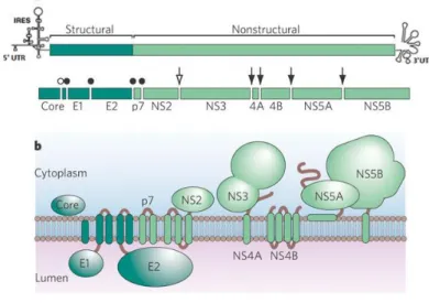
HCV structural proteins Core, E1 and E2 are released after cleavage of polyprotein by host endoplasmic reticulum (ER) signal peptidase and signal peptide peptidases, whereas the non-structural proteins p7, NS2, NS3, NS4A, NS4B, NS5A and NS5B are released after the enzymatic cleavage of HCV polyprotein by viral proteases NS2/3 and NS3/4A6,8 (Figure 1.1).
___________________________________________________________________________________________
Figure 1.1. HCV genome organization and polyprotein processing. A. The structure of the viral genome, including the open
reading frame encoding structural and nonstructural genes, and 5’ and 3’ UTRs. Open circle refers to C-terminal cleavage of Core by signal peptide peptidase; closed circles indicate the signal peptidase cleavage sites; open arrow indicates autocatalytic cleavage of the NS2-NS3 junction; and black arrows refer NS3/4a proteinase complex cleavage sites. B. The HCV proteins with respect to a cellular membrane. (Adapted from 11).
The core protein is further processed by a signal peptide peptidase into a mature protein of 21 kDa. Mature core protein interacts with viral RNA and forms the nucleocapsid6,10,17. E1 and E2 are
viral envelope glycoproteins with 35 kDa and 72 kDa8, respectively. Both envelope proteins are type I
membrane proteins responsible for receptor binding and entry into target cells8,10,17. p7 protein is a
small (63 amino acids) hydrophobic peptide of only 7 kDa, which functions as an ion channel after oligomerized5,6. NS2 protein has 23 kDa6 and, together with the N-terminal protease domain of the
NS3 protein, forms a catalytically active protease that mediates NS2/3 cleavage18. NS3 protein has 69
kDa and has two distinct enzymatic activities: the N-terminal part of the NS3 protein is a serine-protease mediating the cleavage of NS3/4A, NS4A/4B, NS4B/5A, and NS5A/5B. The C-terminal part of the NS3 protein contains an NTPase and an RNA-helicase important for viral genome replication6,18.
For NS3 to function as protease it needs NS4A as co-factor which is a small 6 kDa protein18. NS4B is a
very hydrophobic 27 kDa protein6. NS4B induces membrane rearrangement with consequent
formation of a membranous structure where viral RNA replication and assembly occurs10,18. NS5A is a
3 interaction with NS5B6,10. Finally, NS5B protein has 68 kDa, it is an RNA-dependent RNA polymerase
(RdRp), that catalyses the replication of HCV RNA6,18.
1.1.2. HCV life cycle
Attachment and entry of HCV to the host cell are the first steps in the virus life cycle19. Virus
entry is a result of a fusion process between viral and cellular membranes4. Membrane fusion is
mediated by specific interactions between viral surface glycoproteins (E1 and E2) and host cell receptors (CD81, SR-B1, LDLR, Occludin, Claudin-1)6,8,20. This interaction triggers decrease in pH of the
endocytic compartment, leading to membrane fusion and nucleocapsid release to the cytosol6,20.
Cell-associated attachment molecules, such as the low-density lipoproteins (LDLs) and glycosaminoglycans (GAGs), are involved in early steps of the process facilitating the described interactions between viral glycoproteins and cellular receptors6,20,21.
After decapsidation, the positive-strand HCV RNA genome is delivered to the cytoplasm. This RNA molecule is translated by cellular ribosomes, and the nascent polyprotein is translocated to and processed in the endoplasmic reticulum (ER)10. By definition, viral structural proteins form the viral
particle while viral nonstructural proteins form the replicase complex5. The replicase complex
initiates the synthesis of intermediate negative-strand RNA, which is used as template for the generation of new positive-strand RNA molecules which are encapsidated in a newly formed virion5,10. Virus production occurs by virion budding into the lumen of the ER which results in the
formation of multivesicular bodies (MVB)10,22. Finally, infectious virions are released from infected
cells by fusion of MVB with host cell cytoplasmic membrane10,22 (Figure 1.2).
___________________________________________________________________________________________
Figure 1.2. Hepatitis C virus life cycle. HCV first attaches itself to the host cell surface by interactions with GAG and/ or the
4
then translocates to the tight junctions where CLDN1 and OCLN act as cofactors and induce endocytosis. The virus-containing endosome is acidified in the cytoplasm and HCV RNA is released into the cytosol and translated in the ER, giving rise to a polyprotein that is cleaved into mature viral proteins. HCV RNA replication occurs and assembly into new virions starts in the ER. The viral envelope is acquired by budding into the ER. HCV particles are thought to be released via the constitutive secretory pathway in association with lipoviroparticles (LVP). This lipidation occurs either during budding (model l) or during egress via interaction between the virion and lipoproteins (model II). (Adapted from 20).
1.2. Vaccines
Vaccination remains the most effective method for controlling and preventing infectious diseases23,24. Most vaccines currently available are based on inactivated or live attenuated
pathogens. Even though these vaccines are highly effective, they present several limitations24. For
instance, attenuated pathogens used in attenuated vaccines can revert to their pathogenic status as observed with oral Polio vaccine. Therefore, this strategy is not recommended to those viruses able to establish chronic infectious. Altogether the need to develop safer vaccines strategies urges24,25.
Advances in recombinant DNA technologies and genetic engineering have contributed to the development of subunit vaccines (SUVs). SUVs are vaccines based on isolated components of the pathogen. Therefore, SUVs are considered safer than full pathogen-based inactivated or live attenuated vaccines. However, immunogenicity of SUVs is generally lower when compared to that of full pathogens, thus requiring higher doses, booster administrations or co-administration of adjuvants. Virus-like particles (VLPs) are a special subset of SUVs with enhanced immunogenicity24.
1.2.1. Virus-like particles
Virus-like particles (VLPs) are multimeric protein complexes mimicking the morphology of native viruses but totally or partially lacking the viral genome14,26,27. VLPs are of small size, between
20 and 200 nm in diameter, allowing entry into the lymphatic vessels and passive drainage to the region of lymph nodes, and optimal uptake by professional antigen presenting cells (dendritic cells, B-lymphocytes and macrophages)28,29.
These stable and versatile nanoparticles display excellent adjuvant properties being highly immunogenic per se24,30. They present high-density B-cell epitopes for antibody production and
T-lymphocyte stimulation. This feature of VLP vaccines is likely to be a major contribution to their increased effectiveness30,31.
VLPs have been used as useful platforms for delivering heterologous virus-derived antigens to the immune system and have come into focus for their diverse applications in vaccination, targeted drug delivery, gene therapy and immune therapy14,25,29.
A VLP-based product candidate is hardly competitive in the market unless its manufacturing process is scalable and cost-effective. Therefore, strong product development efforts as modern molecular biology technologies are required. The design and development of new technologies and
5 platforms for the production of a whole new variety of VLPs and expression system is currently a hot-topic in vaccinology25,26.
1.2.2. HCV vaccines
The development of safe, effective and affordable prophylactic and therapeutic vaccines against HCV has become an important medical priority; however, there are many obstacles to its development14,15. These limitations include the high sequence variability of HCV genome which is
associated to the natural high-error prone of HCV RNA-dependent-RNA-polymerase8,9, leading to the
emergence of mutants resistant immune defenses and therapeutics; and the lack of culture system for virus replication and small-animal models for HCV replication and pathogenesis 10,14.
In recent years, several different strategies of HCV antigen delivery have been investigated for use in vaccines against HCV, with different degrees of success, these include the use of recombinant proteins, virus-like particles (VLPs), recombinant nonpathogenic live vectors and prime-boost approaches14.
In parallel, due to their applicability in gene therapy, many efforts were invested to develop a variety of tools and protocols for industrial production and purification of viral vectors derived from retrovirus. Pseudotyped retroVLPs have been extensively used as scaffolds for candidate vaccines against HIV, HCV, HPV and many other virus14,32. In case of HCV, retroVLPs have been formed by the
overexpression of the E1 and E2 envelope proteins together with murine leukemia virus (MLV), to generate chimeric viral particles25,33. These particles display E1 and E2 envelope proteins in the
correct conformation and maintain a preferential tropism for hepatic cells therefore are commonly used to investigate the early events of HCV infection worldwide14,25.
Several preclinical trials involving VLP-based vaccine candidates are currently underway and have demonstrated highly promising results in phase I, II and II clinical trials14,32.
1.3. Mammalian cells engineering tools 1.3.1. Lentiviral transduction
Lentivirus belong to the Retroviridae family and can be used as a tool to deliver genetic material into mammalian cells34,35.
The retrovirus genome encodes four genes: gag (group specific antigen), pro (protease), pol (polymerase) and env (envelope). The gag gene encodes the three main structural proteins: matrix, capsid and nucleocapsid. The pro gene encodes a protease responsible for cleaving Gag and Gag-Pol during virus maturation. The pol gene encodes two enzymes, the reverse transcriptase and the integrase, that catalyze the reverse transcription of the viral genome from RNA to DNA during the infection process and integrate the proviral DNA into the host cell genome, respectively. The env sequence encodes for both surface and transmembrane subunits of the envelope glycoprotein34,36,37.
6 Additionally to the coding sequences, the retroviral genome presents cis-acting sequences that include two long terminal repeats (LTRs), a packaging signal (Ψ), and a polypurine tract (PPT). The LTRs are regulatory sequences of genome found at both the 5´and 3´ends that contain elements required to drive gene expression, reverse transcription and integration into the host cell chromosome. The packaging signal is required for specific packaging of the viral RNA into newly forming virions. The PPT functions as the site for initiating the positive strand DNA synthesis during reverse transcription36-38.
Lentivirus, additionally to gag, pro, pol and env genes, have accessory genes (Figure 1.3), such as tat, vpu, vif, nef and rev. Rev gene codes for a protein which interacts with rev responsive element (RRE), a cis-acting sequence located in the middle of env gene allowing messenger RNA to be exported from nucleus intact and it is therefore the only accessory gene required for lentiviral vectors production36,38,39.
_____________________________________________________________________________________________________ Figure 1.3. Schematic representation of the lentiviral genome (Adapted from 40).
The capability to transduce non-dividing and dividing cells as well as the wide tropism afforded them by pseudotyping has enabled them becoming a highly efficient tool in therapy34-36,41.
Four generations of lentiviral vectors (LVs) are currently considered. The vectors of third generation presented a higher level of biosafety and, currently, are the most commonly applied for the production of lentivirus42. These vectors require the transfection of four independent expression
cassettes encoding gag-pro-pol, env, rev and transgene functions. Gag-pro-pol genes are expressed from a Cytomegalovirus (CMV) promoter and none of the accessory or regulatory proteins is present in this construct. Only rev accessory gene is maintained but is provided by a non-overlapping plasmid. Vector cassette for transgene expression is driven by a heterologous promoter, as virus LTRs were partially deleted36,39,42.
1.3.2. Recombinase-mediated cassette exchange
Recombinase-mediated cassette exchange (RMCE) is a strategy which enables predictable expression of the gene of interest (GOI) at a pre-characterized genomic locus43. RMCE is a two-step
procedure. First, a tagging cassette flanked by two heterospecific recombination target sites, a wild-type (wt) and a mutant target sequence, is random integrated in the genome. Second, a targeting cassette is integrated and the recognition sites for a site-specific recombinase (SSR) of tagging cassette are targeted to the locus of interest by homologous recombination. The SSR inserts a
7 replacement sequence into this tagged site44,45. Recombinase exchanges the two DNA regions
flanked while maintaining the orientation of the regions46,47. For genomic RMCE, the target resides in
the host genome, whereas the gene replacement cassette is introduced into the host cell, traditionally as part of a “donor plasmid”44,48.
_____________________________________________________________________________________________________ Figure 1.4. Recombinase-mediated cassette exchange. Half arrows mark target sites. These sites flank both the tag and the
incoming cassette, which is provided as part of a donor plasmid. In the absence of this donor plasmid, the target cassette remains unperturbed. Upon its addition, a double-reciprocal cross-over between identical FRTs on each of the reaction partners is initiated by recombinases (Adapted from 49).
The use of site-specific recombination systems, like the Flp/FRT (flipase/ flipase-recognition target sites) system that was originally isolated from the yeast Saccharomyces cerevisiae and Cre/lox from the bacteriophage P1 in mammalian cells allow integration of genes of interest into adequate genomic loci50,51. Differences between these systems go back to the particularities of these tyrosine
recombinases regarding the details of the enzymatic mechanism and structure of their target sites49.
1.3.2.1. Cre-RMCE
The CRE-lox site-specific recombination system has been applied to a variety of genetic systems. Cre recombinase, a 38.5 kDa protein, catalyzes reciprocal site-specific recombination between DNA elements termed lox sites. The lox site, loxP, is a 34 bp element composed of two 13 bp inverted repeats separated by an asymmetric 8 bp spacer region. Each 13 bp inverted repeat serves as a binding site for the recombinase, whereas the 8 bp spacer region participates in strand exchange during the recombination reaction. Because a productive recombination event requires synapsis of lox sites between their spacer regions, it is asymmetry of the spacer region that confers directionality to the recombination reaction44,48,52.
_____________________________________________________________________________________________________ Figure 1.5. Architecture of wild-type lox sites. The arrows bracket the 13 bp palindromic arms that serve as binding sites
for the Cre recombinase. The asymmetric 8 bp spacer (red box) is the region where breaking and rejoining events occur. (Adapted from 44).
Targeting cassette Tagging cassette
8
1.3.2.2. Flipase (Flp)-RMCE
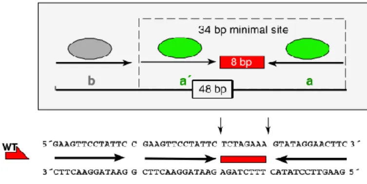
The Flp/FRT system is similar to the Cre/lox system. Flp recombinase consists of a 13 kDa NH2-terminal domain and a larger 32 kDa COOH-terminal domain with the major determinants for DNA binding44. FRT site consists of genuine 48 bp, including three individual 13 bp Flp-recombinase
binding elements, two of which (designated “a,a”) form an inverted repeat around an 8 bp spacer. On one side of this core, the repeat is duplicated after a 1 bp gap. The third 13 bp direct repeat (“b”) is separated from “a” by a single base pair and is not directly involved in catalysis; it rather serves as a modulator, for instance, by acting as an Flp entry site. While inversion and excision reactions are also feasible with a 34 bp minimal variant site consisting of the inverted repeat and spacer only, a integration reaction benefits from or even requires the presence of the extra Flp-recombinase binding element44,43,49.
_____________________________________________________________________________________________________ Figure 1.6. Architecture of a wild-type (WT) 48 bp FRT site and its 34 bp minimal variant. FRT site consists of two inverted
13 bp repeats (“a,a”) (green ovals) that serve as binding sites for Flp-recombinase arranged about an 8 bp spacer. The asymmetric 8 bp spacer (red box) is the region where breaking and rejoining events occur. The third direct repeat (“b”) (gray oval) serves as a Flp entry site. (Adapted from 44).
2. Aims
The main objective of this work is the production of HCV-like particles and the development of a production platform compatible with industrial production requirements.
This work is, then, divided in two parts. In the first part the evaluation two different cell substrates – HEK293 and HuH-7 - for HCV-LPs production, using cassettes constructed with lentiviral backbone that code HCV genes. HCV-LPs production and purification was assessed.
In the second part, a stable cell line using defined gene expression loci by recombinase-mediated cassette exchange (RMCE) method was developed. This method takes advantage of predictable expression of the gene of interest at a pre-characterized genomic locus.
Thus, t
his strategy may open the possibility to generate an engineered HEK293 cell line producer of HCV-LPs.9
3. Material and Methods
3.1. Plasmids used
Primers and templates for all the plasmids constructed in this work are listed in Tables I and II (Attachments).
Lentiviral vectors production
pMD2G codes for the envelope G glycoprotein of the vesicular stomatitis virus (VSV-G) under
the control of cytomegalovirus (CMV) promoter.
pMDLg/pRRE is 3rd generation lentiviral packaging plasmid codifying for Gag-Pro-Pol polyprotein and RRE.
pRSV-REV is a 3rd generation lentiviral packaging plasmid that contains Rev protein.
All these plasmids were kindly provided by Dr. Didier Trono (Addgene, USA) and used for transient lentiviral vector production of pRRLSin derived vectors, by transfection as reported in Dull
et al.42
Lentiviral transgenes
The plasmids described below were constructed using pRRLSin backbone, a self-inactivating (SIN) third-generation lentiviral vector isolated from pRRLSin GFP-S10, kindly provided by Miguel Guerreiro (ACT Unit IBET/ ITQB NOVA, Portugal). This backbone contains an ampicillin resistance marker for plasmid amplification in E.coli, long-terminal repeats (LTRs), a packaging signal (Ψ) for RNA encapsidation in lentiviral particles, RRE element, and a cPPT for efficient nuclear import/export38. After cPPT region each pRRLSin plasmid has a different cassette with specific DNA
elements to drive the expression of different genes of interest and associated mammalian selection markers. Following the mammalian selection gene all pRRLSin vectors have a WPRE regulatory element to increase mRNA stability and a SV40 polyadenylation site to terminate RNA transcription. The different pRRLSin plasmids constructed are:
pRRLSin_Core codes for the HCV Core, under the regulation of the CMV promoter and an
encephalomyocaerditis virus-internal ribosome entry site (EMCV-IRES) drives neomycin resistance gene expression.
pRRLSin_E1E2 codes for both HCV envelope glycoproteins, E1 and E2, under the control of
the CMV promoter and an EMCV-IRES drives zeocin resistance gene expression.
pRRLSin_p7_NS2 codes for HCV non-structural proteins p7 and NS2 under the control of
10
pRRLSin_NS3_NS4a codes for HCV non-structural proteins NS3 and NS4a under the control of
the SV40 promoter and an FMDV-IRES element drives blasticidin resistance gene expression.
pRRLSin_NS4b_NS5a codes for HCV non-structural proteins NS4b and NS5a under the control
of hPGK promoter and an EMCV-IRES drives puromycin resistance gene expression.
Cassette exchange
pZeoCrecontains Cre recombinase gene under a CMV promoter.
pTagLoxP_GFPZeo is a Cre-mediated cassette exchange tagging vector constructed by Ana
Isabel Almeida (ACT Unit IBET/ ITQB NOVA, Portugal), that contains two loxP sites. This vector drives the expression of a GFPZeo gene that was amplified from pSELECT-GFPZeo-LacZ (InvivoGen, USA) by PCR and cloned into pTagLoxP-mcs designed by Ana Coroadinha.
pTarLoxP_E1E2 is a Cre-mediated cassette exchange targeting vector constructed by Hugo
Soares (ACT Unit IBET/ ITQB NOVA, Portugal) from pTarLoxP backbone plasmid. This vector contains a CMV promoter, a β-globin that increase mRNA stability, two loxP sites and E1 and E2 genes amplified from pEPX145-71 (Epixis, France), a SV40 enhancer conjugated with FermH promoter and mEF1 5’UTR as promoter for puromycin expression.
pSVFlpe contains Flp recombinase gene under a SV40 promoter51.
pTagFRT_mCherryis a Flp-mediated cassette exchange tagging vector contains two FRT sites, a wild-type FRT (Fw) site and a spacer mutant FRT site (F5) with hygromycin resistance gene followed by an ATG defective neomycin phosphotransferase gene (Δneo). The construction contains a mCherry gene, amplified from pRSV-CherryPuro by PCR and cloned into NotI and PacI excised pTag, an in-house constructed plasmid.
pTarFRT_Core is a Flp-mediated cassette exchange targeting vector derived from pEmMFG backbone containing a Fw FRT and a F5 FRT, where GFP gene was removed by inverse PCR and replaced by HCV Core gene amplified from pGEM_Core by PCR.
Retrovirus production
pMD2G as described above (letiviral vectors production).
pMLV-GP codes for murine leukemia virus (MLV) under the control of hEF1/HTLV chimeric
11
3.2. Plasmids construction
3.2.1. Cloning procedures
3.2.1.1. Polymerase chain reaction (PCR)
All PCR reactions were performed in a Biometria® T3Personal Thermocycler (Biometria, Germany) with a proof-reading Phusion High-Fidelity DNA Polymerase (Finnzymes, Finland), using PCR conditions appropriate for each fragment. The oligonucleotides used for PCR were custom-made by Sigma-Aldrich (USA) (Tables I and II).
3.2.1.2. DNA fragment enzymatic restriction
Restriction reactions were incubated 3 hours or overnight at 37°C. The enzymes (New England Biolabs, USA) in Tables I and II (Attachments) were used with the appropriate buffers according to the manufacturer’s instructions.
3.2.1.3. DNA fragment purification
All the generated fragments were isolated by agarose gels (NZYTech, Portugal) 0.7% (w/v) prepared in 1x TAE buffer (Tris-Acetate-EDTA) (Qiagen, Germany) and 0.5 µL/mL RedSafeTM Nucleic Acid Staining Solution (INtRON Biotechnology, USA) was added before pouring the gel. Subsequently,
DNA fragments were visualized using GelDocTM XR+ system (BioRad, USA) and purified with NucleoSpin® Gel and PCR Clean-up kit (Macherey-Nagel, Germany), according to the manufacturer’s
instructions.
3.2.1.4. Vector and insert ligation
Vector-insert ligations were performed using In-Fusion® HD Cloning Kit (Clontech Laboratories, USA) following manufacturer’s instructions.
3.2.2. Bacterial strains
Escherichia coli (E.coli) StellarTM (Clontech, USA) and One Shot® Stbl3TM (Invitrogen, USA)
competent cells were used for the production of the DNA plasmids. Transformation procedures were carried under manufacturer’s instructions. The agar and liquid cultures were performed with Luria
Broth media (LB) (Fast-Media® LB from Invivogen, USA) and Terrific Broth media (TB) (Fast-Media® TB
from Invivogen, USA), respectively, supplemented with the appropriated antibiotic. The media was prepared using milliQ water (Milli-Q® from Merck Millipore, USA), according to manufacturer’s instructions.
3.2.3. Plasmid purification and quality control
Final plasmid production and purification was performed in different scales. Small-scale, medium-scale and large-scale production and purification were performed using GeneJET Plasmid
12
Miniprep Kit (Thermo Scientific, USA), ZymoPURETM Plasmid Midiprep (Zymo Research, USA) and Genopure Plasmid Maxi Kit (Roche Applied Science, Germany), respectively, following manufacturer’s
instructions. Working DNA stock solution for each plasmid were generated and stored at -20°C. The DNA concentration was determined in Nonodrop 2000C spectrophotometer (Thermo Scientific, USA). Plasmid purity was determined by the Abs260nm/Abs280nm and Abs260nm/Abs230nm ratios and plasmid
integrity with enzymatic restriction analysis assessed in 0.7% (w/v) agarose gels (NZYTech, Portugal) prepared in 1x TAE buffer (Qiagen, Germany). All plasmids were sequenced using GATC Biotech services (Constance, Germany).
3.3. Cell lines and culture conditions
HEK293 is a Human Embryonic Kidney cell line (ATCC CRL-1573). HEK293T (ATCC CRL-11268) is a HEK293 derived cell line expressing large T antigen from SV40. HuH-7 (JCRB0403) is a Human Hepatoma cell line that was originally take from a liver tumor. All cells were maintained in Dulbecco’s modified Eagle’s medium (DMEM) (Gibco, UK) supplement with 10% (v/v) Foetal Bovine Serum (FBS) (Gibco, UK) and maintained in an incubator at 37°C in a humidified atmosphere containing 8% CO2.
3.4. Cell number and viability
Cell concentration and viability were determined by the trypan blue exclusion method using a 0.1% (v/v) solution prepared in Phosphate Buffer Saline (PBS) (Gilbo, UK) and a Fuchs-Rosenthal hemocytometer (Brand, Germany) observed on an inverted microscope (Olympus, Japan). Cells with damaged membranes stained blue and viable cells remain colorless.
3.5. Lentiviral vector production
For transient lentiviral vector production, the 3rd generation lentiviral packaging system was
used42. HEK293T cells were seeded at 5 x 10⁵ cells/cm2 in 75cm2 t-flask. Polyethylenimine (PEI, linear
25 kDa) (Polysciences, Germany) transfection was carried out 24 hours later with a mixture of pMD2G
(for the envelope), pRSV-REV and pMDLg/pRRE (providing the packaging functions), and pRRLSin derived vectors (section 2.1) providing the gene of interest (transgene) at 1:1.5 ratio of DNA:PEI. The DNA ratio used was 1:0.5:1:2, respectively. 24 hours post-transfection, culture medium was replaced with the original volume (15 mL). Culture medium containing the lentiviral vectors was collected after an additional 24 hours production period. All viral vectors supernatant was filtered through 0.22 µm pore-sized cellulose acetate filter, aliquoted and stored at -80°C until further use.
3.6. Lentiviral transduction
HCV genes were delivered by lentiviral vector infection. HEK293 and HuH-7 cells were seeded at a density 5 x 10⁵ cells/well 24 hours before infection in 6-well plates. Transduction was performed
13 in section 3.5 in the presence of 8 µg/mL of polybrene (Sigma-Aldrich, USA). Cells were incubated at 37°C for 24 hours. 24 hours post-infection, the culture supernatant was exchanged for fresh culture DMEM. Two days after infection, cells were amplified to 75 cm2 t-flasks in DMEM supplemented with
the respective selection antibiotic (according to each plasmid). The cells were maintained in culture for 21 additional days with culture medium exchange every 3-4 days.
3.7. RNA extraction and gene expression quantification by Real-Time quantitative PCR
To analyze HCV components expression total RNA was extracted using QIAamp® RNeasy Mini
Kit (Qiagen, USA) according to the manufacturer’s instructions. Ribosomal protein L22 (RPL-22) was
chosen as a control housekeeping gene. Primer sequences for control and HCV genes are given in Table III (Attachments). The reverse transcription of total RNA was performed according to
Transcriptor High Fidelity cDNA Synthesis Kit (Roche Applied Science, Germany) protocol for cDNA
synthesis using 2 μg of total RNA and oligo dT primer for total mRNA reverse transcription. The reverse transcribed product was aliquoted and stored at -20ºC until further processing. SYBR Green I dye chemistry was used to detect the PCR products using LightCycler® 480 SYBR Green I Master (Roche Applied Science, Germany) according to the manufacturer’s instructions using LightCycler®
480 Real Time PCR System (Roche Applied Science, Germany). Two independent biological replicates
were analyzed using 2-ΔCt method for relative gene expression analysis53.
3.8. HCV-LPs production and purification
For HCV-LP production and purification, cells were seeded in 225 cm2 t-flasks and cultured
until 80% of cellular confluence was reached. Cells were harvested and centrifuged at 300 g for 10 minutes at 4°C. Cells were resuspended in 20 mL of a hypotonic solution supplemented with 1x protease inhibitor cocktail (Roche Applied Science, Germany) and incubated on ice for 30 min. Cells lysis was performed with 5 freeze/thaw cycles with liquid nitrogen. Cell debris were removed by centrifugation (14000 g for 10 min at 4°C). Cleared supernatant was collected and completed with 30 mL PBS. Supernatant was then filter through 0.20 µm for further clarification and centrifuged in a 45
Ti rotor (Beckman Coulter, USA) at 40000 rpm (≈185000 g) for 2 h at 4°C. The pellet was resuspended
in 1.5 mL PBS. Pellet resuspension was added on top of a 30% (v/v) sucrose cushion54 and centrifuged
in a 90 Ti rotor (Beckman Coulter, USA) at 60000 rpm (≈300000 g) for 2 h at 4°C. Virus pellet was resuspended in 1x Tris-buffered saline (TBS) solution.
3.9. Western blot
For protein electrophoresis, NuPAGE® electrophoresis system (Invitrogen, USA) was used. Samples were prepared in denaturing conditions according to manufacturer’s instructions and heated at 70°C for 10 minutes. Samples were resolved on a NuPAGE® 4-12% Bis-Tris gel with
14
NuPAGE® MES SDS Running Buffer, at 180V for 40 minutes. Protein transfer into PVDF membrane
was performed in Trans-Bot® Turbo™ Transfer System (Bio-Rad, California, USA) for 30 minutes, according to manufacturer’s instructions. Membranes were blocked with 5% (w/v) dry milk in 1x PBS, for at least 1 hour at room temperature and incubated overnight with primary antibody diluted in 1% BSA in PBS at room temperature with gentle agitation. The primary antibodies used were mouse monoclonal anti-HCV Core (C7-50) (Santa Cruz, USA) at 1:50 in PBS, mouse monoclonal anti-HCV E1 (BDI198) (Acris Antibodies GmbH, Germany) at 1:50 in PBS and mouse monoclonal anti-HCV E2 (4F6/2) (Austral Biologicals, USA) at 1:500 in PBS. Membranes were washed with 1x PBS for 10 minutes (3 times) and incubated with secondary antibody, anti-mouse IgG HRP conjugate (NA931) (GE Healthcare, UK) at 1:1000 in PBS, for 2 hours at room temperature. Chemiluminescence detection was performed with Amersham™ ECL™ prime western blotting detection reagent (GE Healthcare, UK), according to manufacturer’s instructions, and analyzed under ChemiDocTM XRS system (Bio-Rad, USA).
3.10. Cell transfection with tagging cassette and drug selection
HEK293 clones #1, #2 and #3 previously tagged with the pTagLoxP_GFPZeo cassette were kindly provided by Ana Isabel Almeida (ACT Unit IBET/ ITQB NOVA, Portugal). Cells were seeded in 6 well plates at 5X10⁵ cells/well. After 24 hours, transfection was performed using PEI (Polysciences,
Germany) to insert pTagFRT_mCherry cassette at 1:1.5 ratio of DNA:PEI. Cells were incubated at 37ºC overnight after which medium was exchanged. 48 hours post-transfection, cells were analyzed for mCherry fluorescence by flow cytometry and were cultured for 21 additional days in hygromycin (300 µg/mL) (InvivoGen, USA) containing medium, exchanging culture medium at each 3-4 days. Hygromycin resistante cell populations were cloned by limiting dilution to isolate single cells which were cultured and expanded. Different cell clones with homogeneous mCherry expression and cassette integration pattern were screened by flow cytometry and RT-qPCR.
3.11. Fluorescence signal detection
For GFP and mCherry fluorescence signal detection, HEK293 cells tagged with a
pTagLoxP_GFPZeo cassette and a pTagFRT_mCherry cassette were trypsinised and resuspended in
PBS at a density of 2x10⁶ cells/mL. Cell counts were performed on CyFlow®space (Sysmex Partec,
Germany). The excitation wavelengths for GFP and mCherry were 488 nm and 587 nm and emission were detected at 507 nm and 610 nm55, respectively. The cells were considered fluorescent when the
15
3.12. Tagging copies quantification by Real-Time quantitative PCR
For the quantification of integrated tagging copies into the cell genome, genomic DNA was isolated from 5x10⁶ cells using the QIAamp® DNA Mini and Blood Mini Kit (Qiagen, USA) according to
manufacturer’s instructions. To estimate the number of copies per cell, genomic DNA was quantified using Nanodrop 2000C spectrophotometer (Thermo Scientific, USA) and 200 ng of DNA was used as template for RT-qPCR. Specific primers for hygromycin gene (Table III) were used on LightCycler® 480
Real-Time PCR System (Roche Applied Science, Germany) according to LightCycler 480 SYBR Green I Master (Roche Applied Science, Germany) PCR kit. All data was analyzed using 2-ΔΔCt method for
relative gene expression analysis53. After normalization to the housekeeping gene RPL22, the number
of copies per cell was quantified using a standard curve and compared to a cell line with a single copy integrated genome. Single copy was considered for values between 0.6 and 1.456.
3.13. Retrovirus production
To determine the packaging signal efficiency of the pTagFRT_mCherry cassette incorporated into mRNA of the cells, clones were seeded in 6 well plates at 5X10⁵ cells/well. 24 hours later,
transient transfection was performed using PEI (Polysciences, Germany) at 1:1.5 ratio of DNA:PEI and with 2 µg of pMD2G and 2 µg ofpMLV-GP. After 24 hours, culture medium containing the retrovirus
was collected. HEK293T cells were transduced and analyzed for mCherry fluorescence intensity.
3.14. Cassette exchange
For Cre-mediated cassette exchange, clones #206 and #311were seeded in 6 well plates at 5X10⁵ cells/well. 24 hours later, transfection was performed using PEI (Polysciences, Germany) at
1:1.5 (w/w) ratio of DNA:PEI or calcium phosphate (CaPO4) with 1 µg of targeting plasmid
(pTarLoxP_E1E2) and 3 µg of pZeoCre. 48 hours post-transfection, cells were analyzed for cassette exchange by flow cytometry and were selected with puromycin (1 µg/mL) (InvivoGen, USA). Cells were cultured for 21 days and medium exchanged at each 3-4 days. Puromycin resistance cell populations were cloned by limiting dilution to isolate single cells which were cultured and expanded. One cell clone (#311_F8) with no green expression was expanded and frozen.
For Flp-mediated cassette exchange, #311 parental and clone #311_F8were seeded in 6 well plates at 5X10⁵ cells/well. 24 hours later, transfection was performed equally as described above
with 4 µg of targeting plasmid (pTagFRT_mCherry) and 12 µg of pSVFlpe. 48 hours post-transfection, cells were analyzed for cassette exchange by flow cytometry and were selected with neomycin (500 µg/mL) (InvivoGen, USA). Cell were cultured for 21 days and medium exchanged at each 3-4 days. Neomycin resistance cell populations will be cloned by limiting dilution to isolate single cells, cultured and expanded. One cell clone with no red expression will be expanded and frozen.
16
4. Results
4.1. Evaluation of different cell substrates for HCV-LP production
The production of a HCV-LP vaccine is dependent on the development of a HCV-LP producer cell line14. It is widely known that several factors may affect quality and production yields of viral
particles. Therefore, this work begins by testing two different cell-lines (HEK293 and HuH-7) to produce HCV-LP.
4.1.1. Vectors construction and cells transduction
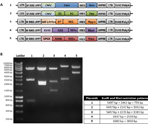
To develop a cell line for stable production of HCV-LP composed of HCV proteins, lentiviral vectors containing HCV genes - Core, E1 and E2, p7 and NS2, NS3 and NS4a, NS4b and NS5a - were constructed (Figure 4.1). Lentiviral vectors were produced by transient transfection of HEK293T cells.
___________________________________________________________________________________________
Figure 4.1. HCV lentiviral vectors. A. Schematic presentation of the HCV lentiviral vectors constructed. LTR, long-terminal
repeat; Ψ, packaging signal; RRE, rev responsive element; cPPT, polypurine sequence; IRES, internal ribosome entry site; WPRE, woodchuck hepatitis virus post-transcriptional regulatory element; and SV40 polyadenylation site. CMV, hEF1/HTLV, SV40 and hPGK are promoters. Neomycin (Neo), zeocin (Zeo), hygromycin (Hygro), blasticidin (Blast) and puromycin (Puro) are selection antibiotics for mammalian (not in scale). B. Vectors restriction analysis. Electrophoresis showing a restriction-specific pattern of the plasmids, when using the EcoRI and XhoI restriction enzymes. 1-pRRLSin_Core; 2-pRRLSin_E1E2; 3-pRRLSin_p7_NS2; 4-pRRLSin_NS3_NS4a; 5-pRRLSin_NS4b_NS5a. Ladder-NZYDNA Ladder III (NZYTech, Portugal); (bp, base pairs).
A .
B .
17 0 0.2 0.4 0.6 0.8 1 1.2 1.4 1.6 1.8 HEK293 HEK293 ΔCore E1E2 p7NS2 HEK293 Core E1E2 HEK293 Core E1E2 p7NS2 HuH-7 HuH-7 ΔCore E1E2 p7NS2 HuH-7 Core E1E2 HuH-7 Core E1E2 p7NS2 mRN A e xp re ssi o n ( 2 -Δ C t ) Core E1 E2 p7 NS2
HEK293 and HuH-7 cells were sequentially transduced with lentiviral vectors transducing HCV genes (Core, E1E2 and p7NS2) and selected with the corresponding antibiotic resistance gene.
4.1.2. Expression of HCV genes and proteins
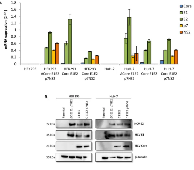
HCV Core, E1 and E2 genes provide all structural components needed for HCV-LP formation. Additionally, evidences suggest that p7 and NS2 are essential for viral particles assembly and function17,27. Before assessing the production of HCV-LP, HCV genes and proteins expression in
HEK293 and HuH-7 transduced cell populations was determined by RT-qPCR and Western blot, respectively.
Figure 4.2. Expression of HCV genes and proteins in lentivirus transduced cell lines. Cell lines used for both analyses were
HEK293 and HuH-7 as negative control (without HCV genes); HEK293 and HuH-7 ΔC E1E2 p7NS2 as Core negative control (without Core); HEK293 and HuH-7 Core E1E2 as p7NS2 negative control (without p7NS2) and HEK293 and HuH-7 Core E1E2 p7NS2. A. Analysis of the HCV genes expression by RT-qPCR. Numbers on the x-axis correspond to cell lines. Numbers on the y-axis indicate the mRNA expression comparedto HEK293 and HuH-7 given by mRNA quantification by RT-qPCR using primers for Core, E1, E2, p7 and NS2 genes. The genes expression was quantified after normalization to a control gene (RPL22) using the 2-ΔCt. Genes expression levels are shown as average ± standard deviation of four technical replicates and two biological replicates. B. Analysis by Western blot of HCV proteins (Core, E1 and E2) expression. Protein extracts
obtained from LV transduced cells. Core (21 kDa), E1 (35 kDa) and E2 (72 kDa) expression detected by an anti-Core antibody, an anti-E1 antibody and anti-E2 antibody, respectively. The β-tubulin (50 kDa) content was also analyzed to verify that equal levels of protein extracts have been loaded and it expression detected by an anti-β Tubulin antibody.
A. .
18 As shown in Figure 4.2A, parental HEK293 and HuH-7 cell lines do not express any of the viral genes while mRNA for all viral components was detected in all transduced cells. It is possible to observe a great variation in gene expression among tested cells, this variation might be explained by the analysis of heterogeneous cell populations rather than homogeneous populations derived from a single-cell clones. Of note is the relative low expression of HCV Core gene in both HEK293 and HuH-7 cell populations. This observation, even though relevant, does not prevent the expression of HCV Core protein in detectable amounts (Figure 4.2B). In parallel, protein expression of viral components was assessed by Western Blot analysis. As visible in Figure 4.2B all viral components (Core, E1 and E2) are expressed at similar levels in all transduced cell populations. HCV p7 and NS2 protein expression could not be assessed since no commercial antibodies are available for the studied HCV strain (H77).
4.1.3. HCV-LP production and purification
After validating the expression of the viral components in transduced HEK293 and HuH-7 cells, we evaluated the production of fully assembled HCV-LP. All cell lines and controls were seeded in 225 cm2 t-flasks and cultured until 80% of cellular confluence was reached. Culture medium was
then replaced with fresh one. HCV-LPs produced during the subsequent 24 hours were harvested and purified by sucrose cushion centrifugation and analyzed for Core, E1 and E2 presence by Western blot.
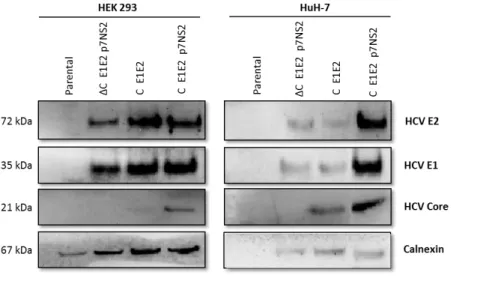
As visible in Figure 4.3 higher amounts of HCV envelope antigens are detected in samples where Core protein is present in higher amounts for both cell lines.
___________________________________________________________________________
Figure 4.3. Characterization of HCV-LP produced. Western blot profile of HCV-LP by Core, E1 and E2 expression. Cell
extracts obtained from LV transduced cells after sucrose cushion centrifugation. Core (21 kDa), E1 (35 kDa) and E2 (72 kDa) expression detected by an anti-Core antibody, an anti-E1 antibody and anti-E2 antibody, respectively. The Calnexin (67 kDa) content was also analyzed to verify that equal levels of protein extracts have been loaded (anti-Calnexin antibody).