i | P a g e
KOVAN MOHAMMED ISMAEL
“MARINE MICROALGAE AS SOURCES OF BIOACTIVE COMPOUNDS WITH ANTI-INFLAMMATORY ACTION”
European master’s in quality in Analytical Laboratories (EMQAL) Under the supervision of:
Prof. Luísa Afonso Barreira (Supervisor)
Prof. Carla Alexandra São Bento Viegas (Co-supervisor) Prof. Dina Simes (Co-supervisor)
University of Algarve Faculty of Science and Technology
ii | P a g e
DECLARATION OF AUTHORSHIP AND COPYRIGHT
I declare that I am the author of this work, which is original. The work cites other authors and works, which are adequately referred to in the text and are listed in the bibliography.
____________________________________ KOVAN MOHAMMED ISMAEL
Copyright: KOVAN MOHAMMED ISMAEL. The University of Algarve has the right
to keep and publicise this work through printed copies in paper of digital form, or any other means of reproduction, to disseminate it in scientific repositories and to allow its copy and distribution with educational and/or research objectives, as long as they are non-commercial and give credit to the author and editor.
iii | P a g e
ACKNOWLEDGMENT
I am unreservedly grateful to the European Commission for the scholarship funded as Erasmus Mundus Category ‘A’ student of Erasmus Mundus Joint Master’s degree in quality in analytical in laboratories (EMQAL).
First & foremost, I offer my unfeigned and sincerest gratitude to my supervisors-Prof. Luísa Afonso Barreira and my co-supervisors Prof. Carla Alexandra and. Prof. Dina Simes for their eternal support, professional guidance, constructive comments, patience, and diligent help. I attribute the level of my master’s degree to them for this thesis, too, would not have been completed without their support.
I’m thankful to Prof. Joao Varela who provided me constructive comments and suggestion while I presented this thesis to Marine Biotechnology Group, Centre of Marine Science (CCMAR), University of Algarve. My sincere appreciations and thanks go to all Marine biotechnology Group, CCMAR, the University of Algarve who supported me in everything I needed laboratory meetings to present our work.
My deepest appreciation goes to our Program coordinator -Prof. Miguel Esteban and the to our edition coordinator prof. Miguel Palma for their commitment, determinations and charismatic leadership qualities for the continual success of the EMQAL program. I’m grateful to Maria Jose for her unlimited support in helping us getting our papers work done during staying at Cadiz University. However, I do thank Farah Kamberovic my EMQAL colleague for helpline me during all the research year in everything.
I do thank all of my 2017/19 EMQAL professors who taught me updated and recent sciences. Last, but not least. Last My cheers also go to my parents may dad Mohammed Ismael and mum Ramzia Ali for their unconditioned support to pursue my education and my life as per my interest and ambition. Above all, my thanks and praise go to Allah the Almighty for the many blessings He bestowed on me and for all things He has done in my life and for my ages. Thanks to His guidance, mercy, healings, unprecedented faithfulness, unfeigned love and protection.
iv | P a g e
ABSTRACT
Inflammation is a defensive mechanism stimulated when the body is attacked by pathogens or irritants, or when cells are damaged. Sometimes, these defensive mechanisms can go wrong, emerging to different inflammatory diseases, such as acute inflammation and chronic inflammation. Despite the existence of several anti-inflammatory drugs on the market, new drugs with fewer side effects and higher efficacy are required for the treatment of inflammatory diseases. Attention has been given to natural bioactive compounds derived from marine organisms, since these are well known to be a source of potential bioactive compounds with different therapeutic applications in several diseases including inflammatory diseases.
The aim of this research project was therefore to find compounds that can serve as new anti-inflammatory drugs or drug leads in microalgae. For that purpose, water, ethanol and ethyl acetate extracts of different microalgae species (Porphyridium sp., Nannochloropsis sp., Tetraselmis sp. CTP4, Isochrysis sp., Phaeodactylum tricornutum, Skeletonema
costatum, Spirulina sp., Haematococcus pluvialis and Tetraselmis chuii) were
characterized for its antioxidant activity as a pre-screening effort to select the most bioactive species/extracts. The most antioxidant extracts (ethanol extracts of
Nannochloropsis sp., Tetraselmis sp. CTP4, and Tetraselmis chuii and water extracts of Nannochloropsis sp. and Porphyridium sp.) were afterwards screened for their
anti-inflammatory activity measuring the inhibition of TNF-α production by LPS-stimulated human macrophage-differentiated THP-1 cells (Mac-THP-1). The best results were obtained with the ethanol extract of Tetraselmis sp. CTP4 (87% inhibition of TNF-α at 50 µg/mL in respect to the LPS control). This extract was therefore fractionated using liquid-liquid extraction (LLE), and the fractions were re-checked for their anti-inflammatory activity, using the previous method in a bioassay-guided fractionation effort. The most active fraction (the hexane fraction) was later analyzed by GC-MS to tentatively identify some of the compounds present in the fraction that could be responsible for its anti-inflammatory properties. Most of the compounds identified were fatty acids, some of which had already been reported to have anti-inflammatory properties. Further studies are needed to identify the exact compound or compounds responsible for the anti-inflammatory effect in the active fraction. Nonetheless, these
v | P a g e
results indicate that microalgae can be a source of compounds with the ability to minimize and reduce inflammation.
Keywords: Microalgae, Inflammation, Antioxidant activity, Bioactive compounds, Solid-liquid extraction, Liquid-liquid Fractionation, GC-MS
vi | P a g e
TABLE OF CONTENTS
DECLARATION OF AUTHORSHIP AND COPYRIGHT _____________________ ii
ACKNOWLEDGMENT ________________________________________________ iii
ABSTRACT __________________________________________________________ iv
TABLE OF CONTENTS ________________________________________________ vi
FIGURE INDEX ______________________________________________________ ix
TABLE INDEX _______________________________________________________ xi
LIST OF ABBREVIATIONS AND ACRONYMS ___________________________ xii
CHAPTER 1: INTRODUCTION _________________________________________ 1
1.1 Marine bioprospecting ____________________________________________ 1 1.2 Inflammation ____________________________________________________ 3 1. 3 Natural sources of alternative anti-inflammatory agents _______________ 5 1.4 Microalgae ______________________________________________________ 7 1.5 Biomedical application of Microalgae _______________________________ 8 1.5.1 Antioxidant activity ____________________________________________ 8 1.5.2 Anticancer activity _____________________________________________ 8 1.5.3 Antiviral activity ______________________________________________ 9 1.5.4 Antibiotic activity _____________________________________________ 9 1.6 Microalgae with cosmeceutical uses _________________________________ 9 1.7 Natural products drug discovery from microalgae ____________________ 10 1.8 THP-1 cells ____________________________________________________ 12 1.9. OBJECTIVE _____________________________________________________ 13
CHAPTER 2: MATERIAL AND METHOD ______________________________ 14
2.1 Chemicals _____________________________________________________ 14 2.2 Algal biomass __________________________________________________ 14 2.3 Preparation of extracts ___________________________________________ 15
vii | P a g e
2.4 Extract fractionation ____________________________________________ 16 2.5 Free Radical Scavenging Activity __________________________________ 17 2.6 Anti-inflammatory assay _________________________________________ 19 2.6.1 Cell cultivation and differentiation _______________________________ 19 2.6.1.1 Cell counting ____________________________________________ 19 2.6.2 Cell viability assessment (MTS assay) _____________________________ 20 2.6.3 Inflammatory assay in Mac THP-1 cells ___________________________ 21 2.6.4 Evaluation of the anti-inflammatory activity through quantification of TNF- by sandwich ELISA _______________________________________________ 22 2.6.4.1 Sandwich ELISA assay for the determination of TNF-α production by Mac THP-1 induced by LPS. ____________________________________________ 22 2.7 Chemical characterization by GC-MS ______________________________ 24 2.7.1 Chemical derivatization ________________________________________ 24 2.7.2 GC-MS analysis ______________________________________________ 24 2.8 Statistical analysis and data interpretation __________________________ 25 2.8.1 Statistical analysis for radical scavenging activity ___________________ 25 2.8.2 Statistical analysis of sandwich ELISA assay _______________________ 25 2.8.3 Statistical analysis for the GC-MS ________________________________ 25 CHAPTER 3: RESULTS AND DISCUSSION _____________________________ 26
3.1 Screening for antioxidant activity __________________________________ 26 3.1.1 Screening of ethyl acetate extracts for their antioxidant activity ________ 27 3.1.2 screening of water extracts for their antioxidant activity ______________ 27 3.1.3 screening of ethanol extracts for their antioxidant activity _____________ 28 3.1.4 Determination of 𝑰𝑪𝟓𝟎 for the extracts with antioxidant activity above 50% _______________________________________________________________ 30 3.2 Anti-inflammatory assays ________________________________________ 32
3.2.1 Cell viability assessment for the extracts __________________________ 32 _______________________________________________________________ 32 3.2.2 Cytokines targeted by anti-inflammatory drugs _____________________ 33 3.2.3 The screening of ethanol extracts for anti-inflammatory activity against THP-1 macrophage cells (Mac THP-THP-1) _____________________________________ 34
viii | P a g e
3.2.4 The screening of water extracts for anti-inflammatory activity against THP-1 macrophage cells (Mac THP-1) ______________________________________ 38 3.3 Fractionation of Tetraselmis sp.CTP4 ethanol extract _________________ 40 3.3.1 Cell viability of the fractions of Tetraselmis sp. CTP4 extract __________ 42 3.3.2 Anti-inflammatory activity of fractions obtained from Tetraselmis sp. CTP4 ethanol extract against THP-1 macrophage cells (Mac THP-1) ______________ 43 3.4 Chemical composition of the hexane fraction obtained from Tetraselmis sp. CTP4 by GC–MS __________________________________________________ 46 4. CONCLUSION ____________________________________________________ 50
ix | P a g e
FIGURE INDEX
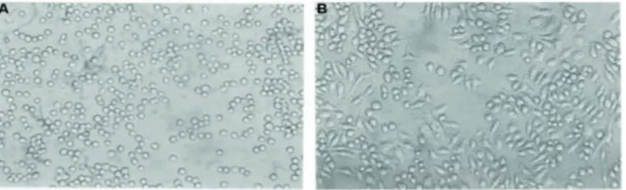
Figure 1: Marine-derived drugs available in the marked8. ______________________ 2
Figure 2:A: THP-cells, B: THP-1 differentiated into macrophages after treatment with
PMA. _______________________________________________________________ 12
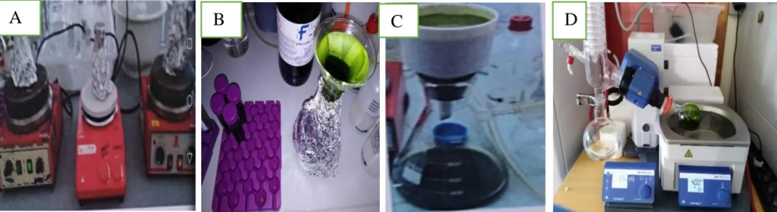
Figure 3:A: Extraction of the biomass using different solvents through stirring at room
temperature. B: filtration of the extract. C: the vacuum filtration system consisting of a funnel coupled with a kitasato flask connected to water flow hose. D: evaporating the extract in a Rotary Evaporator. __________________________________________ 15
Figure 4-A: The process of collecting the fractions. B: Fractions obtained. _______ 17
Figure 5: DPPH radical capturing a hydrogen atom from an antioxidant molecule. 18
Figure 6: Antioxidant assay performed for ethyl acetate extracts at 10,5 and 1 mg/mL
on the same plate. A: Tetraselmis sp.CTP4; B: Skeletonema costatum; C
Nannochloropsis sp oculate; D: BHT and DMSO as positive and negative controls, respectively. _________________________________________________________ 19
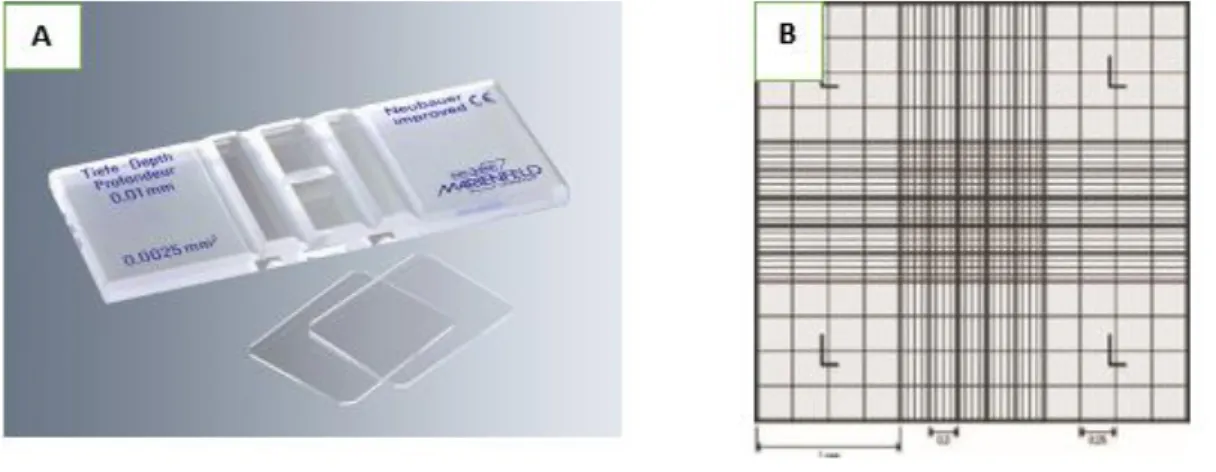
Figure 7: Neubauer counting chamber. The average count of the cells in squares
multiplied by 10000 gives a number of cells per mL (A) (source: www.trade21.com; www.fishersci.co.uk) ___________________________________________________ 20
Figure 8:Radical scavenging activity (𝐈𝐂𝟓𝟎: mg/mL) of ethyl acetate, ethanol, and
water extracts of the different microalgae screened values are mean with standard deviation values not sharing the same letter are significantly different from one another (P<0.05). ___________________________________________________________ 30
Figure 9: MTS cytotoxicity assay screened at 100, 50 and 10 μg/mL for Porphyridium
sp. Water (POC (Water)), Nannochloropsis oculate ethanol (NANNO ethanol) Nannochloropsis oculate water NANNO water ), Tetraselmis sp. CTP4 ethanol (T.
x | P a g e
CTP4 ethanol) and Tetraselmis Chuiie ethanol (T. chuii ethanol). C: negative control (cells without treatment. D: DMSO (DMSO treated with the cells only). __________ 32
Figure 10:Anti-inflammatory activity of extract, Nannochloropsis oculate , Tetraselmis
sp. CTP4 and Tetraselmis chui sp. all are ethanol extracts, in Mac THP-1 cells stimulated with LPS, as determined by levels of TNF-α in the cell culture media. Data are the means ± S.D. of three independent experiments. * p < 0.05, ** p < 0.005, ***p < 0.0005, ****p < 0.0001 vs LPS control. CTR, negative control (cells without
treatment); LPS, LPS pro-inflammatory control; DXM, Dexamethasone positive anti-inflammatory controls: ns, no significance, (50, 20 and 10µg/mL): concentrations tested of each extract. _______________________________________________________ 37
Figure 11: Anti-inflammatory activity of , Nannochloropsis oculate , Porphyridium
cruentum both are water extracts in Mac THP-1 cells stimulated with LPS, as determined by levels of TNFα in the cell culture media. Data are the means ± S.D. of three independent experiments. * p < 0.05, ** p < 0.005, ***p < 0.0005, ****p < 0.0001 vs LPS control. CTR, negative control (cells without treatment); LPS, LPS pro-inflammatory control; DXM, Dexamethasone positive anti-pro-inflammatory control, ns, no significance , (50, 20 and 10µg/mL): concentrations tested of each extract. _____ 39
Figure 12: MTS cytotoxicity assay screened at 50, 20 and 10 μg/mL for hexane,
dichloromethane (DCM), ethyl acetate (EA) and ethanol/water (EtOH/water) fractions. C: negative control cells without treatment. D: DMSO (DMSO treated with the cells only). _______________________________________________________________ 42
Figure 13:Anti-inflammatory activity for (hexane, dichloromethane, ethyl acetate and
ethanol/water) fractions of Tetraselims sp CTP4 extracts in Mac THP-1 cells
stimulated with LPS, as determined by levels of TNF-α in the cell culture media. Data are the means ± S.D. of three independent experiments. * p < 0.05, ** p < 0.005, ***p < 0.0005, ****p < 0.0001 vs LPS control. CTR, negative control (cells without
treatment); LPS, LPS pro-inflammatory control; DXM, Dexamethasone positive anti-inflammatory control, ns, no significance , (50, 20 and 10µg/mL): concentrations tested of each extract. _______________________________________________________ 45
xi | P a g e
TABLE INDEX
Table 1: Radical Scavenging Activity (RSA) (antioxidant activity ) of DPPH free
radicals of water (W), ethanol (EtOH) and ethyl acetate (EA) extracts of the different microalgal species, applied at three concentrations (1, 5 and 10 mg/mL). nd – no activity detected. Values are presented as mean ± standard deviation. ____________ 29
Table 2: % of TNF-α inhibition caused by ethanol extracts at 50, 20 and 10 μg/mL in
Mac THP-1 cells, relative to the LPS control, data are presented in percentage and standard deviation. ____________________________________________________ 36
Table 3: % of TNF-α inhibition caused by water extracts at 50, 20 and 10 μg/mL in
Mac THP-1 cells, relative to the LPS control, data are presented in percentage and standard deviation. ____________________________________________________ 38
Table 4: The yield quantity for both extract and fractions obtained. _____________ 41
Table 5: % of TNF-α inhibition caused by different fractions at 50, 20 and 10 μg/mL in
Mac THP-1 cells, relative to the LPS control, data are presented in percentage and standard deviation ____________________________________________________ 44
Table 6: Chemical composition of the hexane fraction determined by GC-MS. The
percentage of each compound represents its relative abundance in the whole
xii | P a g e
LIST OF ABBREVIATIONS AND ACRONYMS
ABTS: 2.2-Azino-bis (3-ethylbenzo-thiazoline-6-sulfonic acid BHT: Butylated hydroxytoluene
COX: Cyclo-oxygenase DCM: Dichloromethane
DMARDs: Disease-modifying antirheumatic drugs DPPH: 2,2-diphenyl-1-picrylhydrazyl
DXM: Dexamethasone EA: Ethyl acetate
ELISA: Enzyme-linked immunosorbent assay EtOH: Ethanol
GI: Gastrointestinal
HPLC: High pressure liquid chromatography HSV-1: Herpes simplex virus type 1
IL: Interleukins
LCPUFAs: Long chain polysaccarides fatty acides LLE: liquid-liquid extraction
LPS: Bacteria lipopolysaccharides Mac-THP-1: Macrophage THP-1
MTS: (3-(4,5-dimethylthiazol-2-yl)-5-(3-carboxymethoxyphenyl)-2-(4-sulfophenyl)-2H-tetrazolium)
xiii | P a g e
BSA: Bovine serum albumin NF: Nuclear factor
nitric oxide: Nitric oxide
NSAIDs:Non seroidal anti-inflammatory drugs PBS: Phosphate-buffered saline
Phenazine methosulfate: Phenazine methosulfate PMA: Phorbol-12-myristate-13-acetate
PPF: Pigment-protein fraction RSA: Radical scavenging activity RT: Room temperature
SQAG: Sulfoquinovosyldiacylglycerols TNF-α: Tumor necrosis factor
1 | P a g e
CHAPTER 1: INTRODUCTION
1.1 Marine bioprospecting
Since ancient times, natural compounds have been used in disease therapy and still play a major role in modern medication. About half of the drugs existing nowadays that have been approved since 1994 are based on natural products. It is recognized that plants, microorganisms, marine organisms, vertebrates, and invertebrates are essential sources for medicines1-2. In the past 30 years, the interest in marine bioprospecting has expanded among researchers in the globe. The marine environment is different from land-based ecosystems and offers great chemical diversity and high biochemical specificity. Very low octanol-water partition coefficient, more routable bonds and stereogenic centers are some of the chemical properties of small-molecule natural products that make them favorable as lead structures for drug discovery3-4. Marine organisms are therefore known to be as treasures that remain a relatively unexplored source for novel bioactive compounds that could eventually be transferred into therapeutics.
However, as a consequence of the complex molecular structures of natural products, pharmaceutical companies have lately shifted to using synthetic chemical libraries. Hence, clinical trials for modern natural therapeutic products have decreased by 30% between 2001-20085. In addition to structural complexity, drug discovery from natural products faces many other challenges, such as the difficulty of organisms' collection, limited sample quantity, and problems associated with purification and identification of the active agents. When identified, the molecules are often complex which may be too difficult and expensive to produce synthetically. Besides, crude fractions of biological materials are not as amenable to high throughput screening as libraries of pure synthetic compounds3-6. Despite this decline, the utilization of natural products as a source of novel structures in drug discovery is still in progress. Since the 1980s, 174 new compounds were approved and commercialized, from which 93 (53%) are compounds from natural sources or isolated from them7.
So far, the Food and Drug Administration (FDA) in the United States has approved three marine-derived drugs, namely Cytarabine 1 (Cytosar-U®, discovered in sea sponges),
2 | P a g e
Vidarabine 2 (Vira-A®, discovered in sponges), and Ziconotide 3 (Prialt®, discovered in cone snails). The applications for these drugs are cancer, viral infections and pain management, respectively8.
Figure 1: Marine-derived drugs available in the marked8.
In 2007, the European Union approved a marine-derived anticancer drug, Trabectedin 4 (Yondelis®). In addition, 13 marine-derived compounds are either in phase I, phase II or phase III clinical trials, the pipeline of preclinical trials is giving a hundred novel marine compounds each year those compounds are potentially variable9. As mentioned, marine-derived drugs have shown anticancer and antiviral activities. However, marine compounds with antibacterial, anticoagulant, antifungal, anti-inflammatory, antimalarial, antiprotozoal, and anti-tuberculosis effects, in addition to a number of other pharmacological activities are necessary for the treatment and prevention of diseases10. With several compounds approved as drugs, and a pipeline of agents in a clinical trial and preclinical evaluation, the marine environment has performed exceptionally well in yielding new drugs. This suggests that the value of marine natural products in new drug discovery will continue to be powerful in the years to come. There are two major methods in the research for the area of the natural products; old - and new methods11. The old method focuses on the chemistry of the compounds and the selection of natural sources going through ethnopharmacological information as well as traditional uses. However, the isolation and identification of compounds are being done before biological activity
3 | P a g e
testing (primarily in vivo). The new method, the so-called Bioactivity-guided isolation is, as the name says, highly focused on bioactivity. Biological assays (mainly in vitro) are being used to target the isolation and purification of the bioactive compounds. Organisms’ selection is going according to ethnopharmacological information and traditional use, but it can be chosen randomly as well. In the new method, natural marine sources are particularly used because marine natural products provide a great chance in the identification of newly bioactive compounds as drugs for different types of diseases treatment.
The Marine Biodiscovery Process includes the biodiscovery pipeline for marine active bioactive compounds which is getting a biological material either via targeted sampling or through access to curated samples. This comprises analyses of the complete organism like sea squirts or sponges, or it could be sediments/cores of sediment from which microorganism can be isolated. In addition, microorganisms can be isolated from water samples to get sufficient biomass for active compound purification; the purified molecules and extracts can then be screened for the biological activity. The development process could be started if a promising result were obtained from the extracts and their purified molecules. However, that does not mean that always, results in a marketable product12.
1.2 Inflammation
Inflammation is a defensive mechanism stimulated when the body is attacked by for example pathogens, damaged cells or irritants. These responses are important for humans in fighting infections and for starting healing and recovering to normal function in the event of injury. Sadly, these defensive mechanisms sometimes can go wrong, and develop different inflammatory diseases. Inflammation is divided into acute inflammation, which is a short-term process taking place in response to tissue injury, or chronic inflammation, which refers to a slow, long-term inflammation lasting for prolonged periods of several months to years. Acute inflammation starts rapidly, becomes severe in a short time and symptoms may last for a few days, for example, acute pneumonia. It is marked by five cardinal signs: pain, redness, loss of function, swelling and heat. The extent and effects of chronic inflammation, however, may vary with the cause of the injury and the ability
4 | P a g e
of the body to repair and overcome the damage. Chronic and low-grade inflammatory diseases include rheumatoid arthritis, atherosclerosis, Alzheimer’s, asthma, psoriasis, multiple sclerosis, and inflammatory bowel diseases, and numerous of these inflammatory diseases are becoming universal13-14.
As stated by the World Health Organization (WHO), about 235 million people suffer from asthma and it is a widely spread chronic disease among children. Approximately 0.3–1.0% of the general population is suffering from rheumatoid arthritis15.The three fundamental groups of drugs used for the treatment of inflammatory diseases are corticosteroids, non-steroidal anti-inflammatory drugs (NSAIDs), and disease-modifying antirheumatic drugs (DMARDs).
These drugs are commonly used and are effective in treating many inflammatory diseases. Corticosteroids show an important role in the therapy of organ transplantation due to their anti-inflammatory and immunosuppressive effects. However, they are related to some serious side effects. Corticosteroids are recognized to cause Cushing’s syndrome, in addition to other negative effects such as hyperglycemia, increased susceptibility to infection, psychiatric disturbances. On the other hand, NSAID, when used for the long term period, can lead to gastrointestinal ulceration and bleeding and platelet dysfunction14-16.
Inflammation is a complicated mechanism and it involves a multifactorial network of chemical signals to mediate the action. The first anti-inflammatory targets include cyclo-oxygenase (COX)-1 and 2 enzymes, cytokines such as tumor necrosis factor (TNF)-α and interleukins (IL-1β, IL-6), and transcription factor as a nuclear factor (NF)-κB and several more. TNF-α and ILs have been recognized for their central role in the pathogenesis of numerous inflammation diseases, especially asthma and rheumatoid arthritis. TNF-α and ILs are intercellular signal proteins secreted by immune cells and have a lot of functions. The TNF-α function is responsible for a diverse range of signaling events within cells, leading to necrosis or apoptosis of the cells during acute inflammation in addition, the TNF-α protein is also important for resistance against infection and cancers. While the functions of ILs (Interleukins) is to regulate cell growth, differentiation, and motility, both are particularly important in stimulating immune responses, such as inflammation. The transcription factor NF-κB is the main regulator of the expression of several genes involved in the activation of inflammation. It is well established that the excessive
5 | P a g e
production of pro-inflammatory mediators is implicated in several inflammatory diseases. Therefore, inhibition of the overproduction of these mediators is a crucial, exciting target in the treatment of these conditions17-14-18. Herein, it is important to keep searching for new natural compounds possessing anti-inflammatory effects and reducing the side effects since the available drugs have a numerous side effect it’s important to keep searching for new drugs with anti-inflammatory properties and less side effects. The most common side effects produced by chemically synthesized drugs are gastrointestinal (GI) and renal effects associated with NSAIDs. However, sometimes the induced gastric or intestinal ulceration can be accompanied by anemia from the resultant blood loss19.
1. 3 Natural sources of alternative anti-inflammatory agents
It is worldwide recognized that natural products play a major role in the discovery pipeline for making and advancing of drugs for treating human diseases. Thus, natural products provide a great chance in the identification of bioactive compounds as drugs for the treatment of inflammatory diseases. Cyclosporine20 is a natural product that has been synthesized and developed as a first-line immunosuppressive medicine for the treatment of transplant rejection. The discovery of this compound in 1972 was accidental when it was isolated from the fungus Tolypocladium inflatum in Hardangervidda, Norway. Cyclosporine was approved for therapeutic use in 1983 and has since been an important medication, apart from transplants, in the treatment of inflammatory diseases like rheumatoid arthritis and psoriasis. The drug possesses many actions associated with the immunosuppressive activity, but the main action is the selective inhibitory effect on IL-2 and IL-4 gene transcription. This example enlightens the powerful action of natural products as a source for drug discovery14-20.
Lately, many anti-inflammatory pharmacologically active compounds from marine organisms have been identified. These compounds have been isolated and purified from numerous marine sources including sponges, mollusks, bryozoans, sea combs, algae, echinoderms, ascidians, and bacteria21-22-23-24. The Red alga Gracilaria verrucosa possessed anti-inflammatory activities by inhibiting lipopolysaccharide (LPS)-induced nitric oxide (NO) production, TNF-α, and IL625. Fatty acids can be pro-or anti-inflammatory effects in macrophages by modulating the expression of the genes.
6 | P a g e
Specifically, n-3 long-chain polyunsaturated fatty acid (n-3 LCPUFA) have been reported to reduce the inflammatory gene expression via inhibition of nuclear factor κB signaling26.
Astaxanthin, the major carotenoid pigment found in the marine world of algae and aquatic animals, have shown inflammatory properties. Astaxanthin has exhibited anti-inflammatory activities by suppressing the NF-κB activation. It inhibited the production of pro-inflammatory mediators such as TNF-α and IL-1β via doing in vivo studies27. Numerous of microalgae species were studied for the purpose of searching for anti-inflammatory bioactive compounds. For instance, sulfolipids from Porphyridium
cruentum have shown anti-inflammatory activity, the LCPUFAs existing in the crude
sulfoquinovosyldiacylglycerols SQAG fraction.28 The LCPUFAs act as anti-inflammatory compounds through the precursor compounds responsible for the amplification of the inflammation process and increasing the vascular permeability to allow self-propagating responses29. Porphyridium sp. polysaccharides derived from the red alga proved to have an anti-inflammatory effect, through interfering with the production of tumor necrosis TNF-α -induced inflammation, in human coronary artery endothelial cells30.The unrefined polysaccharides of Chlorella Stigmatophora and
Phaeodactylum Tricornutum.extracts had anti-inflammatory activity in the carrageenan-in Vivo studies carrageenan-in rats . Violaxanthcarrageenan-in, isolated from Chlorella ellipsoidea showed an anti-inflammatory effect when tested on lipopolysaccharide (LPS)-stimulated RAW 264.7 mouse macrophage cells. Violaxanthin effectively inhibited LPS-mediated nuclear factor-κB (NF-κB) p65 subunit translocation into the nucleus, suggesting that the violaxanthin anti-inflammatory activity may be based on the inhibition of the NF-κB pathways31. The pigment-protein fraction (PPF) from Nannocloropsis oculate functioned as an anti-inflammatory extract for virus infection 32.
7 | P a g e
1.4 Microalgae
Microalgae are a very diverse assemblage of organisms and this diversity is reflected in the wide variety of chemical compounds of potential commercial interest that they produce. Microalgae are prokaryotic or eukaryotic photosynthetic microorganisms that can grow rapidly and live in harsh conditions due to their unicellular or simple multicellular structure. Prokaryotic microalgae include Cyanobacteria (Cyanophyceae), while green algae (Chlorophyta) and diatoms (Bacillariophyta) are examples of eukaryotic microalgae. Over the past few decades, microalgae have gained great interest due to their potential for many different production purposes. They are marine photosynthetic microorganisms capable to convert carbon dioxide into biochemicals that can be further processed into high-value bioactive compounds, biofuel and food 33. Microalgae can grow rapidly and occur in both aquatic and terrestrial environment, signifying a huge variety of species living in a wide range of condition and habitats. It is estimated that more than 50,000 species of microalgae exist, but only around 30,000, were studied and/or analyzed 34.
The main obligation for the growth of algae is sunlight, water, and nutrients. Microalgae possess the capability to fix CO2 using solar energy with a performance 10 times better than that of terrestrial plants. The requirements of algae are very less than those of terrestrial plants for growing and they do not compete with agriculture or food for precious resources such as water, nutrient, and arable land35. The secondary metabolites produced by microalgae when exposed to severe conditions are being extracted from microalgae with the potential application in biomedicine and pharmacology in the modern field of microalgal biotechnology. The natural active compounds that occur in the microalgal biomass are accountable for distinct biological activities, such as cytotoxic, antibiotic, antioxidant, antifungal, anti-inflammatory and anthelminthic activity35-36. These days, there is different usage for microalgae, for example, microalgae can be utilized to improve the dietary benefit, they play a vital role in aquaculture and they can be used in cosmetic products36. The secondary metabolites delivered by microalgae are vitamins, proteins, lipids, pigments, and carbohydrates – for health, food, cosmetics and biofuel industries37.
8 | P a g e
1.5 Biomedical application of Microalgae
1.5.1 Antioxidant activity
Antioxidants are compounds that provide protection for living cells against free radicals. Free radicals are molecules produced when the body breaks down food or is exposed to tobacco smoke and radiation. The anti-oxidant gives electrons to the free radical and neutralize it. The natural secondary metabolites being produced by microalgae have been reported to possess antioxidant activity. As a result, microalgae have come to be a good source for the natural antioxidants, published by recent studies38.
1.5.2 Anticancer activity
Marine microalgae anti-cancer compounds were under investigation for a long time. The studies were done in the microalgal extracts or fractions obtained using the methods of extraction such as liquid-liquid partitioning or solid-phase extractions. It is uncommon to see dereplication methodologies, fractionations based on high throughput techniques (e.g., HPLC or gas chromatography) or a complete structural elucidation of the compounds that have been found. Despite the low data date available so far, the studies performed on Chlorella sorokiniana and Chaetoceros calcitrans show low activities compared to the commercially available marine anti-cancer drugs. Most of the pharmaceuticals on the market are active at the level of 0.6–7 ng/mL while the fractions from C. sorokiniana and C. calcitrans display significant activity at 1–3 µg/mL. Even if fractions and pure compounds cannot be directly compared in terms of activity, the anti-cancer activities of C. sorokiniana and C. calcitrans extracts seem very promising and appear preferential for further investigation and purification of the active molecules. Despite the low yield (0.0001%) of the active compound in the crude aqueous ethanol extract of the ascidian Ecteinascidia turbinate39, this led to the isolation and development of the anti-cancer drug (trabectedin ET-743).
9 | P a g e
1.5.3 Antiviral activity
Most of the compounds isolated form microalgae and used as antiviral are carotenoids from Haematococcus pluvialis and Dunaliella salina. Pressurized liquid extraction (PLE), an environmentally friendly technique, was used to obtain those compounds. The evaluation of the antiviral activity was done against herpes simplex virus type 1 (HSV-1) at different stages during viral infection. Results demonstrated that the use of PLE allows obtaining antiviral compounds from microalgae used as carotenoids sources, which gives both microalgae biomass an added40-41.
1.5.4 Antibiotic activity
Antimycobacterial activity was found in the hexane crude extracts of six microalgae:
Chlamydomonas mexicana, Porphyridium cruentum, Isochrysis galbana, Rhodomonas sp., Aphanocapsa marina, and Nitzschia palea. All of the hexane extracts were able to
inhibit 90% of the growth of Mycobacterium tuberculosis H37Rv at a concentration of 100 µg/mL-1. The hexane crude extract of I. galbana possessed the highest antimycobacterial activity showing a percentage inhibition that was equal to that of the anti-tuberculosis drug isoniazid and having a minimum inhibitory concentration of 50 μg µg/mL-1. These findings demonstrate that microalgae are an excellent source for the search of novel antimycobacterial compounds42.
1.6 Microalgae with cosmeceutical uses
Numerous secondary metabolites isolated from algae are recognized for their skin benefits43. A worldwide tendency for products considered healthy, environmentally and ecologically sustainable, gained importance in cosmetics industries to fund the research and enhancement of new products with compounds or extracts of natural sources. Algae are natural sources of compounds against oxidative stress and have several efficient protection roles against reactive oxygen species and free radicals, producing compounds
10 | P a g e
that work in cosmetics towards the harmful effects of UV radiation, and similar acts of organic and inorganic filters that are nowadays being used in the market43. The production of both chlorophyll and carotenoids have been increased in C. vulgaris, Nostoc, and
Spirulina platensis when cultivated in the presence of UV radiation43. Therefore, these compounds might assist in the protection against oil oxidation in formulations, largely in emulsions with a huge quantity of oily phase, since they possess antioxidant properties.
Fucus vesiculosus extract is used to decrease the dark circles appear on the skin zone
under the eye via stimulating the expression of heme oxygenase-l, a molecule that terminates the heme construction on the skin by removing heme catabolites. The extract with anti-inflammatory activity and antioxidant properties can make a better appearance of eye bags, and collagen production is stimulated which can assist to reduce fine lines and wrinkles. Besides, it can diminish or prevent skin aging by using make-up and sunscreens44. Several secondary metabolites of specific microalgae can avoid blemishes, repair damaged skin, treat seborrhea, inhibit several inflammation processes and speed up the healing process, and maintain skin moisture45.
Furthermore, red algae extracts are being used in hair care, emollient, sun protection, skincare, refreshing or regenerate care products, anti-aging creams, and anti-irritant in peelers45-46. Algae are mainly incorporated in cosmetic formulations as thickening, water-binding and antioxidants agents. However, more than a single contribution could be attributed to each species, as reported in.47.
1.7 Natural products drug discovery from microalgae
In this study, a bioassay-guided isolation strategy was applied to obtain molecules possessing anti-inflammatory effects isolated from marine microalgae, which could provide the market with a new natural compound to be used as an anti-inflammatory drug in the future.
The steps needed for the preparation of the material before extraction includes the collection of the microalgal biomass, species identification, and storage of the samples. There are different extraction methods available, however, the selection of the extraction method is based on the nature of the source material and the purpose of the isolation.
11 | P a g e
Extraction is a process of getting one or more compounds from a solid matrix or pull out of a solute from a liquid to another liquid (liquid-liquid extraction). Solid samples are most of the time being cut into small fragments or ground into fine particles in order to accelerate solvents penetration. Stirring or shaking can be used to advance the diffusion rate. The solvents that were used int this project for the preparation of the extracts were water, ethanol and ethyl acetate the reason behind selecting different solvents is to obtain different compounds presents in the microalgae since the extraction depends on the polarity index of solvents and compounds. The produced extracts can then be tested in the different bioassays depending on the objective. Often, a simple antioxidant test activity is done before testing with other more complex and expensive, such as the anti-inflammatory assays. Afterwards, based on the antioxidant results, the most promising extracts can be selected for further bioactivity (anti-inflammatory) assays. Anti-inflammatory assays can be performed with macrophages stimulated with a pro-inflammatory drug (i.e. LPS). A pre-incubation with the extracts is used to test for anti-inflammatory action through monitoring the inhibition of TNF-α production. Often, human THP-1 cells are used and differentiated into macrophage THP-1 cells (Mac-THP-1). The production of TNF-α is afterward measured using sandwich enzyme-linked immunosorbent assay (ELISA) which is an analytical biochemistry assay, first described by Engvall and Perlmann in 1972. The assay uses a solid-phase enzyme immunoassay to detect the presence of a ligand in a liquid sample using antibodies directed against the protein to be measured. Active extracts can undergo bioassay-guided fractionation until the purification and isolation of active compounds.
12 | P a g e
1.8 THP-1 cells
The THP-1 cell line (Figure 2: A) is a human monocytic leukemia cell line settled by Tsuchiya et al., in 1980. It was isolated from the blood of a patient suffering from acute monocytic leukemia. THP-1 cells are similar to primary monocytes and macrophages in morphology and differentiation characteristics. THP-1 cells have a large, round single-cell morphology and show distinct monocytic markers. Mostly all THP-1 single-cells start to adhere to culture plates and differentiate into macrophages (Figure 2: B) after being exposed to phorbol-12-myristate-13-acetate (PMA, also known as TPA,12-O-tetradecanoyl phorbol-13-acetate)
THP-1 cells have some technical advantages over human primary monocytes or macrophages. For instance, their genetic background is homogeneous which minimizes the degree of variability in the cell phenotype. Another technical advantage is that genetic modification of THP-1 cells by small interfering RNAs (siRNAs), in order to down-regulate the expression of specific proteins, is relatively simple48. THP-1 differentiated to macrophages have been used as a model in vitro at this research for the anti-inflammatory investigation.
13 | P a g e
1.9. OBJECTIVE
The main objective of this master thesis is to search for a new drug lead for anti-inflammatory treatment in extracts of commercialized microalgal biomass (Porphyridium sp., Nannochloropsis sp., Tetraselmis sp. CTP4, Isochrysis sp., Phaeodactylum
tricornutum, Skeletonema costatum, Spirulina sp., Haematococcus pluvialis, and Tetraselmis chuii). To fulfill the main objective, several specific sub-objectives were
underlined:
1. To prepare ethanol, water and ethyl acetate extracts of the above-mentioned species.
2. To test the prepared extracts for antioxidant activity. A pre-screening for the antioxidant activity will help to identify and select the most promising extracts for the more expensive anti-inflammatory assays;
3. To test the most antioxidant extracts for anti-inflammatory activity, namely by measuring the production of TNF- by LPS-stimulated THP-1 macrophages; 4. To fractionate the most active extract and produce a fraction with higher activity
and less complexity than the initial extract;
5. To perform a chemical characterization of the most active fraction to identify known bioactive compounds (e.g., phenolics, vitamins, and polyunsaturated fatty acids);
14 | P a g e
CHAPTER 2: MATERIAL AND METHOD
2.1 Chemicals
Solvents used for extraction and fractionation: dimethyl sulfoxide (DMSO), methanol, dichloromethane (DCM), ethanol (EtOH), ethyl acetate (EA), hexane, were commercial grade with 96% purity (VWR International, Leuven, Belgium). For the GC derivatization, analytical grade solvents were purchased from the same source. Ultrapure, Type 1 water was obtained using a MilliQ® Water Purification System (Darmstadt, Germany). Roswell Park Memorial Institute medium (RPMI-1640) and fetal bovine serum (FBS), L-glutamine (200 mM), penicillin/streptomycin mixture, Phorbol 12-myristate 13-acetate (PMA), were all from Sigma-Aldrich, St. Louis, MO, USA. MTS 3-(4,5-dimethylthiazol-2-yr)-5-(3-carboxymethoxyphenyl)-2-(4-sulfophenyl)-2H-tetrazolium was from the Cell Titer 96® Aqueous Non-Radioactive Cell Proliferation Assay (Promega Corporation, Madison USA). Butylated hydroxytoluene (BHT), phenazine methosulfate (PMS) and 2,2-diphenyl-1-picrylhydrazyl (DPPH) powder were from Aldrich, Sigma (USA), and the TNF- Elisa kit from PeproTech.
2.2 Algal biomass
Algal biomass of the following species: Porphyridium sp., Nannochloropsis sp.,
Tetraselmis sp. CTP4, Isochrysis sp., Phaeodactylum tricornutum, Skeletonema costatum, Spirulina sp., Haematococcus pluvialis, and Tetraselmis chuii, were kindly
provided by NECTON S.A. (Portugal). This is a company specialized in the cultivation and commercialization of microalgae in Portugal since 1996.
15 | P a g e
2.3 Preparation of extracts
The biomass of each microalga was extracted with ethyl acetate, ethanol, and water. The ratio was 1g of biomass from each algal species to 40mL of the three different solvents prepared in separated flasks. Five grams of dried biomass was mixed with 200 mL of ethanol, ethyl acetate, or distilled water. The extractions were performed overnight (16 h), at room temperature (RT) with agitation using magnetic stirring. (Figure 3: A). After extraction, the mixture was transferred to 50 mL Falcon tubes (Figure 3: B) and centrifuged (Thermo Scientific, USA) at 726.1g for 5min in order to separate the extracts from the remaining biomass. The extracts were filtered through a filter of 10-12 µm pore size (Prat Dumas, France). This process was repeated three times. Finally, the supernatants were combined and vacuum-filtered, using 0.45 µm and 0.2 µm filters sequentially (Figure 3:C). The solvents were evaporated in a rotary evaporator (IKA, RV10, IKA, Germany) to reduce the volume of the extract (Figure 3:D). The concentrated extract was transferred to a smaller pre-weighed vial and completely dried under a gentle nitrogen flow. The yield was determined, and the extracts resuspended in dimethyl sulfoxide (DMSO) at a concentration of 20 mg/mL and stored at -20ºC until analysis. .
Figure 3:A: Extraction of the biomass using different solvents through stirring at room temperature. B: filtration of the extract. C: the vacuum filtration system consisting of a funnel coupled with a kitasato flask connected to water flow hose. D: evaporating the extract in a Rotary Evaporator.
16 | P a g e
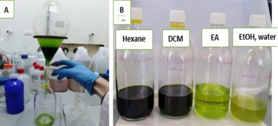
2.4 Extract fractionation
The most active extract, Tetraselmis sp. CTP4 ethanol was selected for fractionation. The fractionation was done by liquid-liquid extraction (LLE), which is a purification technique that is used to separate compounds based on their relative solubilities in two immiscible solvents. Immiscible liquids do not dissolve in each other; they form layers when placed in the same glassware. Immiscibility is a result of two liquids having different polarity. LLE started by mixing the ethanol extract with water (20:80) and extracting sequentially with hexane (H), dichloromethane (DCM), and ethyl acetate (EA) in a separating funnel. The procedure yielded 4 fractions: H, DCM, EA and the remaining ethanol/water (E/W) fraction. For this fractionation, 3 g of the Tetraselmis sp. CTP4 -ethanol extract, already prepared, was mixed with 20 mL EtOH and 80 mL of Milli-Q water and placed in a separation funnel. 50 mL of hexane was added to the mixture and shook gently. After phase separation, the hexane (top) layer was collected in a glass round bottom flask (Figure 4: A). The water layer was re-extracted with another 50 mL of hexane and the process repeated until 150 mL of hexane fraction was collected (three extractions in total). After the extraction with hexane, the ethanol/water fraction was sequentially extracted with 3 x 50 mL of DCM and 3 x 50 mL of EA. The remaining fraction (a mixture of ethanol and Milli-Q water) was collected as well. All the fractions were concentrated in a rotary evaporator, transferred to a 45 mL vial size and dried under gentle nitrogen flow (organic fractions) or freeze-dried (E/W fraction). Part of each fraction was re-dissolved in DMSO at 20 mg/ mL for further anti-inflammatory analysis. However, the rest of the fractions were stored dried at – 20°C. All the fractions (hexane, dichloromethane, ethyl acetate, and water-ethanol) (Figure 4: B) were analysed for anti-inflammatory activity on macrophage differentiated THP-1 cells, as described above. The fraction with the highest activity was chemically characterized by gas chromatography-mass spectrometry (GC-MS) to tentatively identify the major compounds present, and possibly responsible for the observed anti-inflammatory and antioxidant activity visualized at the start point of the assay processing.
17 | P a g e
The fractions yield displayed as a percentage (%) in relative to the starting mass of the extract led for fractionation (3 g) and the calculations were done according to the following equation:
(𝑚1 − 𝑚2 )𝑥100 where m1 (g) corresponds to the weight of the extract residue after solvent removal; m2 (g) is the weight of the dry biomass.
Figure 4-A: The process of collecting the fractions. B: Fractions obtained.
2.5 Free Radical Scavenging Activity
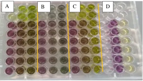
The 2,2-Diphenyl-1-picrylhydrazyl (DPPH) free radical scavenging activity is considered a basic common test for antioxidant activity assessment. DPPH is a stable free radical which has a purple color in solution (Figure 5). When scavenged by antioxidant molecules it is reduced to diphenyl picryl hydrazine, which has a yellow color in solution. This property allows the visual monitoring of the reaction, and free radical scavenging activity can be calculated by measuring the absorption changes at 517nm.
18 | P a g e
Figure 5: DPPH radical capturing a hydrogen atom from an antioxidant molecule.
The DPPH assay was determined using the method described by Moreno et al. (2006) 49. A solution of 120 µM DPPH in methanol was prepared daily. The experiment was done on a 96 well plate (WVR, USA) (Figure 6). The extracts were tested at 1, 5 and 10 mg/mL and butylated hydroxytoluene (BHT) was used as a positive control . DMSO was used as a negative control. Extracts (22 μL) were mixed with 200 μL of DPPH solution (120 μM in methanol) in 96-well microplates. After a 30-min incubation period in the dark, the absorbance was measured at 515 nm using a microplate reader spectrophotometer (Biotech 4 Multi-Detection, USA). The antioxidant activity was calculated as the percentage inhibition relative to a blank containing DMSO in place of the samples, using the following equation:
% 𝑖𝑛h𝑖𝑏𝑖𝑡𝑖𝑜𝑛 = 𝐴𝑏𝑠𝑛𝑒𝑔 𝑐𝑜𝑛𝑡𝑟𝑜𝑙− 𝐴𝑏𝑠𝑡𝑒𝑠𝑡 𝑠𝑎𝑚𝑝𝑙𝑒 𝐴𝑏𝑠𝑛𝑒𝑔 𝑐𝑜𝑛𝑡𝑟𝑜𝑙 ×100
Where Abstest sample = Abs test solution – Abs color control.
IC50, which is the biochemical half maximal inhibitory concentration, was determined for the extracts with radical scavenging activity (RSA)( antioxidant activity) higher than 50% when tested at 10 mg/mL.
19 | P a g e
Figure 6: Antioxidant assay performed for ethyl acetate extracts at 10,5 and 1 mg/mL on the same plate.
A: Tetraselmis sp.CTP4; B: Skeletonema costatum; C Nannochloropsis sp oculate; D: BHT and DMSO as positive and negative controls, respectively.
2.6 Anti-inflammatory assay
2.6.1 Cell cultivation and differentiation
THP-1 cells were kindly provided by Dr. Nuno Santos (CBME, University of Algarve, Faro, Portugal) and were cultivated in RPMI 1640 media containing 1% L-glutamine, 10% heat-inactivated FBS, 1% penicillin-streptomycin (PS), and 1% non-essential amino acids. THP-1 cells were routinely maintained in RPMI 1640 medium at 37 ̊C in a 5% CO2 humidified atmosphere. To differentiate the THP-1 monocytes into macrophages (Mac THP-1), cells were plated in 96 wells plates (2.5×10⁵ per well) in 200 µl/well of RPMI 1640 medium, supplemented with 25 ng/mL of phorbol 12-myristate 13-acetate (PMA), for 48h.
2.6.1.1 Cell counting
To calculate the volume of cell suspension required to plate 2.5𝑥105cells/well, cells counting was made using a Neubauer chamber (VWR International, Leuven, Belgium) (Figure 7) following the equation:
20 | P a g e
𝑁 = A + B + C + D
4 x 𝑑𝑖𝑙𝑢𝑡𝑖𝑜𝑛 𝑓𝑎𝑐𝑡𝑜𝑟 𝑥 10 4
where N represents a number of cells/mL; A, B, C, D correspond to the number of cells in each quadrant of the counting chamber and 104 is a conversion factor.
Figure 7: Neubauer counting chamber. The average count of the cells in squares multiplied by 10000 gives a number of cells per mL (A) (source: www.trade21.com; www.fishersci.co.uk)
2.6.2 Cell viability assessment (MTS assay)
Extracts were tested for cytotoxicity on Mac THP-1 cells before the anti-inflammatory assays. Cell viability was assessed using the MTS method (Cell Titer 96® Aqueous Non-Radioactive Cell Proliferation Assay, USA). Mac THP-1 cells were differentiated as described above for 48h. T After this period, the cells were inspected under a microscope to confirm that the monocytes had differentiated to macrophages by adhering to cell culture plate, and that cells were evenly distributed in every well. Then the old media was removed, and new media, containing the five extracts: water extract of Porphyridium sp., and Nannochloropsis sp. and ethanol extract of Nannochloropsis sp., Tetraselmis sp. CTP4, and Tetraselmis chuii was added. The same process was done to assess the fractions (hexane, dichloromethane, ethyl acetate, and water/ethanol) resulting from the fractionation of the active extract. All were tested at a concentration of 10, 20 and 50 µg/mL. Also, DMSO vehicle at the highest concentration of 0.25% (v/v) was tested to make sure that the vehicle is not toxic. Testing conditions were added to the pre-differentiated cells, and further incubated for 48 hours. Afterwards, 11.9 μL of MTS (3-
(4,5-dimethylthiazol-2-yl)-5-(3-carboxymethoxyphenyl)-2-(4-sulfophenyl)-2H-21 | P a g e
tetrazolium) plus 0.6 μL of phenazine methosulfate (PMS) to a final volume of 120 μL RPMI media were added. The plate was incubated at 37°C, with 5 % CO2 for 1h and the absorbance measured at 490 nm using a spectrophotometer (Biotech 4 Multi-Detection, USA). All the values were compared to the control untreated cells, considered to have 100% of cells viability. The cells viability (%) was calculated after the treatments. The average of the blank absorbance was taken first and subtracted from all the measurements on the 96-well plate, to determine the background noise. The average of the control wells (cells without treatment) was calculated representing 100% cells viability. The cell viability of the DMSO control, positive control, and cells treated with the extracts and the fractions were then calculated by the following formula:
% cell viability = 𝐴𝑏𝑠𝑠𝑎𝑚𝑝𝑙𝑒 − 𝐴𝑏𝑠𝑐𝑜𝑛𝑡𝑟𝑜𝑙 𝐴𝑏𝑠𝑐𝑜𝑛𝑡𝑟𝑜𝑙 × 100
Where 𝐴𝑏𝑠𝑠𝑎𝑚𝑝𝑙𝑒= absorbance of the extracts/fractions treated with the cells 𝐴𝑏𝑠𝑐𝑜𝑛𝑡𝑟𝑜𝑙 = absorbance of untreated cells with the extracts/fractions
2.6.3 Inflammatory assay in Mac THP-1 cells
THP-1 cells were plated and differentiated to macrophages (Mac THP-1) as described in the previous section. After differentiation, cells were treated with extracts/fractions at concentrations of 10, 20 and 50 µg/mL, or not treated corresponding to the basal conditions of Mac THP-1 cells. Also, a positive control to the anti-inflammatory response was performed, by treating the cells with dexamethasone at 2 μM. This pre-treatment was performed for 24 hours. Inflammation was induced by adding 100 ng/mL of LPS to all wells, except for the negative control condition corresponding to cells without treatment, for additional 24 hours. All the extracts were tested in triplicates for each concentration. The conditioned media was collected and centrifuged for 15 minutes at 13.000 rpm and 4ºC to remove the cell debris and kept at -20°C until ELISA.
22 | P a g e
2.6.4 Evaluation of the anti-inflammatory activity through quantification of TNF- by sandwich ELISA
The enzyme-linked immunosorbent assay (ELISA) is a commonly used analytical biochemistry assay, first described by Engvall and Perlmann in 1972. The assay uses a solid-phase enzyme immunoassay (EIA) to detect the presence of a ligand (commonly a protein) in a liquid sample using antibodies directed against the protein to be measured. ELISA has been used as a diagnostic tool in medicine, plant pathology, and biotechnology, as well as a quality control check in various industries50. The types of common ELISA assays include direct, indirect and sandwich ELISA. A sandwich ELISA assay (Pepro tech, USA) was used in this study. A sandwich ELISA quantify antigens detected by two antibodies (i.e. capture and detection antibody), directed against two epitopes on the target molecule. Either monoclonal or polyclonal antibodies can be used as the capture and detection antibodies in a sandwich ELISA system. Monoclonal antibodies recognize a single epitope that allows fine detection and quantification of small differences in antigen. A polyclonal is often used as the capture antibody to pull down as much of the antigen as possible. The advantage of sandwich ELISA is that the sample does not have to be purified before analysis, and the assay can be very sensitive.
2.6.4.1 Sandwich ELISA assay for the determination of TNF-α production by Mac THP-1 induced by LPS.
Sandwich ELISA was used for the determination of TNF-α production by Mac THP-1 induced by LPS. Capture antibodies Rabbit Anti-Human TNF-α (PeproTech, USA) were plated after been prepared in Phosphate-buffered saline (PBS) to a concentration of 1.0 μg/mL. 100 μL of the solution was added to each well and the plate kept at room temperature overnight. Then, aspiration of the liquid took place and the plate was washed 4 times with 300 μL wash buffer/well (wash buffer: 1xPBS with 0.05 % of Tween-20). Followed by the addition of 300 µl/well of blocking buffer (1 % of BSA diluted in 1x PBS and incubated for 1 hour at room temperature. The PBS concentrated 10x was
23 | P a g e
prepared from these compounds (15mMof 𝑁𝑎2𝑃𝑂4, 𝑁𝑎𝐻2𝑃𝑂4,8.1mM 𝑁𝑎𝐻2𝑃𝑂4. 2𝐻2𝑂, and 1.45M NaCl) in Milli-Q water with 7.3-7.4 of pH.
Briefly, the conditioned media from the samples such as negative control (cells without treatment) were diluted 1:20 while the rest of the samples, including the inflammatory positive control (Mac THP-1 cells treated only with LPS), the anti-inflammatory positive control dexamethasone and the conditions with the tested extracts/fractions, were diluted 1:75. A serial dilution from 2000 pg/mL to zerohuman TNF-α standard was performed. Thus, the human TNF-α standard concentrations were 2, 1, 0.5, 0.25, 0.125, 0.0625, 0.03125, 0.0156 pg/mL in Assay diluent ( 0.1% of BSA in 1xPBS containing 0.05%Tween-20). The plate was incubated at room temperature for 2 hours. The plates were washed with 300ul of wash buffer per well 4 times.
The detection antibodies, Biotinylated Rabbit Anti-Human TNF-α solution was prepared to a concentration 50μg/mL in a diluent, and 100 μL was added to all wells. The plates were kept at room temperature for 1 hour followed by washing and addition of 100 μL of avidin-HRP conjugate solution diluted (1:2000) to all wells. The plates were incubated at room temperature for 30 minutes before they were washed with wash buffer 300 μL wash buffer/well, 4 times. Next, 100 μL ABTS (2.2-Azino-bis (3-ethylbenzo-thiazoline-6-sulfonic acid) liquid substrate solution was added to all wells, and the plate was taken to the Microplate Reader spectrophotometer. Kinetic reading of the absorbance at 405 and 650nm was determined for each well at 5 minutes interval. The concentration of TNF- α in the samples was determined using the calibration curve obtained from the Human TNF-α standard. Also, the percentage of TNF-TNF-α inhibition by the extracts and fractions was calculated in respect to the LPS -proinflammatory control, according to the following equation:
% of TNF- α 𝑖𝑛h𝑖𝑏𝑖𝑡𝑖𝑜𝑛 = LPSproinflammatory control − Sample LPSproinflammatory control ×100
Where LPSproinflammatory control = Quantity of the TNF- α production in Mac THP-1 cells treated only with the pro-inflammatory LPS
Sample=Quantity of the TNF- α production in the cells treated pre-treated with extracts/fractions and proinflammatory LPS.
24 | P a g e
2.7 Chemical characterization by GC-MS
2.7.1 Chemical derivatization
Based on the anti-inflammatory results of the fractions, the H fractions were selected for chemical characterization by GC-MS. For this purpose, the fractions were derivatized in order to make it more volatile since GC measures the volatile compounds. The derivatization was done according to a modified protocol of Lepage and Roy51. This method is based on the direct transesterification with acetyl chloride/methanol, followed by direct extraction of the derivatized compounds into hexane. Briefly, 10mg of each fraction was weighed and treated with 1.5 mL of derivatization solution (methanol/acetyl chloride, 20:1, v/v), in reaction vessels. Afterward, 1 mL of hexane was added, and the mixture heated for 1 hour at 100°C. After cooling in ice baths, 1 mL of distilled water was added, and the organic phase removed and dried with anhydrous sodium sulfate. The extract was then filtered and stored at −20 °C until further analysis.
2.7.2 GC-MS analysis
Fractions were analyzed on an Agilent GC-MS (Agilent Technologies 6890 Network GC System, 5973 Inert Mass Selective Detector) equipped with a DB5-MS capillary column (25 m × 0.25 mm internal diameter, 0.25 μm film thickness, Agilent Tech) using helium as the carrier gas. Samples were injected at 300°C and the temperature profile of the GC oven was 60°C (1 min), 30°C min−1 to 120°C, 5°C min−1 to 250°C, and 20°C min−1 to 300°C (2 min). For the identification of the compounds present in the fractions, the total ion mode was used.
25 | P a g e
2.8 Statistical analysis and data interpretation
2.8.1 Statistical analysis for radical scavenging activity
The Abs color control is the Abs recorded for the (DMSO) negative blank control. The IC50 values were calculated for those species in which antioxidant inhibition was more than 50%. IC50. The half-maximal inhibitory concentration is a measure of the potency
of a substance in inhibiting a specific biological or biochemical function. IC50 values were estimated with GraphPad Prism v.7.0, by the sigmoidal fitting of the data.
2.8.2 Statistical analysis of sandwich ELISA assay
The results are presented as a mean ± standard deviation; all analyses were performed in triplicate in three independent experiments. Differences between species and the LPS control were assessed using analysis of variance (one-way ANOVA by Dunnett’s multiple compressions tests). Significant differences were considered when p < 0.05 by means of the statistical program Graph Pad Prism (release 7.0).
2.8.3 Statistical analysis for the GC-MS
Bruker MSWS Software was used with integrated National Institute Standard and Technology (NIST) library to identify the compounds present in the active fraction Components’ relative percentages were calculated based on GC peak areas in respect to the total area of peaks.
26 | P a g e
CHAPTER 3: RESULTS AND DISCUSSION
3.1 Screening for antioxidant activity
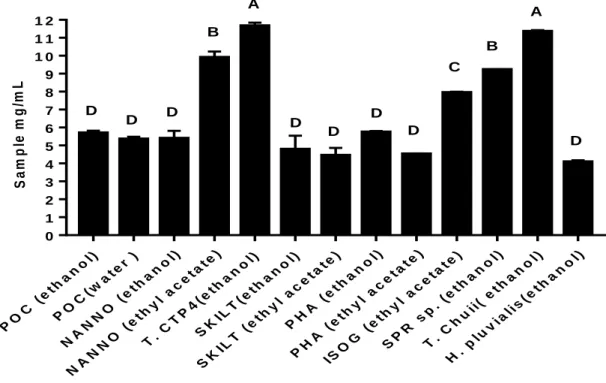
The screening for the antioxidant activity was performed with the ethanol (EtOH), ethyl acetate (EA) and water (W) extracts obtained from Porphyridium sp. (POC),
Phaeodacytlum tricornutum (PHA), Nannochloropsis sp. (NANNO), Tetraselmis sp. CTP4 (T.CTP4), Isochrysis sp. (ISOG), Skeletonema costatum (SKLT), Spirulina sp. (SPR), Tetraselmis chuii (T.Chuii), Haematococcus pluvialis (H Pluvialis).
Thus, extracts were tested for antioxidant activity with the DPPH assay. where the presence of compounds that are able to neutralize the free radical DPPH is determined. All the values obtained were presented as a percentage, compared to the positive control butylated hydroxytoluene (BHT)52. All the samples were analyzed at concentrations of 1, 5 and 10 mg/mL. The response of all the extracts was dose-dependent (Table 1), that is, DPPH scavenging activity was increased by the increasing concentration of the antioxidant compounds present in the samples53.Since all the extracts were dissolved in DMSO prior to the experiment, DMSO was used as a negative control as well, tested at the same concentration as in the extracts. Due to its medium polarity, DMSO is a preferred solvent in extract testing assays for a wide range of compounds, as it can dissolve both polar and nonpolar compounds; in addition, it is miscible with water54. The results for the average antioxidant activity are summarized in Table 1. The best results were accomplished was with the EA extracts, showing the highest antioxidant activity when compared to the positive control BHT and to other water (W) and ethanol (EtOH) extracts.