Document heading doi:10.12980/JCLM.2.2014APJTB-2014-0138 襃 2014 by the Journal of Coastal Life Medicine. All rights reserved.
A
green synthesis of antimicrobial compounds from marine microalgae
N
annochloropsis oculata
Duraiarasan Surendhiran*, Mani Vijay, Abdul Razack Sirajunnisa, Thiruvengadam Subramaniyan, Ammavasai Shanthalin
Shellomith, Kuppusamy Tamilselvam
Bioelectrochemical Laboratory, Department of Chemical Engineering, Annamalai University, Annamalai Nagar, Tamilnadu-608002, India
Journal of Coastal Life Medicine
*Corresponding author: Duraiarasan Surendhiran, Bioelectrochemical Laboratory,
Department of Chemical Engineering, Annamalai University, Annamalai Nagar,
Tamilnadu-608002. India. Tel: +91-9943941851
E-mail: [email protected]
Foundation Project: Supported by University Grants Commission, New Delhi, India. 1. Introduction
There are number of clinically efficacious antibiotics becoming less effective due to the development of antibiotic resistant microorganisms[1,2]. It becomes a greater problem
to treat many diseases caused by resistant pathogenic microorganisms worldwide. In addition, decreased activity of commonly used antibiotic and resistance of pathogens to such antibiotics have anticipated the development of new alternatives[3]. Marine planktons especially algae
are rich source of many interesting bioactive molecules
including lipid which may be useful for the development of antimicrobial drugs[4,5]. Marine microalgae have been an
unique source of chemical compounds of pharmaceuticals, aquaculture, cosmetics, anticancer agents, enzymes, pigments, antioxidants, polyunsaturated fatty acids, dietary supplements, agrochemicals and biofuel[6-12].
There are many reports related to antimicrobial activity of crude extracts of marine macro and microalgae[13,14]. From
the literature survey we learnt that only a few studies have been reported on inhibitory activity of fatty acid methyl esters (FAME) of marine microalgae and there is no report
PEER REVIEW ABSTRACT
KEYWORDS
Nannochloropsis oculata, FAME, Antimicrobial effect, Zone of inhibition, Gas chromatography-mass spectrometer
Objective:To determine the antibacterial and anti-candidal activities of fatty acid methyl esters
(FAME) extracted from marine microalga Nannochloropsis oculata and evaluate the inhibition
activity of DNA isolated from test pathogenic microorganism.
Methods:FAME was synthesized by transesterification of oil using immobilized lipase and
characterized using gas chromatography-mass spectrometer. The FAME profile was determined
using gas chromatography. The antimicrobial effect was tested by disc diffusion method against
Gram-positive bacteria Staphylococcus aureus, Bacillus subtilis, Gram-negative bacteria
Escherichia coli, Pseudomonas aeruginosa and yeast Candida albicans, at varying concentrations of 10, 20 and 30µL/disc.
Results:The results shown that palmitic acid (C16:0), oleic acid (C18:1) and arachidic acid (C20:0)
were dominant in Nannochloropsis oculata oil. The study revealed that FAME was more active
against Gram-negative than against Gram-positive and yeast. DNA inhibition activity results also
confirmed that FAME had the bactericidal effect that was revealed by sheared fragments of DNA.
Conclusions:The results indicated that microalgal FAME could be potentially utilized as a newer
and good source of therapeutic agent in pharmaceutical industry.
Peer reviewer
Dr. Raquírio Marinho da Costa,
Universidade Federal do Pará-Campus
de Bragança, Instituto de Estudos
Costeiros (IECOS). Tel: +55(91)–34251593
E-mail: [email protected];rauquirio@ pq.cnpq.br
Comments
This research work presents an interesting set of data. The obtained results indicate that FAME have a
great potential if used as inhibitor of bacteria growth.
Details on Page 862
Article history:
Received 21Apr 2014
Received in revised form 28Apr, 2nd revised form 5May, 3rd revised form 11May 2014
Accepted 16Jun 2014
on molecular studies. To the best of our knowledge this is the first report on antimicrobial activity of FAME from marine
microalgae produced by an ecofriendly green process using immobilised enzyme system.
The aims of this study were to determine the antibacterial and anti-candidal activities of FAME extracted from marine
microalga Nannochloropsis oculata(N. oculata) and evaluate
the inhibition activity of DNA isolated from test pathogenic
microorganism.
2. Materials and methods
2.1. Microalgal culture
N. oculata, obtained from Central Marine and Fisheries
Research Institute, Tuticorin, Tamilnadu (India), was grown
in sterile Walne’s medium. The filtered sterilized sea water was enriched with required quantity of Walne’s medium containing: NaNO3, 100 g/L; NaH2PO4• 2H2O, 20 g/L; Na2EDTA,
4 g/L; H3BO3, 33.6 g/L; MnCl2• 4H2O, 0.36 g/L; FeCl3• 6H2O, 13 g/L;
vitamin B12, 0.001 g/L and vitamin B1, 0.02 g/L. The trace metal
solution contained: ZnSO4• 7H2O, 4.4 g/L; CoCl2• 6H2O, 2 g/L;
(NH4)6Mo7O24• H2O, 0.9 g/L; and CuSO4• 5H2O, 2 g/L. The medium
was adjusted to pH 8 and autoclaved at 121°C for 20 min.
The filter sterilized vitamins were added after cooling. The contents were later introduced into a 250-mLErlenmeyer flask and finally transferred to 25 L photobioreactor.
Mixing was done by sparging air from the bottom of the photobioreactor; lighting was supplied by four cool-white fluorescent tubes with an intensity of 5000 lux.
2.2. Microscopic study of intracellular lipid
The intracellular lipid present in the microalgae was identified by Nile red staining method. A stock solution of Nile red stain (9-diethlamino-5H-benzo (α) phenoxa-phenoxazine-5-one) was prepared according to Mohamady
et al[15]. About 2.5 mg of Nile red was dissolved in brown
bottle containing 100 mL of acetone and this was stored in dark condition. Each 0.5 mL of microalgae culture broth were centrifuged at 1500 r/min for 10 min and the pellets were
washed with sterile distilled water (equal volume) for several
times. The cell pellets were then mixed with 0.5 mL of Nile red solution incubated for 10 min at room temperature. After washing with distilled water, the stained cells were observed under fluorescence microscopy[16].
2.3. Isolation of microalgal oil
N. oculata oil was extracted using the method of Bligh and
Dyer with slight modifications[17]. The biomass suspension
was mixed with chloroform: methanol (1:2), vortexed for few
minutes and incubated on ice for 10 min. Then, chloroform was added, followed by addition of 1 mol/LHCl and vortexed again for few minutes. Finally, the whole suspension was centrifuged at maximum speed for 2 min. Bottom layer containing lipid was transferred into a fresh previously weighed beaker. The lipid from the aqueous sample was
further extracted using chloroform. The solvent system was evaporated in a rotary evaporator at 30°C. Finally, the lipids
were used for production of FAME.
2.4. Preparation of FAME
The FAME was synthesized by transesterification method
using immobilized lipase. The lipase enzyme was obtained from Hi-Media, India and its activity is 16IU/mg. Before transesterification process the algal oil was heated to 60 °C
for 30 min to reduce viscosity. The immobilized lipase was prepared by adding 1 mg of lipase powder dissolved in 1 mL of sterilized distilled water. Then the lipase enzyme was mixed with sodium alginate solution (2%), the mixer was dripped
into cold sterile 0.2 mol/LCaCl2 using sterile syringe from a
constant distance and was cured at 4°C for 1 h. The beads
were hardened by suspendedagain in a fresh CaCl2 solution
for 24 h at 4 °C with gentle agitation. After immobilization,
the beads were separated through filtration and washed with 25 mmol/L phosphate buffer (pH6.0), in order to remove
excess calcium chloride and enzyme. Then the beads were preserved using 0.9% NaCl solution for future use[18,19]. In
a 20 mL screw cap vial, 5 mL of N. oculata oil was taken and methyl acetate was added (oil to methyl acetate molar
ratio 1:12) along with 2 g of immobilised enzyme beads.
The mixture was then agitated for 24 h, and centrifuged,
transferred into separating funnel and left overnight. The upper layer containing FAME was transferred into a clean
beaker and the content was washed with hot water until clear FAME was obtained. The mixture of FAME and water was
centrifuged to remove water. Finally, the purified FAME was
analysed by gas chromatography-mass spectrometer (GC-MS)
and used for antimicrobial activity.
2.5. Microorganisms for antimicrobial studies
Strains of Gram-negative bacteria [Escherichia coli
(E. coli) and Pseudomonas aeruginosa (P. aeruginosa)],
Gram-positive bacteria [Bacillus subtilis (B. subtilis) and
Staphylococcus aureus (S. aureus)] and yeast Candida
albicans (C. albicans) were obtained from Department of
Microbiology, Raja Muthiah Medical College and Hospital,
Annamalai University, Tamilnadu, India. The bacterial stock cultures were maintained on nutrient agar slant at 4 °C.
The selected bacteria and yeast were cultured (24 h) using
peptone broth and Sabouraud’s dextrose broth respectively for antimicrobial test.
2.6. Antimicrobial assay
In vitro antimicrobial assay was carried out using disc diffusion method[20]. About 20 mL of sterilized M
uller-Hinton agar medium was poured onto sterilized Petri plates.
After solidification, the test microbial suspensions were spread uniformly on the plates using a sterile cotton swab.
The discs were prepared by using Whatman No. 1 filter
paper approximately 5 mm in diameter and sterilized using
an autoclave. Microalgal FAME was loaded onto sterile disc
and placed on the surface of the each plate. The positive controls for bacteria (streptomycin 10µg/disc), for yeast
(amphotericin B100IU/disc) and negative control (diluted
methanol) were used. All the plates were incubated at
37°C for 24 h. After incubation period, zone of inhibition
was formed around the disc which was the evidence of antimicrobial activity[7].
2.7. DNA inhibition activity
The DNA inhibition activity was carried out according to
Surendhiran et al[21]. Most sensitive microorganisms were
selected for analyzing DNA inhibition. The fixed dosage was selected from antimicrobial assay. The FAME extracted was
added to 5 mL nutrient broth containing bacterial culture and then this mixture was incubated at 37°C in a shaker for 24 h. Culture without FAME extract was used as control. Then the DNA inhibition was analysed in molecular level by agarose
gel electrophoresis, with a DNA sample volume of 20µL.
2.8. GC-MS analysis
Fatty acid composition of FAME produced from N. oculata FAMEs were analysed by GC-MS(GC-MS-QP2010, Shimadzu)
equipped with VF-5MS capillary column (30 mm length, 0.25 mm diameter and 0.25µm film thickness). The column temperature of each run was started at 70°C for 3 min, then
raised to 300°C and maintained at 300 °C for 9 min. GC
conditions were: column oven temperature: 70°C; injector
temperature: 240°C; injection mode: split; split ratio: 10; flow
control mode: linear velocity; column flow: 1.51 mL/min; carrier gas: helium (99.9995% purity) and injection volume:
1 µL. MS conditions were: ion source temperature: 200°C;
interface temperature: 240°C; scan range: 40-1000 m/z; solvent
cut time: 5 min; MS start time: 5 min; end time: 35 min and
ionization: EI(-70 eV) and scan speed: 2000 amu/second.
3. Results
3.1. Microscopic identification of intracellular lipid
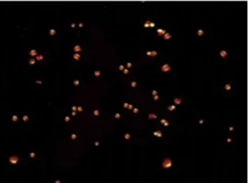
The intracellular lipid molecules were observed under fluorescent microscope at 100伊 with excitation at 450-490 nm and emission at 515 nm. Lipid molecules appeared as yellow dots, whereas cytoplasm was stained in red colour (Figure 1).
Figure 1. Nile red stained cells of N. oculata under fluorescent microscope.
3.2. Antimicrobial activity of FAME
The antimicrobial activity of N. oculataFAME was tested
against bacteria (both Gram-positive and Gram-negative)
and yeast (C. albicans). In this study, we had found that
FAME had the ability to inhibit both bacteria and yeast
which was indicated by zone of inhibition around the disc
(Figure 2). With three different concentrations of FAME(10, 20 and 30µL/disc), the maximum concentration of 30µL/disc resulted in maximum inhibition activity of all tested strains.
Among the different microbial strains tested, E. coli and
P. aeruginosa were found to be more sensitive with the
zone of inhibition of 27 mm and 20 mm (Figure 2a and 2b)
respectively than other microorganisms such as B. subtilis
(16 mm), S. aureus(17 mm). They showed less susceptibility than the Gram-negative bacteria and higher susceptibility than the positive control (Figure 2c and 2d), whereas C.
albicans(19 mm) showed moderate effect towards FAME of N. oculata than the Gram-negative bacteria (Figure 2e).
Different FAMEconcentrations (µL/disc) and their respective zone of inhibition are shown in Figure 3.
Figure 2. Antimicrobial assay by disc diffusion method.
A: 10µL/disc; B: 20µL/disc; C: 30µL/disc; D: Negative control; E: Positive control. a: E. coli; b: P. aeruginosa; c: S. aureus; d: B. subtilis; e: C. albicans.
A A A
A A
D D D
D D
E E E
E E
C C C
C C
B B B
B B
a b c
d e
30
25
20
15
10
5
0
Z
on
e o
f
I
nh
ib
iti
on
(mm
)
E. coli P. aeruginosa B. subtilis S. aureus C. albicans 10µL/disc 20µL/disc 30µL/disc Positive control
Figure 3. Antimicrobial activity of FAME of N. oculata oil at different concentrations.
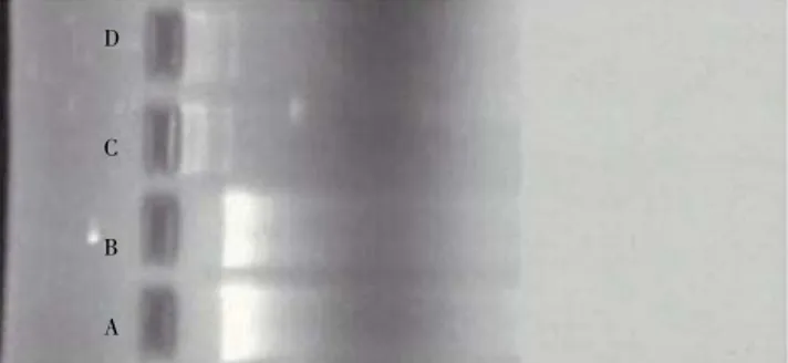
3.4. DNA inhibition activity
The most susceptible bacteria P. aeruginosa and E. coli
towards FAME were selected for the DNA inhibition activity.
After the agarose gel electrophoresis the DNA band was
visualized under UV-transilluminator. DNA isolated from FAME treated bacterial culture was observed as small
fragments and the control appeared as clear band (Figure
4). Formation of small fragments was due to the action of
FAME of N. oculata on DNA of respective microorganisms and
Figure 4.Illustration of sheared and normal DNA isolated from P. aeruginosa
and E. coli.
A: Control DNA isolated from P. aeruginosa (untreated with FAME); B: Control DNA isolated from E. coli(untreated with FAME); C: Sheared DNA of E. coli
isolated after FAME treatment; D: Sheared DNA of P. aeruginosa isolated after FAME treatment.
D
C
B
A
3.5. GC-MS analysis of FAME
By the GC-MSanalysis the major fatty acid composition of FAME produced from N. oculata oil is shown in Table 1.
Table 1
Fatty acid profile of N. oculata oil. Lipid
number Common name Chemical name Mstructureolecular content Fatty acid (%)
C12:0 Lauric acid Dodecanoic acid C12H24O2 9.86
C16:0 Palmitic acid Hexadecanoic acid C16H32O2 19.39
C18:0 Stearic acid Octadecanoic acid C18H36O2 10.76
C18:1 Oleic acid 9-Octadecenoic acid C18H34O2 35.22
C18:2 Linoleic acid 9,12-Octadecadienoic acid C18H32O2 8.15
C20:0 Arachidic acid Eicosanoic acid C18H30O2 16.62
From the retention time, peak values of GC-MS result were
analysed and observed. Lauric acid (C12:0), palmitic acid (C16:0), stearic acid (C18:0), oleic acid (C18:1), linoleic acid (C18:2) and arachidic acid (C20:0), were commonly found
in N. oculata (Table 1). From the data obtained from GC
-MS, the presence of palmitic acid, oleic acid, linoleic acid
and arachidic acid were the reasons for the antimicrobial activity[22-24].
4. Discussion
TThere are some studies on the antimicrobial activity of
FAME. Agoramoorthy et al. reported that FAME from leaves of
blind-your-eye mangrove (Excoecaria agallocha) showed
more activity against Gram-positive bacteria than the
Gram-negative[22]. But in our study, Gram-negative bacteria
were found to be more sensitive to FAME of N. oculata than
Gram-positive bacteria. Similar results were also shown by Yuvaraj et al., in which antibacterial activity of crude extracts were intense at minimum inhibitory concentration of seaweed Cladophora glomerata[25]. These results were
due to the differences in the cell wall composition of Gram variables and permeability characteristics of different fatty acid molecules[22]. From the overall experiment, we
inferred that N. oculata FAME possessed good inhibition
effect against both types of bacteria than the commercial antibiotic streptomycin. This finding was in agreement with
MubarakAli et al[11]. But in case of C. albicans it was less
effective than the positive control amphotericin B.
In this present study, formation of small fragments was due to the action of FAME of N. oculata on DNA of respective
microorganisms and denatured the DNA. This was in agreement with our previous study on DNA inhibition activity
of genistein isolated from Acalypha fruticosa plant[21]. From
these results we had concluded that FAME of N. oculata
directly affected the DNA synthesis particular susceptible
bacteria.
Many reports are available on antimicrobial activity of lipids or free fatty acids of marine macroalgae (seaweeds)
but in this investigation, modification was done in the transesterification of N. oculata oil and their antimicrobial activity was increased[11]. Moreover, the large scale
production of macroalgae bioactive compound synthesis is very difficult since controlled culture condition has to be maintained[26]. In case of microalgae, they can be cultivated
easily under desirable condition and bioactive compounds could be synthesized for pharmaceutical.
Conflict of interest statement
We declare that we have no conflict of interest.
Acknowledgements
The authors thank the University for aiding with all facilities to carry out the research work. We also express our gratitude to the University Grants Commission, New Delhi,
India for their financial support.
Comments
Background
There are many reports related to antimicrobial activity of crude extracts of plants and marine macro and microalgae.
Only a few studies are available on the inhibitory activity of FAME of marine microalgae and no report on molecular
studies. Therefore it is very important to evaluate the antimicrobial activity of FAME from marine microalgae (N.
oculata as well as other species).
Research frontiers
The present work deals with the use of FAME from
microalgae as anti-bacterial and anti-candidal and evaluates the inhibition of DNA isolated from test pathogenic
microorganisms. The authors showed that FAME from
microalgae may have an inhibitory activity on both G ram-negative and positive bacteria and yeast (mainly G
ram-negative bacteria) and these bioactive compounds could be
synthesized for pharmaceutical purposes.
Related reports
et al., 2010; Priyadharshini et al., 2012).
Innovations and breakthroughs
Although there are some reports in the literature about the use of crude extracts of micro and macro algae, few data are available on the use of FAME from these organisms (mainly
microalgae) as an inhibitory compound for bacteria growth.
So, the information presented here suggests that microalgae could have a great potential in pharmaceutical industry.
Applications
The present study indicates that FAME from microalgae
could be efficiently used as bacteria growth inhibitor agent.
In addition, the facility to maintain and produce microalgae in mass intensive or extensive cultures could propitiate the production of these compounds in the near future.
Peer review
This research work presents an interesting set of data. The obtained results indicate that FAME have a great potential if
used as inhibitor of bacteria growth.
References
[1] Monnet DL, Archibald LK, Phillips L, Tenover FC, McGowan
JEJr, Gaynes RP. Antimicrobial use and resistance in eight US
hospitals: complexities of analysis and modeling. Infect Control Hosp Epidemiol 1998: 19: 388-394.
[2] Rosaline XD, Sakthivelkumar S, Rajendran K, Janarthanan S.
Screening of selected marine algae from the coastal Tamil Nadu,
South India for antibacterial activity. Asian Pac J Trop Biomed
2012; 2(Suppl 1): S140-S146.
[3] Omar HH, Shiekh HM, Gumgumjee NM, El-Kazan MM, El-Gendy
AM. Antibacterial activity of extracts of marine algae from the
Red Sea of Jeddah, Saudi Arabia. Afr J Biotechnol 2012; 11(71): 13576-13585.
[4] Tuney I, Cadirci BH, Unal D, Sukatar A. Antimicrobial activities of the extracts of marine algae from the Coast of Urla (Izmir,
Turkey). Turk J Biol 2006; 30: 171-175.
[5] Lazarus S, Bhimba V. Antibacterial activity of marine microalgae against multidrug resistant human pathogens Int J Appl Bioeng
2008; 2(1): 32-34.
[6] Volk RB, Furkert FH. Antialgal, antibacterial and antifungal activity of two metabolites produced and excreted by cyanobacteria during growth. Microbiol Res 2006; 161: 180-186.
[7] Demirel Z, Yilmaz-Koz FF, Karabay-Yavasoglu UN, Ozdemir G,
Sukatar A. Antimicrobial and antioxidant activity of brown algae from the Aegean Sea. J Serb Chem Soc2009; 74(6): 619-628.
[8] Srinivasakumar KP, Rajashekhar M. In vitro studies on bactericidal activity and sensitivity pattern of isolated marine microalgae against selective human bacterial pathogens. Indian J Sci Technol2009; 2(8): 16-23.
[9] Anandhan S, Sorna Kumari H. Biorestraining potentials of marine macroalgae collected from Rameshwaram, Tamilnadu. J Res Biol
2011; 5: 385-392.
[10] Guedes CA, Amaro HM, Barbosa CR, Pereira RD, Malcata
FX. Fatty acid composition of several wild microalgae and
cyanobacteria, with a focus on eicosapentaenoic, docosahexaenoic and α-linolenic acids for eventual dietary uses. Food Res Int
2011; 44: 2721-2729.
[11] MubarakAli D, Praveenkumar R, Shenbagavalli T, Nivetha TM,
Ahamed AP, Al-Dhabi NA, et al. New reports on anti-bacterial and anti-candidal activities of fatty acid methyl esters (FAME)
obtained from Scenedesmus bijugatus var. bicellularis biomass.
RSC Adv 2012; 2: 11552-11556.
[12] Priyadarshani I, Rath B. Bioactive compounds from microalgae and Cyanobacteria: utility and applications. Int J Pharm Sci Res
2012; 3(11): 4123-4130.
[13] Sethubathi GVB, Prabu VA. Antibacterial activity of cyanobacterial species from Adirampattinam Coast, Southeast coast of Palk Bay.
Curr Res J Biol Sci 2010; 2(1): 24-26.
[14] Priyadharshini S, Bragadeeswaran S, Prabhu K, Ran SS.
Antimicrobial and hemolytic activity of seaweed extracts Ulva fasciata(Delile 1813) from Mandapam, Southeast coast of India.
Asian Pac J Trop Biomed2011; 1(Suppl 1): S38-S39.
[15] Mohammady NGE, Ricken CW, Lindell SR, Reddy CM, Taha HM,
Lau CPL, et al. Age of nitrogen deficient microalgal cells is a key factor for maximizing lipid content. Res J Phytochem2012; 6(2): 42-53.
[16] Elumalai S, Prakasam V, Selvarajan R. Optimization of abiotic conditions suitable for the production of biodiesel from Chlorella vulgaris. Indian J Sci Technol2011; 4(2): 91-97.
[17] Bligh EG, Dyer MW. A rapid method of total lipid extraction and purification. Can J Biochem Physiol 1959; 37: 911-917.
[18] Vimalarasan A, Pratheeba N, Ashokkumar B, Sivakumar N,
Varalakshmi P. Production of biodiesel from cyanobacteria
(Oscillatoria annae) by alkali and enzyme mediated
transesterification. J Sci Ind Res2011; 70: 959-967.
[19] Kavardi SSS, Alemzadeh I, Kazemi A. Optimization of lipase immobilization. IJE Trans C Aspects2012; 25(1): 1-9.
[20] Sánchez VM, Chiheb H, Zbakh H, Riadi H, Bouziane H.
Antibacterial activity of benthic marine algae extracts from the
Mediterranean coast Of Morocco. J Microbiol Biotechnol Food Sci
2012; 2(1): 219-228.
[21] Razack S, Surendhiran D, Karthiga J, Nirmala S. Isolation of genistein from Acalypha fruticosa and studying its antibacterial activity by inhibition of bacterial DNA and protein. J Herb Med Toxicol2011; 5(1): 87-96.
[22] Agoramoorthy G, Chandrasekaran M, Venkatesalu V, Hsu MJ.
Antibacterial and antifungal activities of fatty acid methyl esters of the blind-your-eye mangrove from India. Braz J Microbiol
2007; doi: 10.1590/S1517-83822007000400028.
[23] Yuvaraj N, Kanmani P, Satishkumar R, Paari KA, Pattukumar
V, Arul V. Extraction, purification and partial characterization of Cladophora glomerata against multidrug resistant human pathogen Acinetobacter baumannii and fish pathogens. World J Fish Mar Sci 2011; 3(1): 51-57.
[24] Zheng CJ, Yoo JS, Lee TG, Cho HY, Kim YH, Kim WG. Fatty acid synthesis is a target for antibacterial activity of unsaturated fatty acids. FEBS Lett2005; 579: 5157-5162.
[25] Mendiola JA, Jaime L, Santoyo S, Reglero G, Cifuentes A, Ibanez
E, et al. Screening of functional compounds in supercritical fluid extracts from Spirulina platensis. Food Chem 2007; 102: 1357-1367.