Anais da Academia Pernambucana de Ciência Agronômica, v.17, n.1, p. 99-104, 2020.
EMBRYONIC DEVELOPMENT AND HATCHING OF Meloidogyne enterolobii (NEMATODA: MELOIDOGYNIDEAE)
ROMERO MARINHO DE MOURA1
VANESSA LOPES LIRA1
¹Universidade Federal de Pernambuco, Centro de Biociências, Recife, Pernambuco.
Autor para correspondência: [email protected]
Resumo: Foi estudado e documentado o desenvolvimento embrionário do fitonematoide Meloidogyne enterolobii, agente causal da meloidoginose da goiabeira. Os espécimes utilizados foram originários de uma população pura do nematoide, mantida em condições de casa de vegetação. Para as observações, foram utilizadas massas de ovos novas (brancas) e velhas (marrons), usando-se procedimentos nematológicos padrões. Os resultados revelaram que os diferentes estádios de desenvolvimento embrionário do nematoide estudado seguiu um padrão similar daqueles já descritos na literatura para outros Tylenchomorpha.
Termos para indexação: Desenvolvimento embrionário em Nematoda, meloidoginose da goiabeira, nematoide das galhas, embriogênese.
Abstract: It was studied and documented the embryonic development of the plant parasitic nematode Meloydogyne enterolobii, the guava root-knot nematode. The specimens used were collected from a pure culture of this nematode species maintained under greenhouse conditions. For the analysis of the events, it was used young (whites) and old (browns) egg messes, following standard procedures. The results pointed out that the different stages of the embryonic development of the studied nematode followed the similar pattern described in the literature for others Tylenchomorpha.
Index terms: Embryo development in Nematoda; guava root-knot nematode; root-knot nematode, embryogênese.
INTRODUCTION Nemic embryology is a subject
which has stimulated much research, especially because the cells designed to form particular organs are laid down in the very early cleavages. This type of development is termed determinate cleavages and, in substance, means that each blastomere may be identified in the egg as the stem cell of a particular organ or
part of an organ. In other words, the fate of each cell or blastomere is foreordained from the first division (CHITWOOD, 1950). The objective of this investigation was to study and document the embryogenesis of Melodogyne enterolobii Yang &
Hirchmann, (Syn. Meloidogyne
mayaguensis Rammah & Hirschmann, the guava root-knot nematode; a highly virulent
Anais da Academia Pernambucana de Ciência Agronômica, v.17, n.1, p. 99-104, 2020. plant pathogen of this Myrtaceae. This
pathogen is spread wide over the Brazilian Northeastern orchards. Melodogyne enterolobii reproduces by mitotic parthenogenesis, and, therefore, lays unfertilized eggs. It has been observed that it multiplies freely in tomato plants (Solanum lycopersicum L.) causing one of the main root-knot diseases of this
Solonaceae (Fig.1. A-B). The specific objective of this study was to describe the embryogenesis of M. enterolobii, so far no documented. The expected results were programmed to be organized in such way to form a documentary, composed by well-defined microphotographs of the different embryonic stages of the parasite.
MATERIALS AND METHODS For this investigation, it was used a
pure population of M. enterolobii, preserved and periodically multiplied in tomato plants, cultivar Santa Cruz, under greenhouse conditions, with temperature ranging around 28 ± 3ºC. This facility belongs to the Pernambuco State Agriculture and Husbandry Research Company - IPA (Empresa Pernambucana de Pesquisa Agropecuária - IPA). The nematode population was regularly multiplied in tomato plants, Solanum lycopersicum, cultivar Santa Cruz, highly susceptible to the parasite. The plants were obtained using mix sterilized potting substrate, composed of loam soil and sand, in the proportion of 3:1, deposited in sterilized clay pots, following standard procedures. One forty-five days or older infected plant was periodically removed from the substrate for the nematode embryogenesis analysis and documentation. Shortly after the plant removing, the root system was washed up carefully, by slow dipping in clean water, twice, to avoid egg masses losses. In the laboratory, infected
rootlets were cut off from the root system and kept under refrigerator conditions, inside a plastic Petry dish containing wet cotton. For the following step, it was used a stereoscopic microscope and four to six egg masses were picked up individually and placed in a drop of tap water resting on a clean microscope slide. The drop containing the egg masses was then covered with a cover slip, slightly pressed, to release de eggs, sealed with nail polish and observed under light microscope, using different magnifications. Two types of egg masses were analyzed each time; the new ones, that are white, and the old ones, brown. The equipment used in this investigation was a light microscope Leica ICC50 HD, equipped with an automatic photographic digital camera, connected to a microcomputer. The chronology of the embryonic process was not evaluated and for the identification of the different developmental stages it was considered Chitwood, 1950; Bird, 1972; Orion et al., 1994.
RESULTS AND DISCUSSION In the phylum Nematoda, the first
stage of the embryo development is called non-segmented egg stage or one celled egg stage (CHITWOOD, 1950) (Figure 1.C). The embryogenesis itself starts when the
nucleus of the one celled egg stage becomes visible and the cytoplasm constricts (Figure 1.D). Then, the first cleavage takes place and the two celled stage is formed. These cells are also called blastomeres (Figure
Anais da Academia Pernambucana de Ciência Agronômica, v.17, n.1, p. 99-104, 2020. 1.D). The following stages came up as the
new cleavages occur and they are named three, four, five, six and multiple celled stage (Figure 2.A-E). During the cleavages polar bodies may be observed (Figure 2.C). After six cleavages the following stages are the multiple celled stages (Figure 2.F). Approximately after 10 cleavages the embryo looks like a “soccer-ball”, composed by a layer of small cells arranged in a spherical shape, with an empty inner. This is the blastula stage that is formed by isodiametric undifferentiated small cells (Figure 3.A-B). It is important to stress at this point that during the embryogenesis the embryo increases in number of cells but not in volume. This means that the volume of the blastula is almost the same as the initial one celled stage. In the used M. enterolobii population the one celled egg stage measured 70mµ x 35µm ± 2µm (n=20). This length is a little shorter the average for root-knot species and should be confirmed with more measuring. For Meloidogyne incognita (Kofoid & White) Chitwood the more recent measures found were 80µm x 35µm obtained by means of scanning electron microscopy (Orion et al., 1994). The cell layer of the soccer-ball like blastula is the blastoderm and the empty inner the blastocoel. It was not possible to visualize liquid inside of the blastocoel. Shortly after, it was observed, in a longitudinal section view, the periphrerical blastoderm cells and bigger central cells that represent the tadpole stage (Figure 3.C). These inner cells divide and rearrange themselves along to the egg longitudinal axis (Figure 3.D). Orion et. al., 1994 called the following steps elongation stages and basically is the process of transformation of the entire mass of cells into a vermiform embryo (Figure 4. A-B).
At this stage, the cells are very much active physiologically, presenting constant
Brownian movement. Slowly,
undifferentiated events take place leading to the maturation of the first juvenile stage, also called J1, a vermiform moving organism (Figure 4.A). When completely formed, J1 exhibit the stylet and moves slowly inside the egg shell. The event that follows is the first molting or cuticle change (exsheathment) in J1. After molting, J1 evolves and becomes the second juvenile stage or J2. This is a more vigorous moving vermiform organism but morphologically quite similar to J1. The basic morphological difference between J1 and J2 is the presence of a second cuticle over J2 body. This cuticle is reminiscent of the J1 stage. This extra cuticle may be visualized when the J2 assumes certain position moving inside the egg (Figure 5.B). At this stage, J2 repeatedly punctures the egg shell with the stylet trying to hatch, probably due to the heat of the microscope light. After hatching, J2s do not exhibit the extra cuticle (Figure 5. C-D-E). The J2 is the infective stage. Inside of the egg shell and protected by the egg mass (Figure 1.B), J2 may survive for years in the absence of external inducing hatching factors. According to the literature, the chronology of the embryonic development (from one celled stage to the second stage juvenile) varies according to the nematode species (HUNG; JENKINS, 1969; WANG, 1971; FASSULIOTIS, 1975; INSERRA; VOVLAS, 1983) and on the environmental conditions, specially the temperature (BIRD, 1972). The embryonic developmental events here observed and documented in M. enterolobii were consistent with those described in the literature for other plant parasitic nematodes, especially root-knot species (BIRD, 1972; ORION et al., 1994) and similar to what was presented by Chitwood (1950) using Turbatrix aceti (Muller) Peters, the vinegar nematode, a bisexual species.
Anais da Academia Pernambucana de Ciência Agronômica, v.17, n.1, p. 99-104, 2020.
Figure 1. Meloidogyne enterolobii: A. heavily galled root system of tomato cv. Santa Cruz . B. An egg mass of M. enterolobii on a tomato rootlet. C. An egg shortly after laying (70x35 µm). D. The begging of the first cleavage. E. Two celled stage. Photos/credit: R. M. Moura.
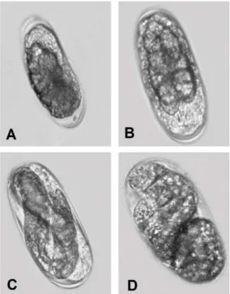
Figure 2. Embryogenesis of Meloidogyne enterolobii. A. Three celled stage. B. Four celled stage. C. Four celled stage and a possible polar body (arrow). D. Five celled stage. E. Six celled stage. F. Multiple celled stage. Photos/credit: R.M. Moura.
Anais da Academia Pernambucana de Ciência Agronômica, v.17, n.1, p. 99-104, 2020.
Figure 3. Embryogenesis of Meloidogyne enterolobii. A. Early blastula stages. B. Blastula stage (soccer-ball form); C-Tadpole stage, with large cells in the central region (endoderm) sorronded by smaller peripherical cells that cosnstitute the ectoderm. D. Begning of the elongation stage. Photos/credit: R. M. Moura.
Figure 4. Embryogenesis of Meloidogyne enterolobii. A-B. Early elongation stages. C-D. Late elongation stages with the embryo showing active Brownian movement. Photos/credit: R. M. Moura.
Anais da Academia Pernambucana de Ciência Agronômica, v.17, n.1, p. 99-104, 2020.
Figure 5. Meloidogyne enterolobii. A. First stage juvenile (J1). B. Secound stage juvenile (J2) (one extra cuticle may be seen). C. Hatching of a J2. D. Anterior end of a heatched J2. E. Posterior end of a hatched the J2. Photos/credit: R. M. Moura.
REFERENCES
BIRD, A. The influence of the temperature on embryogenesis of Meloidogyne javanica. Journal of Nematology, 4: 206-213. 1972.
CHITWOOD, B.G. Nemic Embryology. In. B. G. Chitwood; M.B. Chitwood. Introduction to Nematology. Baltimore, University Park Press, 1950. p. 202-212.
FASSULIOTIS, G. Feeding, egg-laying and embryology of the Columbia lance nematode, Hoplolaimus columbus. Journal of Nematology, 7: 152-158. 1975.
HUNG, C-L.; JENKINS, W. R. Oogenesis and embryology of two plant parasitic nematodes, Pratylenchus penetrans and P. zeae. Journal of Nematology, 1: 352-356. 1969.
INSERRA, R.N.; VOVLAS, N. The biology of Rotylenchulus macrodoratus. Journal of Nematology, 12: 97-102. 1980.
ORION, D.; WERGIN. W. P.; CHITWOOD, D. J.; ERBE, E. F. Low temperature scanning electron microscope observations of the Meloidogyne incognita egg mass: the gelatinous matrix and embryo development. Journal of Nematology, 26: 402-411. 1994.
WANG, L. H. Embryology and life cycle of Tylenchorhynchus claytoni Steiner, 1937 (Nematoda: Tylenchoidea). Journal of Nematology, 3: 101-107. 1971.