Photodegradation Mechanisms on Poly(ε-caprolactone) (PCL)
Danyelle Campos Françaa, Dayanne Diniz Moraisa, Elieber Barros Bezerraa ,
Edcleide Maria Araújoa, Renate Maria Ramos Wellenb*
Received: September 20, 2017; Revised: February 26, 2018; Accepted: June 05, 2018
Photodegradation of PCL was performed exposing injected specimens to ultraviolet radiation
(UV-B) up to 9 weeks. Photodegradated PCL specimens were tested by Differential Scanning Calorimetry
(DSC), Infrared Spectroscopy (FTIR), mechanical properties, Optical Microscopy (MO) and Scanning Electron Microscopy (SEM).Upon exposure decrease in elongation at break and increase of elastic
modulus were verified, higher degree of crystallinity was observed for longer exposure what can be
due to the chemi-crystallization mechanism. From IR spectra new carbonyl group (C=O) peaks were
observed. Presence of micro cracks on specimens’ surface was identified by MO. From morphological
analyses is suggested that the photodegradation in PCL takes place by bulk erosion mechanism.
Keywords: PCL, Phododegradation, Chemi-crystallization, Bulk erosion.
*e-mail: [email protected]
1. Introduction
Synthetic plastics have been used for various purposes, especially in the packaging industrial sector; however, the majority of these materials presently cause serious problems with the waste management 1-4. Focusing on potential solutions
for this problem, polymer researchers have paid attention to biodegradable polymers, from petroleum and renewable sources, since they can be biologically degraded and therefore being considered as environmental friendly materials 5-12.
Among the biodegradable polymers, a special attention
has been given by the scientific community to
poly(ε-caprolactone) (PCL), that is a linear aliphatic semicrystalline polyester, synthesized by ring-opening polymerization of the cyclic lactone in presence of a catalyst. PCL has high ductility due to its chemical structure and low glass transition temperature of -60 ºC, it is thermally stable until 320 ºC, as a thermoplastic it is easily processed and recycled by conventional polymer processing, i.e., extrusion and injection
12-15. PCL has been successful used in a wide range of industrial
applications, including packing, food containers, agricultural
films, biomedical applications, devices for automobile and
electronic industries, and so on 16-18.
Nevertheless, to the best of our knowledge few works are dealt about the photodegradation of PCL. Tsuji et al.
19 suggested that photodegradation of PCL films proceeds
via a bulk erosion mechanism with serious damages to
the elongation at break, they observed that the effects are
deeper in the amorphous phases; and the UV irradiation
significantly increased the degree of crystallinity of PCL
due to thickening of crystallites. Photodegradation of PCL was delayed upon blending with PVC as observed by Christensen et al. (2008) 20.Bei et al. (2000) 21 verified in
poly(ε-caprolactone)-poly(ethylene glycol) block copolymer
(PCE) that the pure poly(ethylene glycol) (PEO) cannot be photodegraded alone, but the carbonyl groups in PCL segment catalyze the breakage of PEO chains by a series of UV initiated free radical reactions.
In this work inject specimens of PCL were photodegraded by exposure to UV-B radiation, and their morphology, thermal and mechanical properties properly analyzed by optical microscopy (OM), scanning electron microscopy
(SEM), differential scanning calorimetry (DSC), infrared
spectroscopy (FTIR) and mechanical properties. According to the DSC results further presented the chemi-crstallization process is suggested taking place and being the responsible for increasing the degree of crystallinity, and the photodegradation in PCL was evidenced by SEM images as occurring by bulk erosion mechanism.
2.Experimental
2.1 Materials
Poly (ɛ-caprolactone) (PCL) CAPA 6500, melting flow
rate (MFR) = 28 g/10 min (190 °C/2.16 kg), was purchased from Perstorp Winning Formulas.
aDepartamento de Engenharia de Materiais, Universidade Federal de Campina Grande, 58429-140,
Campina Grande, PB, Brasil
bDepartamento de Engenharia de Materiais, Universidade Federal da Paraíba, 58051-900, João
2.2 Processing
Neat PCL was processed in a twin screw extruder
Coperion-Werner&Pfleiderer ZSK 18. The temperature
inside the extruder was kept between 80 °C and 100 °C and the screw speed was set up at 250 rpm. After extrusion, the material was granulated and oven dried for 24 hours at 40 ºC. Injected specimens for mechanical experiment were produced in Arburg Allrounder 270C machine at 80-100 ºC, according to ASTM D638 standard.
2.3 Radiation exposure
The radiation tests were performed using a Comexim (type C-UV) appropriate for non-metallic materials, operating
with 40 W F40 UV-B fluorescent lamps with peak radiation
at 313 nm. The samples were exposed to continuous radiation for periods up to 9 weeks, forwarding characterization experiments were performed.
3. Characterization
3.1 Differential scanning calorimetry (DSC)
DSC analyses carried out in a DSC-Q20 TA Instruments, samples weighting approximately 3 mg were heated from 25 ºC to 250 ºC, followed by cooling to 10 °C and reheating to 250 °C. The heating/cooling rate was 10 ºC/min, under
nitrogen flow of 50 mL/min.
3.2 Mechanical tests
Mechanical properties in tension were measured according to ASTM D638; tests were conducted in an EMIC DL 2000 testing machine operating at 50 mm/min of elongation rate and 2000 kgf cell load. Tests were conduct at ambient temperature (~ 23 ºC) and presented results are an average of seven tests.
3.3 Fourier transformed infrared spectroscopy
(FTIR)
FTIR analyses were performed in a Perkin Elmer Spectrometer Spectrum 400 FT Mid-IR, Attenuated Total
Reflectance (ATR) mode was applied and scans ranging
from 4000 to 650 cm-1 were computed.
3.4 Polarized optical microscopy (OM)
Photographs of PCL before and after exposure were
recorded on a Carl Zeiss Polarized Optical Microscope
equipped with a digital camera, AxioCAM MRc.
3.5 Scanning electron microscopy (SEM)
Scanning electron microscopy images were acquired
in a VEGAN3 TESCAN machine with 30 KV. Fractured
surface from mechanical test was covered with gold to avoid accumulation of charges.
4. Results and Discussion
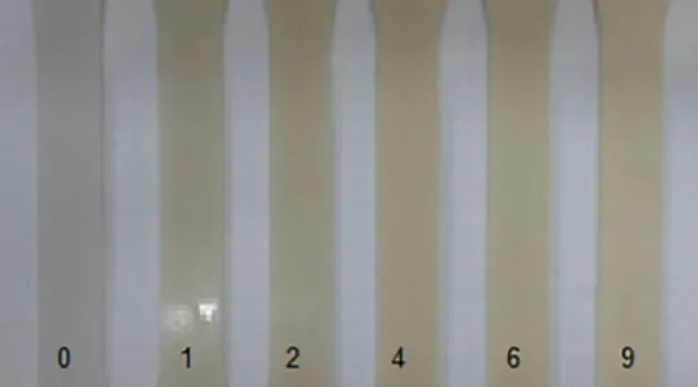
Photographs of PCL specimens unexposed and exposed to UV-B radiation up to 9 weeks are illustrated in Figure 1. After exposure, there is light-yellowing on exposed specimen surface and micro cracks developed with exposition to radiation (Figure 1 from 1 week). According to the literature 15
the yellowing is evidence that photodegradation is taking place, its origin may be accessed to radiation absorption by chromophoric groups and formation of free radicals that absorb oxygen and start chemical and physical changes like chain scission, damaging the mechanical properties.
The texture of the specimen exposed surface becomes rougher and crumbles for longer exposure times. The presence of cracks and roughness in samples exposed to radiation has already been reported for other polymers like poly(3-hydroxybutyrate) (PHB), polyvinyl chloride (PVC), polystyrene (PS), polypropylene (PP) 1,13,14,22,23.
Wellen et al.24 studied the photodegradation and
photostabilization of two commercial grades of PHB. The authors obtained the specimens by injection molding and the specimens were exposed to ultraviolet radiation (UV-A) for up to 12 weeks and then characterized by tensile testing, surface appearance, size exclusion chromatography (SEC) and scanning electron microscopy (SEM). According to the authors the exposure to UV-B radiation caused great damage on the surface color, reduction of molecular size and mechanical properties. However, it is important to note that for PHB changes in color can be not caused by chromophoric groups but by cracks on the specimen surface that increase
its roughness and the diffuse reflectance of visible light. Regarding the superficial changes originated upon
exposition to radiation, according to Rabello and White22 an
important process called chemi-crystallization is observed in some semi-crystalline polymers, for instance in PP. In chemi-crystallization the chain scission occurs due to the degradation mechanism but these broken molecules reorganize in a crystalline phase, provided that they have enough mobility. The main characteristic of this process is the spontaneous crack formation on external surfaces by the contraction of the specimen layers, these cracks contribute to the embrittlement of ductile semi-crystalline
polymers promoting serious deterioration on polymer after short exposure.
Figure 2 shows DSC scans for melt crystallization and melting of PCL unexposed and exposed up to 9 weeks, and Tables 1 and 2 present thermal parameters computed from these scans. It is observed that the crystallization peak shifted
lightly to higher temperatures, the degree of crystallinity (∆Xc) decreased upon exposure. An interesting fact observed for 9 weeks of exposure is that the crystallinity was higher than for shorter intervals of exposure, somehow PCL molecular chains rearranged and crystallized, it may be seen as an
effect of chemi-crystallization.
The melting peak temperature did not show significant changes, the degree of crystallinity (∆Xm) measured from
melting enthalpy presented a similar trend as that verified
for melt crystallization, the degree of crystallinity decreased
Figure 2. DSC scans for Melt Crystallization (left) and Melting (right), acquired during cooling and re-heating cycles, respectively
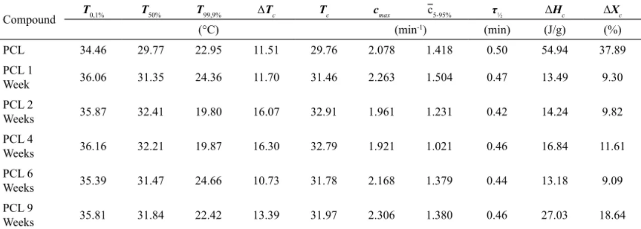
Table 1. Melt crystallization parameters computed from DSC scans during cooling for PCL exposed to UV-B radiation up to 9 weeks.
Compound T0,1% T50% T99,9% ΔTc Tc cmax c5-95% τ½ ΔHc ΔXc
(°C) (min-1) (min) (J/g) (%)
PCL 34.46 29.77 22.95 11.51 29.76 2.078 1.418 0.50 54.94 37.89
PCL 1
Week 36.06 31.35 24.36 11.70 31.46 2.263 1.504 0.47 13.49 9.30
PCL 2
Weeks 35.87 32.41 19.80 16.07 32.91 1.961 1.231 0.42 14.24 9.82
PCL 4
Weeks 36.16 32.21 19.87 16.30 32.79 1.921 1.021 0.46 16.84 11.61
PCL 6
Weeks 35.39 31.47 24.66 10.73 31.78 2.168 1.379 0.44 13.18 9.09
PCL 9
Weeks 35.81 31.84 22.42 13.39 31.97 2.306 1.380 0.46 27.03 18.64
T0.1% (°C): temperature for 0.1% molten/crystallized fraction. T50% (°C): temperature for 50% molten/crystallized fraction. T99.9% (°C): temperature for 99.9% molten/crystallized fraction. Tp, Tc (°C): peak melting/crystallization temperatures. cmax (min
-1): maximum melting/ crystallization rate. τ½ (min): half melting/crystallization time (time to reach 50% molten/crystallized fraction from the start of the event). ΔHm, ΔHc: (J/g): latent heat of melting/crystallization. ΔXm (%), ΔXc (%): change in crystallinity during the event (the literature lists two slightly different reference values for the latent heat of fusion of PCL. Mandelkern27,28 reports the value 142.4J/g in two of his textbooks, another textbook by Van Krevelen and Nijenhuis29 quotes the value 148.1J/g. In the present work we decided to use the value 145J/g, an approximated mean value, as the melting enthalpy of 100% crystalline PCL).
upon exposure and then it increased for longer exposure, i.e.,
9 weeks, it also may be an effect of chemi-crystallization.
Polymer studiers have reported decrease in crystallinity
upon degradation for other resins like PLA, PET and PEEK
25,26. Santonja-Blasco et al.25 investigated the effects of three
types of degradation, thermo-degradation, photodegradation and biodegradation in PLA. At high temperature i.e., thermal degradation at 220 °C, random chain scission was faster followed by photodegradation and biodegradation. Photodegraded specimens presented around the half of molar mass compared to soil degraded ones, i.e. biodegraded. Chain defects such as the anhydride groups formed during photodegradation decreased the crystallization rate, but the extreme damages as higher chain scission and lower
crystallinity were verified for thermal-degradation.
Vaughan and Stevens26 analyzed the effect of gamma
in amorphous state as well as after annealing. The gathered
results were similar to those obtained for PEEK, i.e. as the
radiation doses increased, the melt crystallization peak shifted to higher temperatures and the degree of crystallinity decreased.
Figure 3 shows results for elastic modulus (a), elongation at break (b), maximum stress (c) and tensile strength (d) for PCL before and after exposure to UV-B radiation.
Initially, PCL behaves as a ductile polymer, with low elastic modulus and high elongation at break (>418%). After
exposure, its elastic modulus increased by 48% and remained almost unchangeable over the exposure time.
For elongation at break, a decrease of 80% was observed
after the first week of exposure, in two weeks it decreased
94% in relation to unexposed PCL and 71% to PCL exposed
for 1 week. Further exposure did not provide significant
changes, specimens presented similar mechanical properties. These events, increase in elastic modulus and higher decrease in elongation at break that occurred after exposure
to radiation, modified PCL properties, making it a rigid and
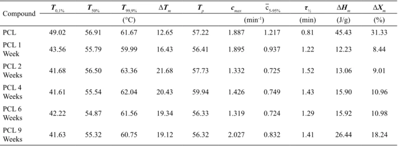
Table 2. Melting parameters computed from DSC scans during the second heating for PCL up to 9 weeks.
Compound T0,1% T50% T99,9% ΔTm Tp cmax c5-95% τ½ ΔHm ΔXm
(°C) (min-1) (min) (J/g) (%)
PCL 49.02 56.91 61.67 12.65 57.22 1.887 1.217 0.81 45.43 31.33
PCL 1
Week 43.56 55.79 59.99 16.43 56.41 1.895 0.937 1.22 12.23 8.44
PCL 2
Weeks 41.68 56.50 63.36 21.68 57.73 1.332 0.725 1.52 13.06 9.01
PCL 4
Weeks 41.61 55.54 62.04 20.43 59.94 1.426 0.749 1.43 15.90 10.96
PCL 6
Weeks 42.22 54.87 61.56 19.34 56.33 1.319 0.724 1.29 15.92 10.98
PCL 9
Weeks 41.63 55.32 60.75 19.12 56.32 2.027 0.832 1.41 26.44 18.24
brittle polymer, micro cracks, crazy development as well as chemi-crystallization are processes possibly linked to these changes 8.
Figure 4 shows FT-IR spectra of PCL unexposed and exposed. According to the literature 30,31, PCL presents infrared
absorption bands which are relevant for polymer characterization and its changes. The spectra in Figure 4 presented in 2944 cm-1 and 2861 cm-1 referring bands to asymmetric and
symmetrical stretching of CH2 groups, respectively. The most intense peak at 1725 cm-1 is characteristic of carbonyl group
C=O, the peak at 1300 cm-1 to the C-O and C-C groups of
Figure 4. (a) FT-IR spectra of PCL unexposed and exposed to UV-B radiation and (b) zoom of Figure 5 (a) illustrating absorption wavenumber of carbonyl group
main chain, at 1243 cm-1 a symmetric elongation of C-O-C
and at 1168 cm-1 a symmetrical elongation of C-O-C groups.
After exposure, the relative peak of carbonyl group (C=O), visualized at 1725 cm-1 broadened what may have
been caused by formation of new radicals (C=O) localized
in different regions in PCL macromolecules, for instance
in terminal groups, this change is suggested as degradation evidence due exposure to UV-B radiation.
Optical microscopy images are presented in Figure 5, it is observed that before exposure PCL had a dense and non-porous surface, Figure 5 (Unexposed). After exposure,
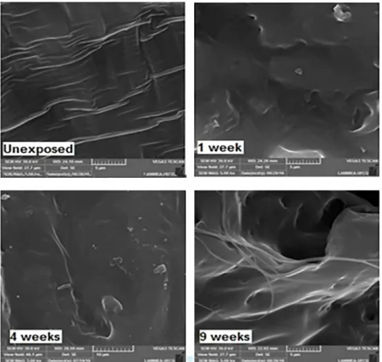
Figure 6. SEM micrographs of PCL fracture surface exposed to UV-B radiation. Exposure times up to 9 weeks Figure 5 for 1 week, presence of micro cracks on PCL surface
is verifiable and they increase with exposure. It can be clearly
seen in the micrographs (Figure 5 for 2 weeks exposure) presence of crystals inside and near of the cracks 32.
Regarding the cracks and roughness, the researcher groups Grigull et al., 2015 32 and Narkis et al., 1985 33, agree that
these, probably, can be originated from chemi-crystallization as well as from degradation mechanism Norrish II. It is plausible assuming the observed decreasing in mechanical properties may be accelerated by these processes.
Figure 6 shows SEM images for PCL specimens upon UV-B radiation. Unexposed PCL presents ductile fracture with evidences of plastic deformation. After exposure the
fracture surface changes and a fibrous morphology is observed.
This behavior has been reported for PCL by Tsuji et al. 2006
19, Castilla-Cortazár et al. 2012 34 and França et al. 2016 17
who related this morphological change due to degradation mechanisms called bulk erosion that is when the degradation occurs throughout the whole material, for this case, both the surface and the bulk of the material degrade. These authors
observed initially surface erosion followed by the fibrous
morphology, being both linked to PCL degradation, similarly
to what was verified in the present work in Figure 6.
5. Conclusions
Photodegradation of PCL was analyzed by exposing injected specimens to UV-B radiation up to 9 weeks. From the gathered results was possible assuming that PCL is
strongly affected by radiation, its mechanical properties
such as elastic modulus and elongation at break changed considerably. FTIR spectra presented alterations related to the carbonyl groups; formation of micro cracks could be observed by optical microscopy. The degree of crystallinity increased for longer exposure time, as probable consequence
of chemi-crystallization. In SEM images erosion and fibrous morphologies were verified what can be linked to loses in
6. Acknowledgments
The authors are grateful to CNPq and Capes (Brasilia/
DF, Brazil) for the financial support and the Fraunhofer
Institute for Manufacturing Technology and Advanced Materials-IFAM in Bremen for experiments with Polarized Optical Microscopy (MO).
7. References
1. Bastioli C, ed. Handbook of Biodegradable Polymers. Shawbury: Rapra Technology; 2005.
2. Mohanty AK, Misra M, Drzal LT, eds. Natural Fibers, Biopolymers, and Biocomposites. Singapore: Taylor & Francis; 2005.
3. Biron M. The Plastics Industry: Economic Overwiew. In: Biron M. Thermosets and Composites - Material Selection, Applications, Manufacturing and Cost Analysis. 2nd ed. Amsterdam: William Andrew; 2014. p. 25-104.
4. Emblem A. Plastics Properties for Packing Materials. In: Emblem A, ed. Packing Technology - Fundamentals, Materials and Processes. Oxford: Woodhead Publishing; 2012. p. 287-309.
5. Van Hung N, De Schryver P, Tam TT, Garcia-Gonzalez L, Bossier P, Nevejan N. Application of poly-ß-hydroxybutyrate (PHB) in mussel larviculture. Aquaculture. 2015;446:318-324.
6. Li X, Qi C, Han L, Chu C, Bai J, Guo C, et al. Influence of dynamic compressive loading on the in vitro degradation behavior of pure PLA and Mg/PLA composite. Acta Biomaterialia. 2017;64:269-278.
7. Sungkapreecha C, Iqbal N, Gohn AM, Focke WW, Androsch R. Phase behavior of the polymer/drug system PLA/DEET. Polymer. 2017;126:116-125.
8. Laycock B, Nikolic M, Colwell JM, Gauthier E, Halley P, Bottle S, et al. Lifetime prediction of biodegradable polymers.
Progress in Polymer Science. 2017;71:144-189.
9. Wachirahuttapong S, Thongpin C, Sombatsompop N. Effect of PCL and Compatibility Contents on the Morphology, Crystallization and Mechanical Properties of PLA/PCL Blends. Energy Procedia. 2016;89:198-206.
10. Hsu ST, Tan H, Yao YL. Effect of excimer laser irradiation on crystallinity and chemical bonding of biodegradable polymer.
Polymer Degradation and Stability. 2012;97(1):88-97.
11. Badia JD, Gil-Castell O, Ribes-Greus A. Long-term properties and end-of-life of polymers from renewable resources. Polymer
Degradation and Stability. 2017;137:35-57.
12. Wang Y, Pan J, Han X, Sinka C, Ding L. A phenomenological model for the degradation of biodegradable polymers. Biomaterials. 2008;29(23):3393-3401.
13. Abdel-Rehim HA, Yoshii F, Kume T. Modification of polycaprolactone in the presence of polyfunctional monomers by irradiation and its biodegradability. Polymer Degradation
and Stability. 2004;85(1):689-695.
14. Zhao Q, Tao J, Yam RCM, Mok ACK, Li RKY, Song C. Biodegradation behavior of polycaprolactone/rice husk ecocomposites in simulated soil medium. Polymer Degradation
and Stability. 2008;93(8):1571-1576.
15. . De Paoli MA. Degradação e Estabilização de Polímeros. 2ª versão on-line. Chemkeys; 2008. Available from: < http://www. chemkeys.com/blog/wp-content/uploads/2008/09/polimeros. pdf>. Access in: 30/1/2018.
16. Copinet A, Bertrand C, Govindin S, Coma V, Couturier Y. Effects of ultraviolet light (315 nm), temperature and relative humidity on the degradation of polylactic acid plastic films. Chemosphere. 2004;55(5):763-773.
17. França DC, Bezerra EB, Morais DDS, Araújo EM, Wellen RMR. Hydrolytic and Thermal Degradation of PCL and PCL/ Bentonite Compounds. Materials Research. 2016;19(3):618-627.
18. Bhullar SK, Rana D, Lekesiz H, Bedeloglu AC, Ko J, Cho Y, et al. Design and fabrication of auxetic PCL nanofiber membranes for biomedical applications. Materials Science and Engineering: C. 2017;81:334-340.
19. Tsuji H, Echizen Y, Nishimura Y. Photodegradation of biodegradable polyesters: A comprehensive study on poly(l-lactide) and poly(ɛ-caprolactone). Polymer Degradation and
Stability. 2006;91(5):1128-1137.
20. Christensen PA, Egerton TA, Franchetti SM, Jin C, White JR. Photodegradation of polycaprolactone/poly(vinyl chloride) blend. Polymer Degradation and Stability. 2008;93(1):305-309.
21. Bei J, He W, Hu X, Wang S. Photodegradation behavior and mechanism of block copoly(caprolactone-ethylene glycol).
Polymer Degradation and Stability. 2000;67(2):375-380.
22. Rabello MS, White JR. The role of physical structure and morphology in the photodegradation behaviour of polypropylene.
Polymer Degradation Stability. 1997;56(1):55-73.
23. Rabello MS, White JR. Crystallization and melting behaviour of photodegraded polypropylene - II. Re-crystallization of degraded molecules. Polymer. 1997;38(26):6379-6387.
24. Wellen RMR, Canedo EL, Rabello MS, Fechine GJM. Photodegradation and Photostabilization of Poly(3-Hydroxybutyrate). Materials Research. 2016;19(4):759-764.
25. Santonja-Blasco L, Ribes-Greus A, Alamo RG. Comparative thermal, biological and photodegradation kinetics of polylactide and effect on crystallization rates. Polymer Degradation and
Stability. 2013;98(3):771-784.
26. Vaughan AS, Stevens GC. On radiation effects in poly(ethylene terephthalate): a comparison with poly(ether ether ketone). Polymer. 1995;36(8):1541-1547.
27. Mandelkern L. Crystallization of Polymers. Volume 1: Equilibrium Concepts. 2nd ed. Cambridge: Cambridge University Press; 2002. p. 249.
28. Mandelkern L, Alamo RG. Thermodynamic Quantities Governing Melting. In: Mark JE, ed. Physical Properties of Polymers Handbook. 2nd ed. New York: Springer; 2007. p. 165-186.
29. Van Krevelen DW, Te Nijenhuis K. Properties of Polymers. 4th ed. Amsterdam: Elsevier; 2009. p. 121.
30. Elzein T, Nasser-Eddine M, Delaite C, Bistac S, Dumas P. FTIR study of polycaprolactone chain organization at interfaces.
31. Lin-Vien D, Colthup NB, Fateley WG, Grassselli J. The
Handbook of Infrared and Raman Characteristic Frequencies of Organic Molecules. London: Academic Press; 1991.
32. Grigull VH, Mazur LP, Garcia MCF, Schneider ALS, Pezzin APT. Estudo da degradação de blendas de poli(hidroxibutirato-co-hidroxivalerato)/poli(l-ácido lático) em diferentes condições ambientais. Engevista. 2015;17(4):444-462.
33. Narkis M, Sibony-Chaouat S, Siegmann A, Shkolnikt S, Bell JP. Irradiation effects on polycaprolactone. Polymer. 1985;26(1):50-54.
34. Castilla-Cortázar I, Más-Estellés J, Meseguer-Dueñas JM, Escobar Ivirico JL, Marí B, Vidaurre A. Hydrolytic and enzymatic degradation of a poly(e-caprolactone) network.