Fisioter. Mov., Curitiba, v. 31, e003122, 2018 DOI: http://dx.doi.org/10.1590/1980-5918.031.AO22 Licensed under a Creative Commons attribution
Using the spirometry to indicate respiratory
exercises for elderly with Parkinson’s disease
O uso da espirometria na indicação de exercícios
respiratórios para idosos com Doença de Parkinson
Daniela Guimarães, Gabriel Duarte, Karen Trippo, Graziella Furtado, Jamary Oliveira Filho, Daniel Dominguez Ferraz*
Universidade Federal da Bahia (UFBA), Salvador, BA, Brazil
[R]
Abstract
Introduction: Respiratory dysfunction is the main cause of death in Parkinson's disease (PD) patients
and bronchoaspiration pneumonia is the most common clinical respiratory complication. Objective: To
assess respiratory function of elderly with PD in mild to moderate phase of the disease. Methods: A cross-sectional study was carried. Elderly in 2 to 3 PD Hoehn & Yahr stage have participated. A single researcher has evaluated maximal inspiratory pressure (MIP), maximal expiratory pressure (MEP), thoracoabdominal
amplitude, forced vital capacity (FVC) and expiratory volume in the first second (FEV1). Results: Sixty
elderly have participated and their all spirometry and manovacuometry parameters presented significant
differences (p < 0.05) comparing with predicted values, except for FVC (p = 0.25). Only umbilical level did
not reach normal values on cirtometry parameters. Patients classified as a restrictive disorder presented significant decrease in thoracic expandability. However the participants classified as an obstructive disorder showed significant decrease in expiratory muscle strength and peak expiratory flows. Conclusion: Elderly in mild or moderate phase of PD presented reduction in respiratory parameters. Spirometry showed to be
* DG: BS, e-mail: [email protected]
GD: BS, e-mail: [email protected]
2
an important tool to evaluate respiratory function and to indicate the modality of respiratory exercise. Our
results suggest the indication of thoracic flexibility exercises for patient with PD classified as restrictive disorder and strength exercise of respiratory muscles for those classified as obstructive disorder.
Keywords: Aged. Parkinson Disease. Respiratory Function Tests. Exercise.
Resumo
Introdução: Disfunções respiratórias são as principais causas de morte em pacientes com Doença de Parkinson (DP) e a pneumonia por bronco-aspiração é a complicação respiratória mais comum. Objetivo: Avaliar a função respiratória de idosos com doença de Parkinson (DP) em fase leve a moderada da doença. Métodos: Foi realizado um estudo transversal. Participaram idosos nos estágios 2 a 3 da classificação de Hoehn & Yahr. Um único pesquisador avaliou a pressão inspiratória máxima (PIM), pressão expiratória máxima (PEM), amplitude toracoabdominal, capacidade vital forçada (CVF) e volume expiratório forçado no primeiro segundo (VEF1). Resultados: Sessenta idosos participaram e todos os seus parâmetros de espirometria e manovacuometria apresentaram diferenças significativas (p < 0,05) em comparação com os valores previstos, exceto a CVF (p = 0,25). Somente o nível umbilical não atingiu valores normais nos parâmetros da cirtometria. Pacientes classificados como distúrbios restritivos apresentaram diminuição significativa na capacidade de expansão torácica. No entanto, os participantes classificados como transtornos obstrutivos mostraram diminuição significativa na força muscular expiratória e dos fluxos expiratórios máximos. Conclusão: Os idosos em fase leve ou moderada da DP apresentaram redução nos parâmetros respiratórios. A espirometria mostrou ser uma ferramenta importante para avaliar a função respiratória e para indicar a modalidade do exercício respiratório. Nossos resultados sugerem a indicação de exercícios de flexibilidade torácica para pacientes com DP classificados como distúrbios restritivos e exercícios de força da musculatura respiratória para aqueles classificados como desordem obstrutiva.
Palavras-Chave: Idoso. Doença de Parkinson. Testes de Função Respiratória. Exercício.
Introduction
Parkinson’s disease (PD) is a degenerative and chronic disease of the central nervous system, which results in death of substantia nigra dopaminergic neurons and causes a decrease in dopamine of
nigrostriatal pathway [1, 2]. It mainly affects the
population above 65 years old and it is estimated
a prevalence of 100 to 200 cases per 100,000 inhabitants [3], approximately 1 to 2% of the world
population [4].
PD is among the major diseases affecting the
elderly [5] affecting 1 in 100 people over 75 years and 1 in every 1000 people over 65 years [6]. Due
to the increase in life expectancy around the world, it is estimated that more than 40 million people will
have PD around 2020 [6, 7].
The main clinical signs of PD are bradykinesia, resting tremor, rigidity, postural changes and balance
deficit [1, 2, 8]. However, repercussions of PD affect
musculoskeletal system and others, including
respiratory system [8].
Trunk flexion posture, corporal changes caused
by aging, reduction of motion chest and decreased extension trunk movement may promote respiratory dysfunctions in this population [5]. Weakness of respiratory muscle strength, especially expiratory muscles which decrease proportionally during disease evolution, also participates in respiratory
dysfunctions process [8]. So, elderly with PD
presents low pulmonary volumes, decreased thoracic expandability and cough effectiveness
deficit [5].
3
initial respiratory adaptations or problems in
respiratory function [8 - 10].
Respiratory dysfunction is the main cause of death in PD patients and bronchoaspiration pneumonia is the most common clinical respiratory complication [5, 9]. Although respiratory changes are the main cause of mortality in individuals with PD, there are still few studies that aim to evaluate respiratory function and thus guide the composition of physiotherapeutic prevention and treatment
programs [10, 11].
However, it is not common to evaluate respiratory function and to treat respiratory dysfunctions of patients with PD. So, the objective of this study was to evaluate the respiratory function of elderly in the mild to moderate phase of PD.
Methods
We performed an observational, quantitative and cross-sectional research. The study was carried out in accordance with the Declaration of Helsinki and approved by an Ethics Human Research Committee
(ref no. 1.016.971). The study was conducted at the Centro de Referência Estadual de Atenção à Saúde do Idoso (CREASI) (State Reference Health Care
Centre for Elderly), a public reference outpatient clinic for the elderly in Salvador, Bahia, Brazil. Data
were collected in the period between June/2015 and August/2016.
Elderly (≥ 60 years-old) with idiopathic PD according to the London Brain Bank Criteria [12]
participated in the study. All participants complied with the inclusion and exclusion criteria. The inclusion criteria were: age 60 years or older, regular use of PD medication, 2, 2,5 or 3 points according to the Hoehn & Yahr classification, independence
for basic activities (Barthel ≥ 90 points) and instrumental activities (Pfeffer ≤ 5 points) of
daily life, no cognitive deficit (Mini Mental State
Examination ≥ 24 points) and gait capacity
without assistance. The exclusion criteria were: neurodegenerative diseases excluding PD, dementias, limiting osteomyelitis, chronic diseases (hypertension, diabetes mellitus, chronic pain), unstable cardiovascular diseases (acute and chronic heart failure, recent myocardial infarction, unstable angina and uncontrolled arrhythmias), use of alcohol and other toxic substances.
Epidemiological and clinical variables were collected to characterize the sample. It was used
the Unified Parkinson's Disease Rating Scale (UPDRS) [13] and the modified Hoehn & Yahr classification [14] to assess severity and disease
progression and general health condition. The elderly were submitted to respiratory function evaluation through spirometry and manovacuometry, according to the Guidelines for Pulmonary Function
Testing [15, 16], and cirtometry, both performed by
a single physiotherapist trained.
The functional parameters evaluated were forced vital capacity (FVC), forced expiratory volume in
the first second (FEV1), forced expiratory volume ratio in the first second and forced vital capacity also called Tiffeneau index (FEV1/FVC) and expiratory flow peak (EFP).
The manovacuometry was performed through an analogue manovacuometer. The functional parameters were maximal inspiratory pressure (MIP) and maximum expiratory pressure (MEP).
The cirtometry was performed using a common tape measure staggered in centimeters at the level of the axillary regions, just below the axillary fold; xiphoid, having as reference point the lower border of the xiphoid appendix; and umbilical, with the
tape on the umbilical scar [17, 18]. The evaluator
monitored respiratory movements with the tape, and it was considered the greatest difference measure between the inspiration and expiration of each level,
called the amplitude coefficient (AC).
The frequency distribution format of the quantitative variables was evaluated using histograms and the Kolmogorov-Smirnov test. The quantitative variables were summarized in mean and standard deviation, and the categorical variables in absolute and relative frequencies. In statistical inference,
Pearson correlation coefficient was used to calculate
the linear correlations, ANOVA test with Bonferroni post-test was used to compare three independent variables and paired T-test was used to compare two dependent variables. We used the Statistical Package for the Social Sciences (SPSS – version 22).
Results
Sixty elderly have participated in the study. Table
1 shows epidemiological and clinical characteristics
4
Table 1 - Epidemiological and clinical characteristics of elderly with PD attended at CREASI in Salvador/BA, 2016
Variable Mean ± SD
Age 69 ± 5
BMI 25.8 ± 3.9
HY 2.5 ± 0.4
UPDRS 29.3 ± 14.9
Note: BMI = Body Mass Index; HY = Hoehn and Yahr; UPDRS = Unified Parkinson's disease rating scale; SD = Standard Deviation.
Table 2 shows cirtometry results according to the AC reference value, at axillary, xiphoid and umbilical levels. The reference value adopted for normality was
4 to 7 cm [5, 19]. The participants presented reduced
values only in umbilical level.
Table 2 - Cirtometry values of elderly with PD attended at CREASI in Salvador/BA, 2016
Variable Mean ± SD
AC
Axillary 6.03 ± 2.06
Xifoidian 5.10 ± 2.48
Umbilical 1.70 ± 3.52
Note: SD = Standard Deviation; AC = Amplitude Coefficient.
A significant difference (p = 0.022) was observed
in axillary level in relation to sex. Males presented a better result (6.52 ± 2.26).
Table 3 shows the spirometry and manovacuometry values performed and predicted, in relation to sex, weight, age and height. All variables presented
significant (p < 0.05) reduced values comparing to
predicted ones, except FVC.
Table 3 - Spirometry and manovacuometry values of
elderly patients with PD attended at CREASI in Salvador/BA, 2016
Variable Mean ± SD
Expected Performed % p
FVC 3.42 ±
0.76
3.61 ± 1.51
105.71 ±
37.12 0.248
FEV1 2.65 ± 0.57 2.15 ± 0.71 82.10 ± 23.13 < 0.001
FEV1/FVC 77.79 ± 2.00 63.25 ± 18.60 81.31 ± 23.78 < 0.001
EFP 8.55 ±
1.94
3.07 ± 1.62
37.13 ±
19.68 < 0.001
MIP -90.44 ±
13.69
-62.75 ± 23.50
67.73 ±
24.05 < 0.001
MEP 96.10 ± 17.96 64.08 ± 23.86 67.41 ± 23.91 < 0.001
Note: SD = standard deviation, FVC = Forced Vital Capacity, FEV1= Forced Expiratory Volume in the first second, EFP = Expiratory Flow Peak, MIP = Maximum Inspiratory Pressure, MEP = Maximum Expiratory Pressure.
A significant difference (p = 0.021) was found between FEV1 percentage in elderly with scores 2 and 3 of Hoehn & Yahr classification. No difference
was found between these stages and patients in stage
2.5 of Hoehn & Yahr classification.
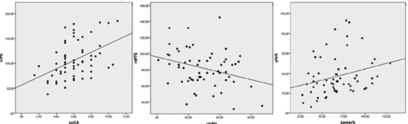
A regular and significant (p ˂ 0.001) correlation
between FVC and AC at the axillary level, and between
FEV1 and UPDRS (ρ = 0.303, p = 0.018) can be observed in figure 1. A significant weak correlation (ρ = 0.269, p = 0.038) was found between EFP
and MEP.
Note: % = Percentage of Forced Vital Capacity; axiAC = Amplitude Coefficient at the Axillary Level; % 1 = Percentage of Forced Expiratory Volume in the First Second; Revised = Unified Parkinson's Disease Rating Scale; efp% = Expiratory Flow Peak Percentage; Pemax% = Maximum Expiratory Pressure Percentage.
5
Other combinations were performed among the variables studied, but no correlation was found.
In relation to respiratory disorder obtained
through spirometry, 45% of the participants were
considered normal (Figure 2).
Note: Normal, n = 27 (45%); Restrictive Disorder, n = 8 (13.3%); Obstructive Disorder, n = 25 (41.7%).
Figure 2 - Proportion of respiratory disturbance
classification by spirometry in elderly with PD attended at CREASI in Salvador/BA, 2016.
A significant difference was found between axillary (p = 0.026) and xiphoid level (p = 0.001) among normal participants and elderly classified as a
restrictive disorder. However, this difference was not
observed between normal elderly and those classified as an obstructive disorder (p = 1.00) at both levels.
There was also a significant difference in EFP percentage (p < 0.001) and MEP (p = 0.023) among normal elderly and those classified as an obstructive
disorder. This difference was not found among normal
elderly and the participants classified as a restrictive disorder (EFP: p = 0.339; MEP: p = 0.718).
Discussion
The results of this study suggest that elderly with PD in the mild to moderate stages of the disease present a reduction in respiratory parameters. Elderly with
restrictive disorder present a significant decrease
in thoracic expansibility, while the participants
classified as an obstructive disorder present a significant reduction in expiratory muscle strength and maximal expiratory flow. Our results suggest that
it is important to treat respiratory function in elderly with PD in the mild to moderate stages with exercise. Spirometry evaluation may help exercise indication.
So, thoracic flexibility exercises is indicated for
patient with PD classified as restrictive disorder and
strength exercise of respiratory muscles is indicated
for those classified as obstructive disorder.
Thoracoabdominal mobility depends, among other factors, on respiratory muscles used [20]. The participants in our study presented normal chest expansion and reduced abdominal expansibility. This abdominal expansion during inspiration is characterized by diaphragmatic breathing [20], because the diaphragm is the main muscle used for this purpose. A diaphragm dysfunction may cause this decrease in abdominal mobility. In addition, elderly with PD present alterations in respiratory drive, respiratory muscle flexibility and respiratory activation and coordination [9], which may also explain the reduction of abdominal expansion in our participants. Therefore, diaphragmatic involvement seems to occur before worsening of chest rigidity, making this reduction of abdominal mobility more evident than thoracic expansibility in early stages of PD.
Differences in the patterns of thoracoabdominal mobility between sexes in PD are not described in the
literature. In our study men had significantly better
chest expansion compared to women. However, studies with healthy individuals have reported that there is no difference in thoracoabdominal movement
in relation to sex during resting breathing [21, 22].
Patients with PD seem to present a worse thoracic mobility comparing with controls of the same age.
Ramos et al. [5] have evaluated 10 individuals with 1.5 to 5 of modified Hoehn & Yahr scale, and they
also found a decrease in thoracic mobility of their
participants. Cardoso and Pereira [10] demonstrated that individuals with PD had significantly lower
respiratory measures than healthy controls with similar characteristics.
Trunk extension limitation and thorax amplitude reduction decrease pulmonary compliance, making pulmonary expansion harder and promoting decrease pulmonary volumes [23]. All pulmonary measurements performed in this study were
significantly lower than predicted in all variables,
except for FVC. Olowabi et al. [24] also demonstrated
that the values of FVC, FEV1, FEV1/FVC and PEF of elderly with PD in stages 1 to 5 on the Hoehn & Yahr scale were significantly lower than healthy controls.
Changes in lung volumes can be observed in a short
6
stages of Hoehn & Yahr scale. Elderly in moderate
phase presented a significant lower values of FEV1
comparing with participants in the milder phase of PD. Similar results were observed in Olowabi et al.´s study [24]. These authors found that pulmonary
function parameters were significantly lower in
individuals with higher Hoenh & Yahr scores. So, both results indicate that respiratory changes worsen with PD progression.
Chest expansion maybe influence the expiration volume of air mobilized, because a reduced airflow
will eventually restrict pulmonary ventilation. We
found a regular and significant correlation between
thoracic expansibility and FVC, and also between
FEV1 and disease progression. According to Cardoso and Pereira [10], a rigid chest may cause reduction
of thoracic mobility and decrease of pulmonary expansion, limiting pulmonary ventilation and reducing pulmonary volumes.
In our study, obstructive disorders were more common than restrictive ones. Similar results were observed in Olowabi et al. [24] study. However, in
PD restrictive lung status is more common [10]
and occurs mainly due to the stiffness of thoracic wall [24]. These changes contribute to the reduction of pulmonary expansibility that can be evaluated through the spirometry.
According to Trindade et al. [25], the restrictive
disorder is associated with a significant reduction
of vital capacity and a maintenance or increase of
FEV1/FVC. In our results, the participants performed
better values of FVC comparing with the predicted,
but worse values of FEV1/FVC comparing with
predicted ones. These results contributed to a major
reduction of FEV1 in relation to FVC, demonstrating
the narrowing of airways, which characterizes obstructive ventilatory disorder.
Our participants were in the early stages of PD and
do not have a significant rigidity capable of triggering
restriction pictures. It seems that obstructive disorder is an early event in PD [26]. Respiratory muscle
strength of our participants was significantly lower
than predicted ones. The study proposed by Guedes
et al. [27] corroborates with this finding. Their results confirmed that respiratory pressures in PD are lower
than predicted values. This reduction in respiratory muscles strength, especially expiratory muscles, may be associated with aging process, neurological impairments, costal arches stiffening, reducing of rib
cage movement and flexed posture [10, 28]. These
characteristics reduce muscle capacity to generate
forced flows, as well as overcome thoracic rigidity,
contributing to the reduction of pulmonary volumes and respiratory pressures, especially the maximum expiratory pressure, which will worsen with the
evolution of the disease [5, 27].
Strong expiratory muscles are essential to achieve an effective cough, because its contraction is the
responsible for generating high velocity airflow able
to remove undesirable airway materials [29]. Studies have reported that individuals with PD present a decrease in cough effectiveness due to the reduction of contraction force of abdominal musculature during
reflex and voluntary cough. Decrease in sensitivity also impairs the activation of an induced cough reflex.
So, respiratory system of these patients presents a real
difficulty to remove airways secretions, facilitating
aspiration during swallowing and consequently causing aspiration pneumonia [29, 30].
Our results suggest the correlation between strength of expiratory musculature and capacity
to generate maximum expiratory airflows. Thus,
expiratory muscle training may be an important strategy to improve respiratory muscle strength and consequently the ability to cough. Weakness of expiratory muscles induces a lower capacity to
generate forced expiratory maneuvers [27], which
are important for regular airway cleansing.
The main limitation of our study is the small number of participants.
Conclusion
In this study, elderly with PD in the mild to moderate
phase of the disease presented a significant reduction
in most of respiratory parameters. The reduction in abdominal expansion suggests that a diaphragm involvement occurs before thoracic stiffness worsens. The reduction of thoracic expansibility is the main respiratory impairment for elderly with restrictive respiratory disorder, whereas decrease of expiratory
muscular force and maximum expiratory flows is the major respiratory dysfunction for elderly classified
as obstructive disorder.
7 References
1. Haase DCBV, Machado DC, Oliveira JGD. Atuação da
fisioterapia no paciente com doença de Parkinson. Fisioter Mov. 2008;21(1):79-85.
2. Lana RC, Álvares LMRS, Nasciutti-Prudente C, Goulart FRP, Teixeira-Salmela LF, Cardoso FE. Percepção da qualidade de vida de indivíduos com doença de Parkinson através do PDQ-39. Rev Bras Fisioter.
2007;11(5):397-402.
3. Brasil. Ministério da Saúde. Protocolo clínico e diretrizes
terapêuticas da Doença de Parkinson. 2010 [cited 2016 Aug 10]. Available from: https://tinyurl.com/oj7bcwx.
4. Bonjorni LA, Jamami M, Lorenzo VAP, Pessoa BV.
Influência da doença de Parkinson em capacidade física,
função pulmonar e índice de massa magra corporal.
Fisioter Mov. 2012;25(4):727-36.
5. Ramos ML, Neves DR, Lima VP, Orsini M, Machado D, Bastos VHV, et al. Análise de parâmetros pneumofuncionais em pacientes com doença de Parkinson: estudo piloto. Rev Bras Neurol.
2014;50(2):38-43.
6. Morris ME. Movement disorders in people with Parkinson disease: A model for physical therapy. Phys
Ther. 2000;80(6):578-97.
7. Goulart F, Santos CC, Teixeira-Salmela LF, Cardoso F. Análise do desempenho funcional em pacientes portadores de doença de Parkinson. Acta Fisiatr.
2004;11(1):12-6.
8. Ferreira FV, Cielo CA, Trevisan ME. Aspectos respiratórios, posturais e vocais da Doença de Parkinson:
considerações teóricas. Rev CEFAC. 2011;13(3):534-40.
9. Owolabi LF, Nagoda M, Babashani M. Pulmonary function tests in patients with Parkinson’s disease: A case-control
study. Niger J Clin Pract. 2016;19(1):66-70.
10. Guedes LU, Rodrigues JM, Fernandes AA, Cardoso FE, Parreira VF. Respiratory changes in Parkinson’s disease may be unrelated to dopaminergic dysfunction. Arq
Neuropsiquiatr. 2012;70(11):847-51.
11. Parreira VF, Guedes LU, Quintão DG, Silveira EP, Tomich GM, Sampaio RF, et al. Padrão respiratório em pacientes portadores da doença de parkinson e em idosos
assintomáticos. Acta Fisiatr. 2003;10(2):61-6.
12. Gibb WR, Lees AJ. The relevance of the Lewy body to the pathogenesis of idiopathic Parkinson’s disease. J Neurol
Neurosurg Psychiatry. 1988;51(6):745-52.
13. Movement Disorder Society Task Force on Rating Scales
for Parkinson's Disease. The Unified Parkinson's Disease
Rating Scale (UPDRS): status and recommendations.
Mov Disord. 2003;18(7):738-50.
14. Hoehn MM, Yahr MD. Parkinsonism: onset, progression
and mortality. Neurology. 1967;17(5):427-42. 15. Cardoso SRX, Pereira JS. Análise da função respiratória
na doença de Parkinson. Arq Neuro-Psiquiatr.
2002;60(1):91-5.
16. Alves LA, Coelho AC, Brunetto AF. Fisioterapia respiratória na doença de Parkinson idiopática: relato
de caso. Fisioter Pesqui. 2005;12(3):46-9.
17. Caldeira VS, Starling CCD, Britto RR, Martins JA, Sampaio RF, Parreira VF. Precisão e acurácia da cirtometria em
adultos saudáveis. J Bras Pneumol. 2007;33(5):519-26. 18. Costa D, Forti EMP, Barbalho-Moulim MC, Rasera-Junior I. Estudo dos volumes pulmonares e da mobilidade toracoabdominal de portadoras de obesidade mórbida, submetidas à cirurgia bariátrica, tratadas com duas
diferentes técnidas de fisioterapia. Rev Bras Fisioter. 2009;13(4):294-300.
19. Souza RB. Pressões respiratórias estáticas máximas. Diretrizes para testes de função pulmonar. J Pneumol.
2002;28(Suppl 3):S155-65.
20. Pereira CAC. Espirometria. Diretrizes para testes de
função pulmonar. J Bras Pneumol. 2002;28(Suppl 3):S1-82.
21. Basso RP, Regueiro EMG, Jamami M, Di Lorenzo VAP, Costa D. Relação da medida da amplitude tóraco-abdominal de adolescentes asmáticos e
saudáveis com seu desempenho físico. Fisioter Mov. 2011;24(1):107-14.
22. Simon KM, Carpes MF. Avaliação da mobilidade torácica em crianças saudáveis do sexo masculino pela medição do perímetro torácico. Fisioter Pesqui.
2006;13(2):6-12.
23. Caldeira VS, Starling CCD, Britto RR, Martins JA, Sampaio RF, Parreira VF. Precisão e acurácia da cirtometria em
8
24. Verschakelen JA, Demedts MG. Normal
thoracoabdominal motions. Influence of sex, age,
posture, and breath size. Am J Respir Crit Care Med.
1995;151(2 Pt 1):399-405.
25. Araujo JB, Souza MN, Silva AZ, Normando VMF, Pontes LS. Comparative study of respiratory function tests between healthy patients and carriers of idiopathic Parkinson’s disease. Rev
Para Med. 2010;23(4).
26. Trindade AM, Sousa TLF, Albuquerque ALP. A interpretação da espirometria na prática pneumológica: até onde podemos avançar com o uso dos seus parâmetros? Pulmão RJ.
2015;24(1):3-7.
27. Cardoso F, Dabien-Haddad L, Ribeiro A, Camargos S, Amaral S, Sulmonetti N. Função respiratória em doença de Parkinson-Pacientes não expostos a
Levodopa. Arq Neuropsiquiatr. 1998;26(Suppl 1):7.
28. Fleck CS, Gerzson LR, Steidl EMS, Hernandez NM. Caracterização da capacidade funcional, nível cognitivo e força muscular respiratória de idosas com síndrome parkinsoniana. Estud interdiscipl Envelhec.
2014;19(1):109-21.
29. Pitts T, Bolser D, Rosenbek J, Troche M, Okun MS, Sapienza C. Impact of expiratory muscle strength training on voluntary cough and swallow function in
Parkinson disease. Chest. 2009;135(5):1301-8.
30. Troche MS, Brandimore AE, Okun MS, Davenport PW, Hegland KW. Decreased cough sensitivity and aspiration
in Parkinson disease. Chest. 2014;146(5):1294-9.
Received in 10/19/2017 Recebido em 19/10/2017