Modulatory effects of the antioxidant ascorbic acid on the direct genotoxicity
of doxorubicin in somatic cells of
Drosophila melanogaster
Edson José Fragiorge
1, Mário Antônio Spanó
1and Lusânia Maria Greggi Antunes
21
Instituto de Genética e Bioquímica, Universidade Federal de Uberlândia, Uberlândia, MG, Brazil.
2Departamento de Análises Clínicas, Toxicológicas e Bromatológicas, Faculdade de Ciências
Farmacêuticas de Ribeirão Preto, Universidade de São Paulo, Ribeirão Preto, SP, Brazil.
Abstract
In this study two different crosses involving the wing cell markers mwh and flr3 (standard (ST) cross and high
bioactivation (HB) cross, the latter being characterized by a high constitutive level of cytochrome P450 which leads to an increased sensitivity to a number of promutagens and procarcinogens) were used to investigate the modulatory effects of ascorbic acid (AA) combined with the antitumor agent doxorubicin (DXR) inDrosophila melanogaster. We observed that the two different concentrations of AA (50 or 100 mM) had no effect on spots frequencies, while DXR treatments (0.2 or 0.4 mM) gave positive results for all types of spots, when compared to negative control. For marker-heterozygous (MH) flies, a protective effect was observed with the lower concentration of AA (50 mM) that was able to statistically decrease the frequency of spots induced by DXR (0.2 mM), while an enhanced frequency of spots induced by DXR was observed with the higher concentration of AA (100 mM), when compared to DXR treat-ment (p < 0.05). These results suggest that AA may interfere with free radicals generated by DXR and with other pos-sible reactive metabolites. The efficiency of AA in protecting the somatic cells ofD. melanogaster against mutation and recombination induced by DXR is dependent on the dose used and the protection is directly related to the activity of cytochrome P450 enzymes.
Key words:ascorbic acid, doxorubicin,Drosophila melanogaster, genotoxicity tests, somatic mutation and recombination test. Received: June 26, 2006; Accepted: November 23, 2006.
Introduction
Doxorubicin (DXR), bleomycin, cisplatin and cyclo-phosphamide are potent drugs used worldwide against many forms of human cancer. The use of these antitumor drugs can cause physiological side-effects and the possible induction of genotoxicity in non-tumor cells (Gentileet al., 1998).
Doxorubicin is an anthracycline antibiotic that is one of the most frequently used active anticancer agents in clin-ical oncology, especially in the treatment of acute leukemia and lymphomas but also in some solid tumors such as breast, ovarian and endometrial cancers (Minotti et al., 2004). Doxorubicin is also a genotoxic agent that inhibits the activity of the enzyme topoisomerase II, resulting in the accumulation of DNA strand breaks that, if not repaired by the cell, can provoke mutations and chromosomal aberra-tions in tumor and non-tumor cells in mammalian systems (Islaihet al., 2005; Resendeet al., 2006).
Cellular enzymes are capable of converting DXR into free-radical metabolites (Benchekrounet al., 1992; Mene-golaet al., 2001). Furthermore, a significant reduction in total plasma antioxidant capacity was observed in small-cell lung cancer patients treated with DXR (Erholaet al., 1996). It is important to reduce the genotoxicity of DXR in non-tumor cells, a goal that has been achieved experimen-tally by concurrent administration of free radical scaven-gers such as antioxidants (Amara-Mokrane et al., 1996; Antunes and Takahashi, 1998; Gentileet al., 1998; Costa and Nepomuceno, 2006; Antuneset al., 2007).
Ascorbic acid (AA) is involved in various biological processes, including free radical scavenging, and it has been demonstrated that treatment with AA significantly re-duced the genotoxicity of well-known mutagens (Odin, 1997; Cabrera, 2000; Nefic, 2001; Raoet al., 2001; Kayaet al., 2002; Siddiqueet al., 2005). However, administration of large amounts of AA to biological systems has lead to genotoxic effects in several different test systems (Sham-berger, 1984) because AA has both antioxidant and pro-oxidant activities (Bijuret al., 1997).
www.sbg.org.br
Send correspondence to Mário Antônio Spanó. Laboratório de Mutagênese, Instituto de Genética e Bioquímica, Universidade Federal de Uberlândia, Bloco D - Sala 2D52, Campus Umuarama, 38400-902 Uberlândia, MG, Brazil. E-mail: [email protected].
The fruit flyDrosophila melanogasterwith its well development array of genotoxicity test systems has been used in a number of studies on the modulatory effects of various compounds, along with somatic mutation and re-combination tests (SMART) have been employed (Grafet al., 1998; Kayaet al., 2002; Rizkiet al., 2004; Sinigagliaet al., 2004; Romero-Jiménezet al., 2005; Costa and Nepo-muceno, 2006).
In the study described in the present paper two differ-ent crosses involving the wing cell markersmwh andflr3
were used to investigate the modulatory effects of AA com-bined with the antitumor agent DXR in Drosophila: the standard (ST) cross and the high bioactivation (HB) cross, the latter being characterized by a high constitutive level of cytochrome P450 which leads to an increased sensitivity to a number of promutagens and procarcinogens.
Materials and Methods
Chemicals
Doxorubicin (DXR) (Doxina®- Eurofarma Labora-tórios Ltda., São Paulo, Brazil - CAS n. 23214-92-8) was obtained from the Hospital de Clínicas da Universidade Federal de Uberlândia - MG, Brazil. Ascorbic acid (AA) (CAS n. 50-81-7) was purchased from Fluka (Buchs, Swit-zerland). The solutions were always freshly prepared in dis-tilled water immediately before use.
Drosophila stocks and crosses
The markers multiple wing hairs (mwh, 3-0.3) and flare (flr3, 3-38.8) used in the wing spot test are at the tip and roughly in the middle of the left arm of chromosome 3, respectively. Two crosses were carried out to produce the experimental larval progeny: 1] Standard (ST) cross,
flr3/In(3LR)TM3, ri pp sep l(3)89Aa bx34e e BdS females crossed withmwhmales; 2] High bioactivation (HB) cross,
ORR;flr3/In(3LR)TM3, ri pp sep l(3)89Aa bx34ee BdS fe-males crossed withmwh males (Graf et al., 1984, 1989; Graf and van Schaik, 1992).
Somatic mutation and recombination test
From the two crosses eggs were collected for 8 h in culture bottles with an agar-agar base (4% w/v) topped with a thick layer of fermenting live baker’s yeast supplemented with sucrose. The larvae were washed out of the bottles 72±4 h later with tap water and collected in a stainless steel strainer. For chronic feeding, a series of vials were prepared with 1.5 g mashed potato flakes (Yoki Alimentos S.A., Brazil) and 5 mL of different concentrations of AA (50 or 100 mM) alone and AA (50 or 100 mM) in association with DXR (0.2 or 0.4 mM). Negative (distilled water) and posi-tive (DXR 0.2 or 0.4 mM) controls were included in both experiments. Equal batches of 72 h larvae were then distrib-uted into the vials where they fed for the remainder of their
larval life (~ 48 h) pupated and hatched as adult flies. Each treatment was done in duplicate.
Each cross produce two types of progeny, i.e.
marker-heterozygous (MH) (mwh flr+/mwh+flr3) and bal-ancer-heterozygous (BH) (mwh flr+/mwh+TM3, BdS) flies. The dominantBdSmarker allows the wings of these two ge-notypes to be distinguished. The hatched flies were stored in 70% (v/v) ethanol and the wings mounted on slides with Faure’s solution and analyzed under a compound micro-scope at 400x magnification (Grafet al., 1984). Frequency and size of single and twin spots were recorded.
Statistical analysis
The data were evaluated according to the multiple-decision procedure of Frei and Würgler (1988; 1995). The frequencies of each type of mutant clones per fly were com-pared with the concurrent negative control series using the conditional binomial test of Kastenbaum and Bowman (1970), with significance levels set atα=β= 0.05. For the analysis of the modulatory effects of AA on DXR-induced genotoxicity, the data were evaluated according to the Mann-Whitney nonparametric U-test and Wilcoxon rank sum text (Frei and Würgler, 1995).
Results
All compounds were tested in two different experi-ments. The data, based on scoring 40 wings, were pooled after verifying that the two independent experiments were in agreement with good reproducibility. In this wing cell as-say, the following types of spots were evaluated separately: small single spots with only one or two affected cells, large single spots with more than two affected cells, twin spots and total of spots observed for both MH and BH flies. For the statistical evaluation the results were compared with the corresponding control.
Standard (ST) cross
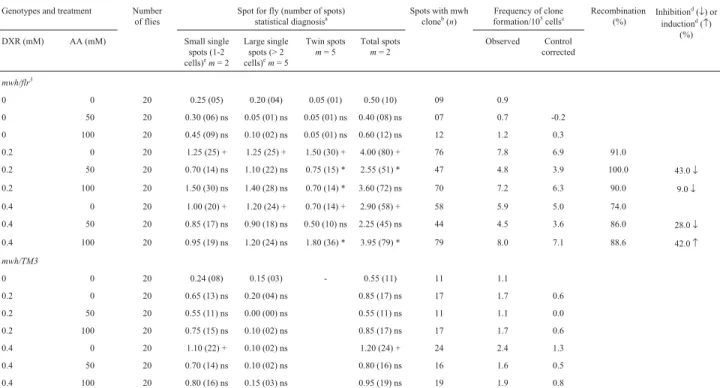
For BH individuals neither AA nor DXR (with excep-tion of DXR 0.4 mM) induced significant increase in the spot frequencies when compared to negative control (p > 0.05). The combined co-treatment with AA and DXR was ineffective in the inhibition of small single spots, large simple spots and total spots when compared to DXR alone (p > 0.05).
Comparison of the frequencies of wing spots in the BH and MH descendents indicated that recombination was a major response, indicating, under these experimental con-ditions, genotoxic (from 74 untill 100% recombinagenic) activity.
High bioactivation (HB) cross
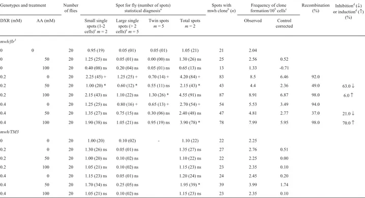
The number of spots and their distribution obtained for MH and BH individuals of the HB-cross are presented in Table 2. Both concentrations of AA were not genotoxic but DXR showed genotoxic activity by increasing the fre-quencies of all types of spots (p < 0.05). The co-treatments with the lower concentrations of AA and DXR produced a statistically significant reduction (63%) in the frequencies of small single spots, large single spots and total of spots (p < 0.05), while co-treatments with DXR (0.2 mM) and AA (100 mM) led to a statistically significant reduction
only in the frequencies of twin spots. The same was not observed in the other treatment with AA and DXR, and the series treated with AA 100 mM and DXR 0.4 mM showed a positive increase (70%) in the total of spots when compared to DXR alone.
For BH flies no positive effect were observed for AA or DXR, alone or in combination, when compared to nega-tive control (p > 0.05). However, AA 50 mM did alter the DXR 0.4 mM genotoxicity, increasing the frequencies of total spots when compared to DXR 0.4 mM (p < 0.05).
Comparison of the frequencies of wing spots in the BH and MH descendants indicated that, for the HB cross, the recombinagenic activity ranged from 37 to 98%.
Discussion
In this investigation, we used the wing somatic muta-tion and recombinamuta-tion test inD. melanogasterbecause it represents a rapid and inexpensive way to evaluate the genotoxicity/antigenotoxicity of single compounds as well as of complex mixtures (Grafet al., 1996). It has been dem-onstrated by the wing spot test that the co-administration of coffee is effective in significantly reducing the frequencies of single and twin spots induced by cyclophosphamide,
Table 1- Summary of results obtained with the Drosophila somatic mutation and recombination test (SMART) in the marker-heterozygous (MH) and
balancer-heterozygous (BH) progeny of the standard (ST) cross after chronic treatment of larvae with ascorbic acid (AA) and doxorubicin (DXR).
Genotypes and treatment Number
of flies
Spot for fly (number of spots) statistical diagnosisa
Spots with mwh cloneb
(n)
Frequency of clone formation/105
cellsc
Recombination (%)
Inhibitiond
(↓) or inductiond
(↑) (%)
DXR (mM) AA (mM) Small single
spots (1-2 cells)e
m= 2
Large single spots (> 2 cells)e
m= 5
Twin spots
m= 5
Total spots
m= 2
Observed Control
corrected
mwh/flr3
0 0 20 0.25 (05) 0.20 (04) 0.05 (01) 0.50 (10) 09 0.9
0 50 20 0.30 (06) ns 0.05 (01) ns 0.05 (01) ns 0.40 (08) ns 07 0.7 -0.2
0 100 20 0.45 (09) ns 0.10 (02) ns 0.05 (01) ns 0.60 (12) ns 12 1.2 0.3
0.2 0 20 1.25 (25) + 1.25 (25) + 1.50 (30) + 4.00 (80) + 76 7.8 6.9 91.0
0.2 50 20 0.70 (14) ns 1.10 (22) ns 0.75 (15) * 2.55 (51) * 47 4.8 3.9 100.0 43.0↓
0.2 100 20 1.50 (30) ns 1.40 (28) ns 0.70 (14) * 3.60 (72) ns 70 7.2 6.3 90.0 9.0↓
0.4 0 20 1.00 (20) + 1.20 (24) + 0.70 (14) + 2.90 (58) + 58 5.9 5.0 74.0
0.4 50 20 0.85 (17) ns 0.90 (18) ns 0.50 (10) ns 2.25 (45) ns 44 4.5 3.6 86.0 28.0↓
0.4 100 20 0.95 (19) ns 1.20 (24) ns 1.80 (36) * 3.95 (79) * 79 8.0 7.1 88.6 42.0↑
mwh/TM3
0 0 20 0.24 (08) 0.15 (03) - 0.55 (11) 11 1.1
0.2 0 20 0.65 (13) ns 0.20 (04) ns 0.85 (17) ns 17 1.7 0.6
0.2 50 20 0.55 (11) ns 0.00 (00) ns 0.55 (11) ns 11 1.1 0.0
0.2 100 20 0.75 (15) ns 0.10 (02) ns 0.85 (17) ns 17 1.7 0.6
0.4 0 20 1.10 (22) + 0.10 (02) ns 1.20 (24) + 24 2.4 1.3
0.4 50 20 0.70 (14) ns 0.10 (02) ns 0.80 (16) ns 16 1.6 0.5
0.4 100 20 0.80 (16) ns 0.15 (03) ns 0.95 (19) ns 19 1.9 0.8
Marker-trans-heterozygous flies (mwh/flr3) and balancer-heterozygous flies (mwh/TM3) were evaluated.
aStatistical diagnoses according to Frei and Würgler [1995].U-test, two-sided, probability levels: +, p < 0.05vs.untreated control; *, p < 0.05vs.DXR
only; ns, not significant.
b
Consideringmwhclones frommwhsingle and twin spots.
cFrequency of clone formation: clones/flies/48,800 cells (without size correction).
d
Calculated as [DXR alone - DXR + AA) / DXR alone] X 100, according to Abraham (1994).
diethylnitrosamine, mitomicyn C, procarbazine and ure-thane (Abraham, 1994) and that turmeric inhibited the genotoxic effect of urethane (El Hansset al., 1999). Fur-thermore, sodium selenite has been shown to be antigenotoxic in combination with potassium dichromate (Rizkiet al., 2001) and AA modulated the genotoxic action of several mutagens (Kayaet al., 2002), showing the suit-ability of this test system for mimicking the normal intake of substances.
Doxorubicin (DXR) was selected in this study be-cause it is an effective clastogenic and potent carcinogenic agent, and DXR has genotoxic effects that do not require enzymatic activation (Dhawanet al., 2003). The successful use of this antitumor agent is restricted by the risk of devel-oping cardiotoxicity. This risk increases exponentially with cumulative dose, and studies have reported that 10% to 26% of patients administered cumulative anthracycline doses above those recommended develop congestive heart failure, and that more than 50% of patients administered these doses will experience measurable functional impair-ment months to years after the end of therapy (Jensen, 2006). Possible clinical options for reducing DXR-induced cardiotoxicity include agents such as antioxidants that
pre-vent oxygen-free radical generation. The patterns of DNA damage in anthracycline-treated cancer cells seem to sup-port the concept that direct oxidative lesions only occur if cancer cells are exposed to elevated concentrations of an-thracyclines. Clinically relevant concentrations have been shown to cause the formation of protein-associated DNA single-strand and double-strand breaks, which might result from anthracycline inhibition of topoisomerase II by form-ing an anthracycline-DNA topoisomerase II complex (Gewirtz, 1999; Minottiet al., 2004).
The genotoxicity of DXR has been discussed for many years. In Drosophila assay systems, DXR induced sex-liked recessive lethals (Clementset al., 1984) and was classified as a strong mutagen inducing all types of spots (Freiet al., 1985). The spots can be due to different geno-toxic events, either mitotic recombination or mutations such as deletion, point mutation, specific types of trans-locations, etc. The significant induction of twin spots indi-cates that DXR, a strong direct-acting mutagen, is capable of inducing mitotic recombination. According to Lehmann
et al.(2003) DXR is a preferential inducer of homologous recombination when compared with mutational events in
D. melanogastersomatic cells. In our present study, DXR
Table 2- Summary of results obtained with the Drosophila somatic mutation and recombination test (SMART) in the marker-heterozygous (MH) and
balancer-heterozygous (BH) progeny of the high bioactivation (HB) cross after chronic treatment of larvae with ascorbic acid (AA) and doxorubicin (DXR)
Genotypes and treatment Number
of flies
Spot for fly (number of spots) statistical diagnosisa
Spots with mwh cloneb
(n)
Frequency of clone formation/105
cellsc
Recombination (%)
Inhibitiond
(↓) or inductiond
(↑) (%)
DXR (mM) AA (mM) Small single
spots (1-2 cells)e
m= 2
Large single spots (> 2 cells)e
m= 5
Twin spots
m= 5
Total spots
m= 2
Observed Control
corrected
mwh/flr3
0 0 20 0.95 (19) 0.05 (01) 0.05 (01) 1.05 (21) 21 2.04
0 50 20 1.25 (25) ns 0.05 (01) ns 0.00 (00) ns 1.30 (26) ns 25 2.56 0.52
0 100 20 0.40 (08) ns 0.20 (04) ns 0.05 (01) ns 0.65 (13) ns 13 1.33 -0.71
0.2 0 20 2.25 (45) + 1.25 (25) + 0.70 (14) + 4.20 (84) + 83 8.5 6.46 92.0
0.2 50 20 1.00 (20) * 0.60 (12) * 0.55 (11) ns 2.15 (43) * 43 4.4 2.36 49.0 63.0↓
0.2 100 20 2.15 (43) ns 1.10 (22) ns 1.30 (26) * 4.55 (91) ns 87 8.91 6.87 98.0 6.0↑
0.4 0 20 1.25 (25) ns 0.80 (16) + 0.65 (13) + 2.70 (54) + 54 5.53 3.49 94.0
0.4 50 20 1.35 (27) ns 0.75 (15) ns 0.30 (06) ns 2.40 (48) ns 47 4.81 2.77 37.0 21.0↓
0.4 100 20 1.90 (38) ns 1.05 (21) ns 0.95 (19) ns 3.90 (78) * 78 7.99 5.95 98.0 70.0↑
mwh/TM3
0 0 20 1.00 (20) 0.10 (02) - 1.10 (22) 22 2.25
0.2 0 20 1.30 (26) ns 0.05 (01) ns 1.35 (27) ns 27 2.76 0.51
0.2 50 20 1.00 (20) ns 0.10 (02) ns 1.10 (22) ns 22 2.25 0.00
0.2 100 20 1.05 (21) ns 0.10 (02) ns 1.15 (23) ns 23 2.35 0.10
0.4 0 20 1.15 (23) ns 0.05 (01) ns 1.20 (24) ns 24 2.45 0.20
0.4 50 20 1.70 (34) ns 0.25 (05) ns 1.95 (39) * 39 3.99 1.74
0.4 100 20 1.05 (21) ns 0.10 (02) ns 1.15 (23) ns 23 2.35 0.10
Marker-trans-heterozygous flies (mwh/flr3) and balancer-heterozygous flies (mwh/TM3) were evaluated.
a
Statistical diagnoses according to Frei and Würgler [1995].U-test, two-sided, probability levels: +, p < 0.05vs.untreated control; *, p < 0.05vs.DXR only; ns, not significant.
b
Consideringmwhclones frommwhsingle and twin spots.
cFrequency of clone formation: clones/flies/48,800 cells (without size correction).
d
Calculated as [DXR alone - DXR + AA) / DXR alone] X 100, according to Abraham (1994).
e
treatment gave positive results for all types of spots, al-though the DXR concentrations used in our investigation were lower than those used by Freiet al.(1985) and, ac-cording to Lehmannet al.(2003), DXR preferentially in-duced recombination rather than other genotoxic events. The data presented in our paper do not show a dose-response effect of DXR. The frequencies of spots at the highest concentration of DXR (0.4 mM) were no higher than those observed at the lower one (0.2 mM). The same pattern was found by Spanóet al.(2001) for different con-centrations of 9,10-dimethylantracene on the wing spot test in Drosophila. The protection afforded by antioxidants against DXR genotoxicity appears not to depend on the treatment schedule, since there was no modification in pro-tection for time intervals of 30 or 60 min in the antioxidant pretreatment and administration of DXR in rats (Praha-lathanet al., 2006).
We found that after treatments with DXR it was clear that the frequencies of wing spots on BH flies were lower than those found on MH flies. Nevertheless, in the BH flies the small single spots induced by DXR (0.4 mM) in the ST-cross were statistically significant compared with those of the respective negative control. The large majority of the spots induced by DXR on the wings of MH flies are ascrib-able to mitotic recombination, but a small number of them have other causes and may be due to point mutation or chro-mosome breaks.
The results of DXR photodegradation in plasma, urine and cell culture medium as measured by HPLC indi-cates that DXR is very unstable in cell culture medium when exposed to light (Le Botet al., 1988). Therefore, it is important for the correct interpretation of the effects of DXR activity on biological systems, especially when cul-ture medium is used, to investigate DXR genotoxicity in the absence of light, as performed in the present study.
Ascorbic acid (AA) is known to have both anti-mutagenic and anticarcinogenic activities. According to Halliwell (2001) AA, a water-soluble glucose derivative, has considerable antioxidant activityin vitro, in part be-cause of its ease of oxidation and bebe-cause the semidehy-droascorbate radical derived from it is of low reactivity, but there are conflicting effects of AA reported on the induc-tion of DNA strand breaks, micronuclei and frequency of chromosomal aberrations. It has been previously reported that AA successfully inhibited the chromosome aberrations induced by DXR in rat bone marrow cells (Antunes and Takahashi, 1998), micronuclei in cytokinesis-blocked hu-man lymphocytes and the chromosomal aberration assay in V79 Chinese hamster cells induced by the mycotoxin patulin (Alves et al., 2000) and mercury-induced geno-toxicity in human blood cultures (Raoet al., 2001). How-ever, administration of high doses of AA appears to have mutagenic effects in different test systems. In human blood lymphocytes culture treated with lower concentrations of AA there was a significant reduction in chromosomal
aber-rations induced by DXR but at higher doses AA did not present the same protective effect and was cytotoxic (Antu-nes and Takahashi, 1999).
We observed that the two different concentrations of AA tested (50 and 100 mM) had no effect on spots frequen-cies. Tripathyet al.(1990) observed that larvae exposed to 100 and 300 mM showed inconclusive results for the induc-tion of the small single spots and negative results for large single spots. Kayaet al.(2002) showed that AA (25, 75 and 250 mM) did not induce significant increases in the fre-quency of mutant clones in the Drosophila wing spot test. The authors concluded that the differences between the control and the AA-treated series were not of biological significance and AA was considered negative.
The ability of AA to modulate the genotoxic action of several mutagens has also been investigated in the wing spot test ofD. melanogaster. Nevertheless, when co-treat-ment experico-treat-ments with AA were carried out, different re-sults were found (Kayaet al., 2002). In our present study, combined co-treatment with the lower concentration of AA plus DXR led to a statistically significant reduction in the frequencies of twin and total of spots in MH flies in the ST-cross and in the frequencies of small single spots, large single spots and total spots in the HB-cross, while treat-ments with DXR (0.4 mM plus 100 mM AA in both crosses) increased the frequencies of total spots when com-pared to DXR alone.
Similar results were obtained by Cederberg and Ra-mel (1989) who reported that AA co-treatment had a modi-fying effect in the wing spot test on genotoxicity induced by the antitumor agent bleomycin. Grafet al.(1998) reported that AA and catechin were able to protect againstin vivo
nitrosation products of methyl urea in combination with so-dium nitrite, while Kayaet al.(2002) found that AA was ef-fective in reducing the genotoxicity of K2Cr2O7virtually to
the control level in the Drosophila wing spot test. On the contrary, co-treatment experiments by Kayaet al., (2002) indicate that different concentrations of AA (25, 75 and 250 mM) did not show any antigenotoxic effect on the genotoxicity of 4-nitroquinoline 1-oxide (4-NQO) and Kayaet al.(2002) found that when co-treatments experi-ments with cobalt chloride (CoCl2) were carried out,
in-stead of reducing the genotoxicity of CoCl2AA acts as a
co-mutagen by inducing a significant increase in the fre-quency of mutant clones over the values obtained with CoCl2alone.
cytotoxic and mutagenic effects of oxidative stress. Kono-packaet al.(1998) demonstrated the modifying effect of treatment with AA on the clastogenic activity of gamma rays in mice as measured by the micronucleus assay in bone marrow polychromatic erythrocytes and exfoliated bladder cells. Depending on AA concentration, the number of mi-cronucleated polychromatic erythrocytes can be enhanced or reduced.
In conclusion, under the present experimental condi-tions, our data shows that DXR induced mitotic recombina-tion in somatic cells of Drosophila and that 50 mM of AA protected against DXR genotoxicity while 100 mM of AA enhanced the frequencies of DXR-induced spots. These re-sults suggest that AA may interfere with free radicals gen-erated by DXR and with other possible reactive metabolites which elevate spot frequencies inD. melanogaster.
References
Abraham SK (1994) Antigenotoxicity of coffee in the Drosophila assay for somatic mutation and recombination. Mutagenesis 9:383-386.
Alves I, Oliveira NG, Laires A, Rodrigues AS and Rueff J (2000) Induction of micronuclei and chromosomal aberrations by the mycotoxin patulin in mammalian cells: Role of ascorbic acid as a modulator of patulin clastogenicity. Mutagenesis 15:229-234.
Amara-Mokrane YA, Lehucher-Michel MP, Balansard G, Duménil G and Botta A (1996) Protective effects of
α-hederin, chlorophyllin and ascorbic acid towards the in-duction of micronuclei by doxorubicin in cultured human lymphocytes. Mutagenesis 11:161-167.
Antunes LMG, Bueno RBL, Dias FL and Bianchi MLP (2007) Acetylsalicylic acid exhibits anticlastogenic effects on cul-tured human lymphocytes exposed to doxorubicin. Mutat Res 626:155-161.
Antunes LMG and Takahashi CS (1998) Effects of high doses of vitamins C and E against doxorubicin-induced chromo-somal damage in Wistar rat bone marrow cells. Mutat Res 419:137-143.
Antunes LMG and Takahashi CS (1999) Protection and induction of chromosomal damage by vitamin C in human lymphocyte cultures. Teratog Carcinog Mutagen 19:53-59.
Benchekroun MN, Sinha BK and Robert J (1992) Doxorubicin-induced oxygen free radical formation in sensitive and doxorubicin-resistant variants of rat glioblastoma cell lines. FEBS Letters 322:295-298.
Bijur GN, Ariza ME, Hitchcock CL and Williams MV (1997) Antimutagenic and promutagenic activity of ascorbic acid during oxidative stress. Environ Mol Mutagen 30:339-345. Cabrera G (2000) Effect of five dietary antimutagens on the
genotoxicity of six mutagens in the microscreen prophage-induction assay. Environ Mol Mutagen 36:206-220. Cederberg H and Ramel C (1989) Modifications of the effect of
bleomycin in the somatic mutation and recombination test in
Drosophila melanogaster. Mutat Res 214:69-80.
Clements J, Phillips M and Todd N (1984) Mutagenicity of adriamycin in Drosophila melanogaster. Mutat Res 135:175-179.
Costa WF and Nepomuceno JC (2006) Protective effects of a mix-ture of antioxidant vitamins and minerals on the genoto-xicity of doxorubicin in somatic cells of Drosophila melanogaster. Environ Mol Mutagen 47:18-24.
Dhawan A, Kayani MA, Parry JM, Parry E and Anderson D (2003) Aneugenic and clastogenic effects of doxorubicin in human lymphocytes. Mutagenesis 18:487-490.
El Hamss R, Analla M, Campos-Sánchez J, Alonso-Moraga A, Muñoz-Serrano A and Idaomar M (1999) A dose dependent anti-genotoxic effect of turmeric. Mutat Res 446:135-139. Erhola M, Kellokumpu-Lehtinen P, Metsä-Ketelä T, Alanko K
and Nieminen MM (1996) Effects of anthracyclin-based chemotherapy on total plasma antioxidant capacity in small cell lung cancer patients. Free Radic Biol Med 21:383-390. Frei H, Würgler FE, Juon H, Hall CB and Graf U (1985) Aris-tolochic acid is mutagenic and recombinogenic in Drosophila genotoxicity tests. Arch Toxicol 56:158-166. Frei H and Würgler FE (1988) Statistical methods to decide
whether mutagenicity test data from Drosophila assays indi-cate a positive, negative, or inconclusive result. Mutat Res 203:297-308.
Frei H and Würgler FE (1995) Optimal experimental design and sample size for the statistical evaluation of data from so-matic mutation and recombination tests (SMART) in Drosophila. Mutat Res 334:247-258.
Gentile JM, Rahimi S, Zwiesler J, Gentile GJ and Ferguson LR (1998) Effect of selected antimutagens on the genotoxicity of antitumor agents. Mutat Res 402:289-298.
Gewirtz DA (1999) A critical evaluation of the mechanisms of ac-tion proposed for the antitumor effects of the anthracycline antibiotics adriamycin and daunorubicin. Biochem Phar-macol 57:727-741.
Graf U, Würgler FE, Katz AJ, Frei H, Juon H, Hall CB and Kale PG (1984) Somatic mutation and recombination test in
Drosophila melanogaster. Environ Mutagen 6:153-188. Graf U, Frei H, Kägi A, Katz AJ and Würgler FE (1989) Thirty
compounds tested in the Drosophila wing spot test. Mutat Res 222:359-373.
Graf U and van Schaik N (1992) Improved high bioactivation cross for the wing somatic mutation and recombination test inDrosophila melanogaster. Mutat Res 271:59-67. Graf U, Spanó MA, Guzmán Rincón J, Abraham SK and Andrade
HH (1996) The wing somatic mutation and recombination test (SMART) in Drosophila melanogaster: An efficient tool for the detection of genotoxic activity of pure com-pounds or complex mixtures as well as for studies of anti-genotoxicity. Afr Newslett on Occup Health and Safety 6(Suppl 1):9-13.
Graf U, Abraham SK, Guzmán-Rincón J and Würgler FE (1998) Antigenotoxicity studies in Drosophila melanogaster. Mutat Res 402:203-209.
Halliwell B (2001) Vitamin C and genomic stability. Mutat Res 475:29-35.
Islaih M, Halstead BW, Kadura IA, Li B, Reid-Hubbard JL, Flick L, Altizer JL,et al.(2005) Relationships between genomic, cell cycle, and mutagenic responses of TK6 cells exposed to DNA damaging chemicals. Mutat Res 578:100-116. Jensen BV (2006) Cardiotoxic consequences of
Kaya B, Creus A, Velázquez A, Yanikoglu A and Marcos R (2002) Genotoxicity is modulated by ascorbic acid: Studies using the wing spot test in Drosophila. Mutat Res 520:93-101.
Kastenbaum MA and Bowman KO (1970) Tables for determining the statistical significance of mutation frequencies. Mutat Res 9:527-549.
Konopacka M, Widel M and Rzeszowska-Wolny J (1998) Mod-ifying effect of vitamins C, E and beta-carotene against gamma-ray-induced DNA damage in mouse cells. Mutat Res 417:85-94.
Le Bot MA, Riche C, Guedes Y, Kernaleguen D, Simon S, Begue JM and Berthou F (1988) Study of doxorubicin photodegra-dation in plasma, urine and cell culture medium by HPLC. Biomed Chromatogr 2:242-244.
Lehmann M, Franco A, Vilar KSP, Reguly ML and Andrade HHR (2003) Doxorubicin and two of its analogues are preferential inducers of homologous recombination compared with mu-tational events in somatic cells ofDrosophila melanogaster. Mutat Res 539:167-175.
Minotti G, Menna P, Salvatorelli E, Cairo G and Gianni L (2004) Anthracyclines: Molecular advances and pharmacologic de-velopments in antitumor activity and cardiotoxicity. Pharmacol Rev 56:185-229.
Menegola E, Broccia ML and Renzo FD (2001) Teratogenic ef-fects of doxorubicin in rats at midgestation and at term. Teratog Carcinog Mutagen 21:283-293.
Nefic H (2001) Anticlastogenic effect of vitamin C on cisplatin in-duced chromosome aberrations in human lymphocyte cul-tures. Mutat Res 498:89-98.
Odin AP (1997) Vitamins as antimutagens: Advantages and some possible mechanisms of antimutagenic action. Mutat Res 386:39-67.
Prahalathan C, Selvakumar E, Varalakshmi P, Kumarasamy P and Saravanan R (2006) Salubrious effects of lipoic acid against
adriamycin-induced clastogenesis and apoptosis in Wistar rat bone marrow cells. Toxicology 222:225-232.
Rao MV, Chinoy NJ, Suthar MB and Rajvanshi MI (2001) Role of ascorbic acid on mercuric chloride-induced genotoxicity in human blood cultures. Toxicol In Vitro 15:649-654. Resende FA, Barcala CAMA, Faria MCS, Kato FH, Cunha WR
and Tavares DC (2006) Antimutagenicity of ursolic acid and oleanolic acid against doxorubicin-induced clastogenesis in Balb/c mice. Life Sci 79:1268-1273.
Rizki M, Amrani S, Creus A, Xamena N and Marcos R (2001) Antigenotoxic properties of selenium: Studies in the wing spot test in Drosophila. Environ Mol Mutagen 37:70-75. Rizki M, Kossatz E, Creus A and Marcos R (2004) Genotoxicity
modulation by cadmium treatment: Studies in the Drosophila wing spot test. Environ Mol Mutagen 43:196-203.
Romero-Jiménez M, Campos-Sánchez J, Analla M, Muñoz-Ser-rano A and Alonso-Moraga A (2005) Genotoxicity and anti-genotoxicity of some traditional medicinal herbs. Mutat Res 585:147-155.
Shamberger RJ (1984) Genetic toxicology of ascorbic acid. Mutat Res 133:135-159.
Siddique YH, Beg T and Afzal M (2005) Antigenotoxic effects of ascorbic acid against megestrol acetate-induced genotoxi-city in mice. Hum Exp Toxicol 24:121-127.
Sinigaglia M, Reguly ML and Andrade HHR (2004) Effect of vanillin on toxicant-induced mutation and mitotic recombi-nation in proliferating somatic cells of Drosophila melanogaster. Environ Mol Mutagen 44:394-400.
Spanó MA, Frei H, Würgler FE and Graf U (2001) Recom-binagenic activity of four compounds in the standard and high bioactivation crosses of Drosophila melanogasterin the wing spot test. Mutagenesis 16:385-394.
Tripathy NK, Würgler FE and Frei H (1990) Genetic toxicity of six carcinogens and six non-carcinogens in the Drosophila wing spot test. Mutat Res 242:169-180.