Escola de Ciências da Saúde
Daniel Filipe Miranda Mendanha
U
NDERSTANDING THE RELEVANCE OF ASTROCYTIC VESICULAR RELEASE IN GLIOBLASTOMADissertação de Mestrado Mestrado em Ciências da Saúde
Trabalho efetuado sob a orientação do: Doutor João Filipe Oliveira
e co-orientação do: Doutor Bruno M. Costa
Nome: Daniel Filipe Miranda Mendanha
Endereço electrónico: mendanha@ecsaude.uminho.pt Telefone: 913184484
Número do Cartão de Cidadão: 14398719
Título da dissertação:
Understanding the relevance of astrocytic vesicular release in glioblastoma
Orientador:
Doutor João Filipe oliveira Co-Orientador:
Doutor Bruno M.Costa
Ano de conclusão: 2016
Designação do Mestrado: Ciências da Saúde
É AUTORIZADA A REPRODUÇÃO INTEGRAL DESTA DISSERTAÇÃO APENAS PARA EFEITOS DE INVESTIGAÇÃO, MEDIANTE DECLARAÇÃO ESCRITA DO INTERESSADO, QUE A TAL SE COMPROMETE.
Universidade do Minho, 8 de Junho de 2016 Assinatura:
iii
Os resultados apresentados nesta tese são conjugação de 2 anos de dedicação, esforço e trabalho, que apenas foi possível devido ao apoio de diversas pessoas que de uma forma ou outra contribuíram para a realização deste trabalho. Por isso agradeço a todas as pessoas que estiveram direta ou indiretamente envolvidos na realização desta tese.
Começo por agradecer às pessoas que me deram a oportunidade de evoluir, aprender e crescer como cientista, os meus orientadores, Dr. João Oliveira e o Dr. Bruno Costa. Agradeço toda a disponibilidade e ensinamentos prestados durante este ano. Fico contente por ver singrar 2 pessoas tão jovens e com abordagens distintas, neste complicado mundo da ciência.
Agradeço à Sónia Gomes, por tudo que me ensinaste, pela disponibilidade e preocupação demonstrada, e por todas as palavras de incentivo mesmo quando a motivação faltava. Acredito sinceramente que vais ter um grande futuro à tua frente. Agradeço também às restantes meninas do grupo, Vanessa, Gabriela e Joana, pela ajuda, interesse e por me integrarem tão bem num grupo de trabalho.
Agradeço também às meninas do grupo do 1º piso. Agradeço à Marta Pojo e à Ana Oliveira por toda a ajuda e ensinamentos práticos, que foram essenciais no começo e evolução desta tese. Agradeço a vossa disponibilidade, que mesmo depois da saírem do laboratório, estiveram sempre disponíveis para me ajudar. Agradeço também à Joana, pelas palavras de motivação, paciência e toda a ajuda, que permitiram conclusão desta tese. À Céline e às diversas pessoas que passaram em ambos os grupos de trabalho, agradeço os debates construtivos sobre os resultados que vos apresentava ao longo do ano e todas as vossas sugestões.
Agradeço aos meus colegas de mestrado, por todos os almoços e pausas a conversar e que me permitam espairecer a mente quando ela mais necessitava.
Acabo por agradecer à instituição, ICVS, que me permitiu entrar neste mestrado e ofereceu todas as condições necessárias para o desenvolvimento de um trabalho do qual me orgulho e que espero que o leitor aprecie.
Apesar de as palavras não serem suficientes, ficam aqui os mais sinceros agradecimentos a todas estas pessoas que foram uma parte ativa da minha vida nos últimos 2 anos.
v
Glioblastomas (GBMs) are the most common and malignant type of glioma, the majority subtype of primary brain tumors. Despite the therapeutic advances over the past years, the median overall survival of patients is only 15 months after diagnosis. The high degree of heterogeneity between GBMs leads to an unpredictable clinical outcome. The variety of complex components in the tumor microenvironment has been associated with the aggressiveness of GBMs and with the inefficacy of treatments currently available. Astrocytes, the major glial cell type in the brain, are usually associated with the regulation of brain homeostasis, being involved in distinct regulatory processes, as angiogenesis or blood brain barrier (BBB) regulation. In GBM, astrocytes have been shown to secrete factors/proteins that may to regulate tumor growth, invasion, and progression. This secretion is performed, among others, by SNARE-dependent exocytosis, a mechanism impaired in astrocytes of the dnSNARE mouse model. By using this model, we explored the role of astrocytic exocytosis in the growth and invasion of GBM using in vitro and in vivo complementary approaches. We evaluated the effect of conditioned medium (CM), derived from wild – type (WT) and dnSNARE glial cultures, in the viability and migration of a glioma cell line. Moreover, using a syngeneic orthotopic glioma murine model (GL261), we evaluated the influence of astrocytic SNARE-dependent exocytosis in glioma growth in vivo and mice survival. The viability assays in vitro suggested a regulation of glioma cells by WT-derived CM, significantly increasing glioma cell viability, which was not significant when glioma cells were in contact with dnSNARE-derived CM. Concerning the in vivo results, our data suggest that the substances secreted by astrocytes appear to influence GBM behavior, leading to a decrease in mice survival. Interestingly, tumor size was similar in mice of both genotypes, suggesting other cancer hallmarks may be regulated in this GBM-astrocyte interaction. The results discussed in this thesis suggest that by releasing regulatory molecules, astrocytes might support GBM pathophysiology. The identification of the astrocyte-derived regulatory molecules may identify novel therapeutic targets for GBM treatment.
vii
Os glioblastomas (GBMs) são o tipo mais comum e maligno de gliomas, o maior sub-tipo de tumor primário no cérebro. Apesar dos avanços terapêuticos nos últimos anos, o tempo médio de sobrevivência dos pacientes depois de diagnosticados, é de apenas 15 meses. O elevado grau de heterogeneidade entre GBMs encontra-se relacionado com a imprevisibilidade no desenrolar desta doença. A variedade de células no microambiente tumoral tem sido associada com a agressividade apresentada por estes tumores e pela ineficácia dos tratamentos actualmente disponíveis. Os astrócitos, o principal tipo de célula da glia no cérebro, estão associados à regulação homeostática deste, estando envolvidos em processos como a angiogénese e a regulação da barreira hematoencefálica. Num contexto de GBM, os astrócitos secretam fatores/proteínas que podem regular o crescimento, invasão e progressão dos tumores. Esta secreção astrocítica, é mediada, entre outras, pelo complexo SNARE que regula a exocitose, e que se encontra inibido nos astrócitos do modelo de ratinho dnSNARE. Utilizando este modelo, foi explorado o papel da libertação vesicular astrocítica no crescimento e invasão dos glioblastomas utilizando abordagens complementares. O efeito do meio condicionado (MC), derivado de culturas de glia provenientes de animais controlo ou dnSNARE, foi testado na viabilidade e migração de uma linha celular de glioma. Complementarmente, usando um modelo ortotópico de glioblastoma (GL261), avaliamos a influência da exocitose mediada pelo complexo SNARE em astrócitos, no crescimento tumoral e na sobrevivência dos animais de ambos os genótipos. Os ensaios de viabilidade in vitro sugerem uma regulação das células de glioma pelo MC derivado de animais controlo, aumentando a viabilidade destas, o que não é significativo quando utilizado o MC derivado de culturas de glia dnSNARE. Relativamente aos resultados in vivo, estes sugerem que as substâncias secretadas pelos astrócitos podem influenciar o comportamento tumoral, levando a uma menor sobrevivência dos animais. Interessantemente, o volume tumoral foi similar entre genótipos, sugerindo que outras características tumorais podem ser mediadas por esta interação entre astrócitos e GBM. Os resultados discutidos durante esta tese sugerem que os astrócitos, libertando moléculas reguladoras, podem contribuir para as características do GBM. A identificação de moléculas derivadas de astrócitos podem apontar para novos alvos terapêuticos, no que diz respeito ao tratamento do GBM.
ix
Acknowledgments / Agradecimentos ... iii
Abstract ... v
Resumo ... vii
Table of Contents ... ix
Abbreviations List ... xi
Figures List ... xiii
Table List ... xiii
Chapter 1 – Introduction ... 1
-1.1 Primary brain tumors ... 4
-1.1.1 Glioma ... 5
-1.1.2 Glioblastoma (GBM) ... 6
-1.1.3 Glioma microenvironment ... 7
-1.2 Astrocytes ... 12
-1.3 Role of astrocytes in gliomas and brain metastases ... 16
-1.4 The dnSNARE model for study of astrocyte modulation of microenvironment ... 20
-1.5 Research goals ... 21
Chapter 2 – Materials and Methods ... 23
-2.1 Cell lines and culture conditions ... 25
-2.2 The dnSNARE mouse model ... 25
-2.3 Mouse genotyping ... 26
-2.4 Primary culture of glial cells ... 28
-2.5 Viability assays... 29
-2.5.1 Trypan blue assay ... 29
-2.5.2 MTT assay ... 29
-2.6 Migration assay... 30
-2.7 Intracranial orthotopic glioma models ... 31
-2.7.1 Survival study... 31
-x
2.8.1 Immunofluorescence ... 32
-2.8.2 Western blot ... 33
-2.9 TCGA data metaanalysis in glioma patients... 35
-2.10 Statistical analyses ... 36
Chapter 3 Results ... 37
-3.1 In vitro studies ... 39
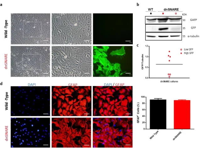
-3.1.1 Characterization of primary glial cultures ... 39
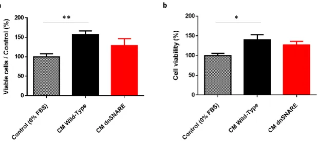
-3.1.2 Regulation of GL261 cells viability by astrocytic CM ... 41
-3.1.3 Regulation of GL261 cells migration by astrocytic CM ... 42
-3.2 In vivo studies ... 44
-3.2.1 Role of astrocytic vesicular release on mice survival ... 44
-3.2.2 Morphological assessment of GBM ... 45
-3.2.3 Role of astrocytic vesicular release on glioma growth ... 48
-3.3 In silico studies ... 49
-3.3.1 SNARE proteins expression in GBM patients ... 49
Chapter 4 – Discussion ... 53
Chapter 5 – Conclusions and Future Perspectives ... 63
Chapter 6 – References ... 67
-xi
ACM - Astrocyte Conditioned Medium AD - Alzheimer´s Disease
ALS - Amyotrophic Lateral Sclerosis Ang2 - Angiopoietin 2
ANOVA - Analysis of Variance BBB - Blood Brain Barrier
BDNF - Brain-Derived Neurotrophic Factor bFGF - basic Fibroblastic Growth Factor BoNTs - Botulinum Neurotoxins BrdU - 5-Bromo-2-DeoxyUridine BSA - Bovine Serum Albumin CM - Conditioned Medium CNS - Central Nervous System CSF-1 - Colony Stimulating Factor 1 Cx43 - Connexin43
DMEM - Dulbecco´s Modified Eagle Medium DNA - Deoxyribonucleic Acid
DOX - Doxycycline EC - Endothelial Cells ECM - Extracellular Matrix
EGFP - Enhanced Green Fluorescent Protein ET - Endothelin
FBS - Fetal Bovine Serum GBM - Glioblastoma
GDF-15 - Growth Differentiation Factor 15 GDNF - Glial Derived Neurotrophic Factor GFAP - Glial Fibrillary Acidic Protein GFP – Green Fluorescence Protein GSC- Glioma Stem Cells
H&E - Hematoxylin and Eosin
HBSS - Hank´s Balanced Salt Solution HIF - Hypoxia Inducible Factor IGF-1 - Insulin-like Growth Factor 1
xii
KPS - Karnofsky Performance Status M1 - Macrophages Type 1
M2 - Macrophages type 2
MCP-1 - Monocyte Chemoattractant Protein-1 M-CSF - Macrophage Colony-Stimulating Factor MMP - Matrix Metalloprotease
MT1-MMP - Membrane type 1 metalloprotease OS - Overall Survival
PCR – Polymerase Chain Reaction PD - Parkinson´s Disease
PFA - Paraformaldehyde PVN - Perivascular Niche ROS - Reactive Oxygen Species RPM - Rotations per Minute RT - Room Temperature SDS - Sodium Dodecyl Sulfate
SNAP - Synaptossomal Associated Protein sRNAs - small Non Coding RNAs
STI1 - Stress-Inducible Protein 1 STX1A – Syntaxin Protein 1
TAM - Tumor Associated Macrophage TEM - Tie-2 Expressing Monocyte
TGF-β - Transforming Growth Factor Beta TLR2 - Toll Like Receptor 2
TMZ - Temozolomide TNC - Tenascin-C
TNF-α - Tumor Necrosis Factor Alpha tTA - Tetracycline Transactivator
VAMP - Vesicular Associated Membrane Protein VAMP 2 - Synaptobrevin 2
VEGF - Vascular Endothelial Growth Factor WHO - World Health Organization
xiii
FIGURE 1.1 THE HALLMARKS OF CANCER ... -4
-FIGURE 1.2 CLASSIFICATION OF GLIOMAS BASED IN THEIR ORIGIN AND MOLECULAR CHARACTERISTICS ... -6
-FIGURE 1.3 TUMOR NICHES OF GLIOBLASTOMA ... -9
-FIGURE 1.4 SECRETORY PATHWAYS AND SECRETED SUBSTANCES BY ASTROCYTES ... -14
-FIGURE 1.5 PUTATIVE ROLES OF ASTROCYTES IN CANCER PROGRESSION ... -19
-FIGURE 2.1 DNSNARE MODEL ... -26
-FIGURE 3.1 CHARACTERIZATION OF PRIMARY GLIAL CULTURES ... -40
-FIGURE 3.2 CM DERIVED FROM WT GLIAL CULTURES INCREASE GLIOMA CELLS VIABILITY ... -42
-FIGURE 3.3 CM DERIVED FROM GLIAL CULTURES DO NOT AFFECT GLIOMA CELLS MIGRATION ... -43
-FIGURE 3.4 ASTROCYTIC VESICULAR RELEASE IMPAIRMENT INCREASES SURVIVAL OF GBM SYNGENEIC ORTHOTOPIC MODEL SURVIVAL ... -45
-FIGURE 3.5 TUMOR DEVELOPED IN DNSNARE MICE PRESENT TYPICAL GBM FEATURES ... -47
-FIGURE 3.6 ASTROCYTIC VESICULAR RELEASE IMPAIRMENT DOES NOT AFFECT IN VIVO GBM GROWTH ... -49
-FIGURE 3.7 SNARE COMPLEX AS A PROGNOSTIC VALUE IN GBM PATIENTS... -51
-SUPPLEMENTARY FIGURE 1 GFP REPORTER IS A GOOD READOUT OF DNSNARE TRANSGENE EXPRESSION ... -79
-SUPPLEMENTARY FIGURE 2 RELATION BETWEEN SNARE PROTEINS AND OS OF GBM PATIENTS ... -81
-Table List
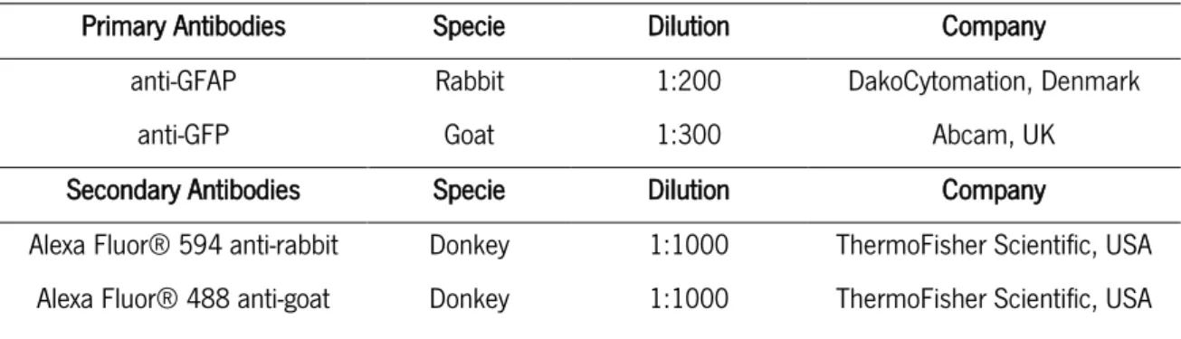
TABLE 1 PCR CONDITIONS FOR GENOTYPE IDENTIFICATION ... -27-TABLE 2 INFORMATION ABOUT THE ANTIBODIES USED FOR IMMUNOFLUORESCENCE ... -33
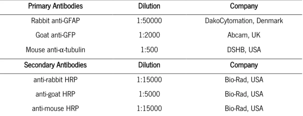
-TABLE 3 DILUTIONS AND INFORMATION ABOUT THE PRIMARY AND SECONDARY ANTIBODIES USED FOR WESTERN-BLOT ANALYSES. ... -35
- 1 -
- 3 -
1 Introduction
In the current century, cancer remains one of the major health problems, affecting people of every socioeconomic status around the world. According to World Health Organization (WHO), 14.1 million of new cancer cases were diagnosed in 2012. Despite the advances in cancer therapies, as chemotherapy and radiotherapy, 8.2 million cancer deaths were accounted in 2012 (Ferlay et al., 2015).
Cancer can be defined as uncontrolled cell growth with invasive/metastatic potential resultant from the accumulation of molecular alterations, such as deoxyribonucleic acid (DNA) mutations, copy number aberrations, chromosomal rearrangements and epigenetic modifications (responsible for gene expression regulation) (McLendon et al., 2008). These events are usually associated with the activation of oncogenes and deactivation of tumor suppressor genes. These genes are responsible for the regulation of cell proliferation, survival and differentiation, being necessary an alteration in both gene types for a neoplastic transformation. The deregulation of key signaling pathways is associated with genetic alterations neoplastic cells, that result in specific cancer features (e.g. proliferation, invasion, angiogenesis, etc.). In addition to the genetic alterations, the cellular microenvironment is also crucial for a neoplastic profile, where, for instance, increased secretion of growth factors can constitutively activate key pathways for cell proliferation (Hanahan and Weinberg, 2011).
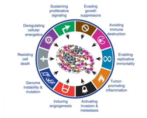
With the continuous growth of knowledge about cancer dynamics, it is well understood that tumors are not homogeneous masses of proliferating cancer cells. In fact, they are complex tissues composed by a large variety of cells, from parenchyma to immune cells, that together with cancer cells establish a large range of interactions. Thus, the advances in cancer therapeutics cannot only pass through the target of neoplastic cells, being necessary to understand and stop the contribution of the tumor microenvironment to tumorigenesis. In this scope, Hanahan and Weinberg proposed the ten cancer hallmarks (Figure 1.1), that include: i) evading growth suppressors; ii) avoiding immune destruction; iii) enabling replicative immortality; iv) tumor promoting inflammation; v) activating invasion & metastasis; vi) inducing angiogenesis; vii) genome instability & mutation; viii) resistance to cell death; ix) deregulating cellular energetics and x) sustaining proliferative signaling (Hanahan and Weinberg, 2011).
- 4 -
Figure 1.1 – The hallmarks of cancer – Cancer cells present specific capabilities that enhance tumor growth and metastatic dissemination (Adapted from (Hanahan and Weinberg, 2011)).
1.1
Primary brain tumors
The central nervous system (CNS) can present several tumor subtypes, which can be grossly separated in benign and malignant entities (Ostgathe et al., 2010). Tumors that have their origin in brain cells are classified as primary brain tumors, in contrast to cancer cells from a different origin that spread into the brain, commonly designated as brain metastases. Primary brain tumors present a low incidence between the primary tumors, only 2 %, nonetheless presented a high mortality rate (Buckner et al., 2007, Louis et al., 2007).
In 2007, the WHO published its 4th edition updating the classification of CNS tumors. According to
this manuscript, more than 100 different types were already described, regarding their origin, localization and histopathological features. By measuring these distinct characteristics WHO created a classification that grades CNS tumors in a malignant scale, being gliomas the most common form of brain tumor in CNS (Louis et al., 2007, Ostrom et al., 2014). An update version of CNS tumors classification was published this year, where for the first time, tumors were classified not only by their histological features but also by the distinct molecular components of tumors, resulting in the classification of new entities (Louis et al., 2016).
- 5 -
1.1.1 Glioma The origin of gliomas is still unclear, being crucial to understand how a series of molecular alterations that began in a couple of cells can result in a devastating disease. Nowadays, two major hypothesis emerge regarding glioma origin. The first one postulates that the accumulation of mutations and alterations in differentiated glial cells, as astrocytes or oligodendrocytes, leads to a dedifferentiation of those cells, acquiring a rapid proliferative and neoplastic profile. The second hypothesis, proposes that carcinogenic cells have their origin in progenitor undifferentiated cells, present in specific niches of the brain, that undergo molecular alterations resulting in neoplastic transformation (known as glioma initiating cells) (reviewed in Gonçalves et al., 2013). In fact, supporting this hypothesis are the glioma stem cells (GSC) that can be found in tumors, presenting a self-renewal capacity and high replicative potential, and which are able to promote the development of gliomas (Nguyen et al., 2012, Sampetrean and Saya, 2013). For the majority of gliomas, no underlying carcinogenic have been identified, being the exposure to high-dose of ionizing radiation the only well-established environmental risk factor established. Although some epidemiological studies in glioma have been published, the data regarding other environmental risk factors are still inconclusive (Bondy et al., 2008, Ohgaki, 2009).
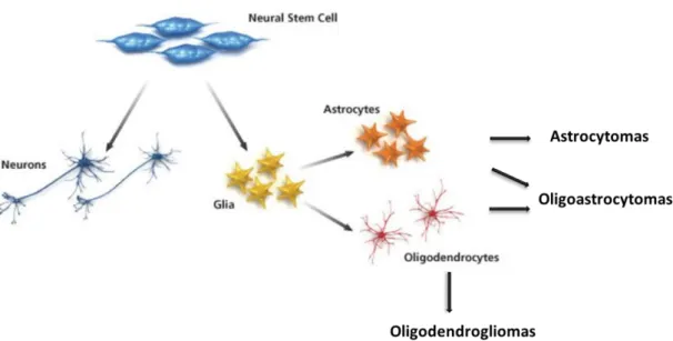
Gliomas represent approximately 80 % of all malignant brain tumors (Ostrom et al., 2014). With several specific pathological and immunohistochemical characteristics, gliomas are usually classified considering the type of glial characteristics they present (Figure 1.2). Therefore, it is usual to separate gliomas in: astrocytomas (similarities with astrocytes); oligodendrogliomas (similarities with oligodendrocytes); oligoastrocytomas (hold mixed characteristics from astrocytes and oligodendrocytes) and ependymomas (present similar features to ependymal cells) (Louis et al., 2007). Considering all the resemblances between these cells, it is usually accepted that glioma subtypes have their origin in the specific glial cell subtypes or their precursors. Being one subtype of primary brain tumors, gliomas are also classified according to their malignancy in four distinct grades (I-IV) by the WHO. Grade I gliomas are designated as benign tumors with a low proliferative potential, having the possibility of cure by surgical resection. Tumors designated as grade II present an infiltrative nature and are already classified as malignant tumors although, together with grade I tumors, they are considered low grade gliomas. Usually, the grade II tumors progress to high grade tumors, as for example, the low-grade diffuse astrocytoma (II) that is able to progress to glioblastoma (IV). Grade III gliomas present histological evidences of malignancy, as is the case of nuclear atypia and mitotic activity. Finally, the most malignant gliomas are classified with grade IV and are usually associated with a rapid pre and post-operative disease evolution
- 6 -
and a fatal outcome for the patient, being designated glioblastomas (Louis et al., 2007, Ostrom et al., 2014).
Figure 1.2 - Classification of gliomas based in their origin and molecular characteristics – Gliomas can be classified considering the histological similarity to the major type of glial cells. Astrocytomas have similar characteristics to astrocytes and oligodendrogliomas present similar characteristics to oligodendrocytes. Oligoastrocytomas, have features that are present in both glial cell types.
1.1.2 Glioblastoma (GBM) Glioblastoma (GBM) is the most common and malignant primary brain tumor, being the most common subtype of glioma (54.7% of all gliomas) (Ostrom et al., 2014). According to the WHO, GBM presents a grade IV classification, being represented by nuclear atypia, increased mitotic activity, microvascular proliferation and tissue necrosis. The tumor mass of GBM is characterized by its poor delineation and for having a high degree of regional heterogeneity. In the peripheral zones of the tumor it is common to find highly proliferating cancer cells, where the center of the tumor is mainly constituted by necrotic tissue (Inda et al., 2014, Hambardzumyan and Bergers, 2015). Despite being a cancer with low metastatic capacity, GBM is a highly invasive tumor especially along myelinated brain structures. The invasiveness of cells is the main reason for the poor efficacy of therapies, due to their ability to escape from surgical resection and radiotherapy (that target the main tumor mass) originating local recurrences (Louis et al., 2007).
- 7 -
In 1940, Hans-Joachim Scherer, a pioneer in glioma research, established for the first time a difference between the primary and secondary GBM (Scherer, 1940). Nowadays, we known that approximately 90 % of GBM cases are primary, meaning that they arise de novo, usually in elderly patients without a clinical or histological evidence of a lower grade glioma. Patients with GBM are usually asymptomatic until a late course of the disease, when the first clinical signs appear (e.g. headache and nausea) derived from the intracranial pressure rise caused by the tumor mass size, complicating the early detection and treatment (Wen and Kesari, 2008). Secondary GBM represent the remaining 10 % of the cases, progress from lower grade astrocytomas, and manifest in younger patients presenting a better prognosis. Histologically, primary and secondary GBM, are virtually indistinguishable, but they present several differences regarding the genetic and epigenetic profile (Ohgaki and Kleihues, 2007, 2013).
Despite the therapeutic advances over the past 20 years, the median overall survival of patients is only 15 months after diagnosis and treatment. In fact, all the treatments available are mostly palliative (Stupp et al., 2005, Wen and Kesari, 2008). The surgical resection is in the first line of treatments against GBM, corresponding to the resection of the maximal volume of tumor mass without putting in risk the CNS system activity of the patient. Usually, the surgical resection is followed by a radiotherapy treatment on a dose schedule of 60 Gy administered in 2.0 Gy per fraction (Malmstrom et al., 2012, Lacroix and Toms, 2014). Several advances in chemotherapy have also emerged in the last decade, being a standard treatment the use of the alkylating agent temozolomide (TMZ) in conjugation with radiotherapy (Stupp et al., 2005). Other therapeutics have also been used in the treatment of this disease, including bevacizumab, a new anti-angiogenic drug, that targets the high vascularization of these tumors by bindings to the vascular endothelial growth factor (VEGF), neutralizing its biological activity and leading to a deficient angiogenesis that slows GBM progression (Friedman et al., 2009).
1.1.3 Glioma microenvironment In the recent years, the interest in glioma microenvironment has arose, and several studies showed the importance of parenchyma cells in the course of the pathology. There is a current recognition that gliomas are complex tumors composed of neoplastic and non-neoplastic cells, being virtually each type individually able to contribute for cancer formation, progression and or response to treatment. The microenvironment of glioma is usually composed by different non-neoplastic cell types including fibroblasts, endothelial cells and immune system cells (Hambardzumyan et al., 2015). However, the interaction between brain-resident and infiltrating cells in the pathology of primary and metastatic brain
- 8 -
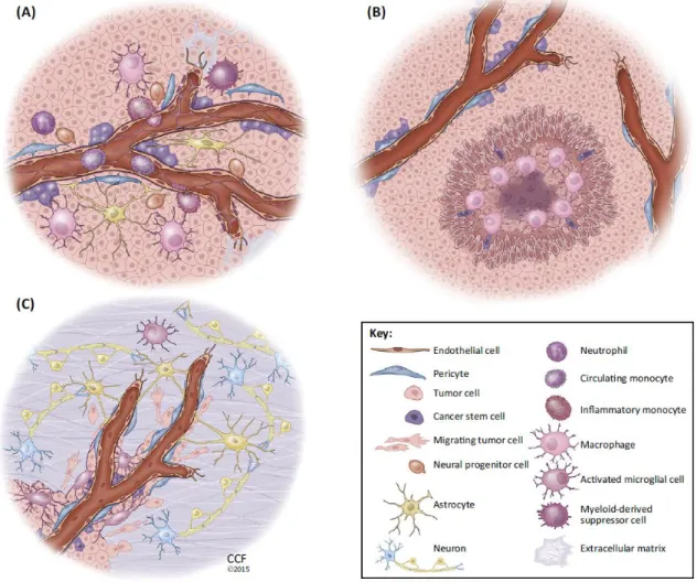
tumors is still poorly understood (Lorger, 2012). To help understanding the complexity of glioma microenvironment, Hambardzumyan and Bergers proposed a compartmentalization of the tumor microenvironment into three anatomically distinct regions, designated tumor niches (perivascular niche (PVN), hypoxic niche, invasive niche; Figure 1.3). In the tumor microenvironmental niches, tumor and stroma cells interact via direct cell contact or paracrine signaling to ensure maintenance, growth and protection of tumor and cancer stem cells (Hambardzumyan and Bergers, 2015).
The PVN presents a multicellular structure composed by several non-neoplastic cells already present in the brain or recruited from the periphery. The main function of this niche is to provide a supportive environment for cancer cells. The second GBM niche is the hypoxic niche, created by the abnormal vascular function in GBM, which leads to a deficient oxygen deliver within the tumor. Usually present in the center of the tumor mass, the niche is characterized by several necrotic areas responsible for the release of hypoxia inducible factors (HIF-1α and HIF-2α), leading to the expansion of GSC and the recruitment of innate immune cells. The last niche is usually found in more peripheral tumor zones, and it is designated as invasive niche. This niche is characterized by a large population of non-neoplastic cells that contribute to the invasiveness of glioma cells. Although astrocytes and pericytes are partially detached in these particular areas, it is possible to see an increased functional vasculature, that can be used to invade different brain areas. Moreover, it is important to understand that niches are non-static and develop several alterations with the course of the disease (Hambardzumyan and Bergers, 2015). Over the last years, studies targeting the microenvironment emerged, mostly due to the failure of treatments targeting the neoplastic cells. However, it is still necessary to understand how each of the different cell types present in the tumor can influence the disease progression.
- 9 -
Figure 1.3. - Tumor niches of Glioblastoma - Glioblastomas can present different niches, that present specific features and different intra-cellular composition. (A) Perivascular GBM niche – present a multicellular structure composed for several non-neoplastic cells (e.g. pericytes, microglia, astrocytes), providing a supportive environment for neoplastic cells. (B) Hypoxic GBM niche – present in the center of a tumor mass with a considerable volume, do not present a larger cellular diversity due to the lack of oxygen. Is usually associated for the release of factors that attract cells of the immune system. (C) Invasive GBM niche – similar to niche (A), also present a rich diversity of non-neoplastic cells, that release enhancer invasion factors. Present a rich and functional vascularization that can be used by glioblastoma cells to invade (Hambardzumyan and Bergers, 2015).
The complex role of the tumor microenvironment in the progression of brain tumors is still poorly understood, and only recently the scientific field has started to decode the interaction between stromal and cancer cells. Microglia and macrophages have been the primary targets of new studies for their important immunological role and, in fact, they present a significant role in the disease progression. However, it is important to understand how the different components of the tumor microenvironment individually and collectively contribute to the tumor. In the case of primary brain tumors, astrocytes, the
- 10 -
main glial cell in the brain, can play a crucial role in the disease development. Since this project focus on the astrocytic modulation of GBM, the next sub-chapters will summarize the functions that have already been attributed to non-neoplastic cells present in the brain tumor microenvironment, and a new sub-chapter will be dedicated to astrocytes and their relations to GBM.
Microglia and tumor associated macrophages (TAMs)
Microglia are a type of mononuclear cells that are distributed throughout the brain and act as immune effector cells of the CNS, being considered the resident macrophages of CNS (Hambardzumyan et al., 2015). In neuropathological conditions, as GBM, the blood brain barrier (BBB) is disrupted, resulting in an infiltration of monocytes from the periphery (Hambardzumyan et al., 2015). After entering the CNS, monocytes are able to differentiate into tumor-associated macrophages (TAMs), which present a high level of similarity with resident microglia. In fact, TAMs and microglia cells together, constitute approximately 30 to 40 % of the cells in GBM (Charles et al., 2011, Hambardzumyan and Bergers, 2015). Microglia and TAMs react to cancer cells, being these cell types already found accumulated around a single metastatic cancer cell, acquiring an amoeboid morphology. The migration of microglia and TAMs into glioma cells results from the release of chemo attractive factors by glioma cells, such as: monocyte chemoattractant protein-1 (MCP-1); glial-derived neurotrophic factor (GDNF) and colony stimulating factor 1 (CSF-1). Glioma cells can also release factors that induce a shift in microglia and macrophages phenotype, as is the case of macrophage colony-stimulating factor (M-CSF) (Pyonteck et al., 2013). Macrophages can be simplistically separated based in two distinct profiles: type 1 macrophages (M1), that present a pro-inflammatory profile and are usually associated with the release of anti-tumorigenic substances; and type 2 macrophages (M2), that have an anti-inflammatory profile, presenting pro-tumorigenic characteristics. TAMs are commonly associated with a M2 profile, although presenting some differences. The regulation and manipulation of this profile switch has been proposed as a possible therapeutic target for gliomas (reviewed in Hambardzumyan et al., 2015).
The accumulation of microglia and TAMs in and around glioma cells results in a direct interaction between them, resulting in the promotion of glioma growth and invasion. In 2009, Markovic and co-workers, established that a reduction in microglia number in a transgenic mice model, was sufficient to attenuate glioma growth (Markovic et al., 2009). Microglia and TAMs around the tumor present a high expression of stress-inducible protein 1 (STI1), which upon secretion promotes glioma growth (da Fonseca et al., 2014).
- 11 -
High grade gliomas are severely invasive and a large number of studies already attributed some of this invasive capacity to the role of microglia and macrophages. The release of factors by glioma cells, regulating the pro-tumorigenic profile of microglia, leads to an increase in interleukyn-6 (IL-6) secretion by microglia, promoting the invasive capacity of glioma cells (Saederup et al., 2010). Transforming growth factor-β (TGF-β) released by microglia can also increase glioma migration through a process involving increased integrin function and expression (Wick et al., 2001). Besides that, TGF-β induces the expression of matrix metalloprotease 2 (MMP2) that acts in the extracellular matrix (ECM) facilitating the glioma invasion (Markovic et al., 2005). In fact, a recent study showed that glioma cells release versican, an endogenous ligand that triggers the toll like receptor 2 (TLR2) signaling. An increased TLR2 expression is able to switch the microglia to a pro-tumorigenic phenotype resulting in an upregulation of matrix metalloprotease 9 (MMP-9) and membrane type – 1 matrix metalloproteinase (MT1-MMP), which in turn can lead to changes in ECM and ultimately to an increase in glioma growth and invasion (Vinnakota et al., 2013, Hu et al., 2014, Hambardzumyan and Bergers, 2015).
A different population of myeloid cells that derives from circulatory monocytes, are the Tie-2 expressing monocytes (TEMs). This subpopulation, which only accounts for approximately 7 % of blood mononuclear cells, has been associated with a high impact in tumor angiogenesis (De Palma et al., 2005, Venneri et al., 2007). Similarly to TAMs, when they reach the tumor, after crossing the BBB, they strongly polarize to a M2 activation state, secreting basic fibroblastic growth factor (bFGF) that induces a pro-angiogenic activity (Lorger, 2012). More recently, it was suggested that TEMs play an important role in glioma anti-vascular therapies, by promoting the invasion of cancer cells after the treatment. The use of Bevacizumab, since 2009, an anti-VEGF agent, did not reach the level of efficacy expected. In 2016, Cortes-Santiago and co-workers, claimed for the first time, that TEMs can be responsible for this inefficacy. They suggested that using an agent against angiopoietin 2 (Ang2) combined with anti-VEGF therapy, may result in the improvement of the treatment and in less glioma recurrences (Cortes-Santiago et al., 2016).
Endothelial cells
The high vascularization characteristic of gliomas, has in its base the endothelial cells (EC) that constitute the blood vessels. These cells have emerged as critical participants in the progression of brain tumors, not only by allowing a constant flux of oxygen and nutrients to tumor cells, but also by the direct communication with glioblastoma stem cells (GSC). The secreted factors by ECs contribute to the GSC maintenance of their stem cell-like characteristics (Charles et al., 2011). In 2010, Charles and his
co-- 12 co--
workers suggested that nitric oxide released by ECs activates the Notch signaling in cancer cells, contributing to the maintenance of the stem cell-like state (Charles et al., 2010). Moreover, the increase of blood vessels or endothelial cells in orthotropic brain tumor was associated with the increase of the self-renewing population, resulting in a faster tumor growth (Bonavia et al., 2011).
Endothelial cells ,together with pericytes and astrocytes, are the main cell components of BBB, a neurovascular unit responsible for the regulation of dynamic exchanges between bloodstream and the brain, giving the brain an immune-privileged position (Abbott, 2013). In primary brain tumors BBB is disrupted mainly by loss of properties by ECs and is responsible for the income of innate immune system cells in the brain that, as previously mentioned, contribute to the progression of brain cancer (Hambardzumyan and Bergers, 2015).
Pericytes
The strong and abnormal angiogenesis is one of the main GBM features, where is common to find disorganized and leaky blood vessels (Hambardzumyan and Bergers, 2015). This feature is usually attributed to the detachment of pericytes from the vessels, caused by the increased concentration of VEGF in the tumor microenvironment. Pericytes are perivascular cells with contractible capacity that support blood vessels and promote vascular maturation, being usually known for the control of dynamic processes in vasculature (Cleaver and Melton, 2003, Bergers and Song, 2005). In the context of tumor development, recruitment of pericytes to tumor core is crucial for structural stability and survival of endothelial cells. Nowadays, it is believed that glioma cells release HIF-1α, a chemoattractant for pericytes progenitor cells, which results in the promotion of angiogenesis and glioma neovascularization, a feature associated to malignant gliomas (Chekenya et al., 2002, De Palma et al., 2005).
1.2
Astrocytes
More than 150 years passed since Rudolf Virchow introduced for the first time the concept of neuroglia, which he designated as the brain connective tissue, assuming years later that the tissue “also contains a certain number of cellular elements” (Virchow, 1856). The second part of 19th century was
rich in advances in cellular histology, with different glial cells being described. In 1893, Michael von Lenhossek proposed for the first time the term “astrocyte”, further spliced in fibrous and proplasmatic regarding their localization, white and grey matter, respectively (Lenhossék, 1893). Our knowledge on the
- 13 -
properties and diversity of neuroglial cells have dramatically increased. Even though, many years already passed and the whole range of actions in the CNS and their role on brain diseases is still far away from totally understood (Kettenmann and Verkhratsky, 2008).
Defining an astrocyte is not a simple task but it has been accepted that astrocytes are heterogeneous cells with a star-shaped morphology, with extending numerous processes that surround neighbor neurons and blood vessels. The most defining characteristic of astrocytes is the expression of glial fibrils, known as intermediate filaments (Wang and Bordey, 2008). Perhaps, because of the lack of ability to form action potentials, astrocytes were for many years saw as the “ugly duck” by the scientific community. However, with brain research development and with several emerging functions being associated to these cells nowadays, thinking of astrocytes merely as neuron supporting cells is immensely reductive.
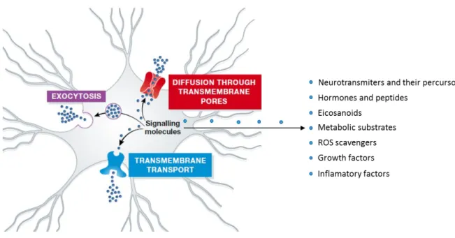
In 1909, less than 20 years after astrocytes been described, Held and his team proves the ability of astrocytes to secret molecules that is known to be crucial for brain homeostasis (Held, 1909). Astrocytes have been already associated to several brain functions, being the synthesis of proteins and adhesion molecules that compose the ECM one of them. However, these cells are also able to synthetize and secrete proteolytic enzymes, as MMPs that play a vital role in ECM degradation and remodeling (Muir et al., 2002). Their role in angiogenesis and BBB induction and maintenance are other physiological aspects attributed to astrocytes action. A better understanding of angiogenesis and of the dynamics between astrocytes and endothelial cells (to regulate BBB stability and permeability) is critical for understanding the process of tumorigenesis and neurogenesis. In fact, astrocytes act as a part of a neuroglial secretory network, that can be defined as gliocrine system of the CNS (Vardjan and Zorec, 2015). With a wide range of substances, some of the astroglia-derived secretory substances are: neurotransmitters and their precursors; hormones and peptides; eicosanoids, scavenger reactive oxygen species (ROS); growth factors; “plastic” factors and pathological molecules, as inflammatory factors (Verkhratsky et al., 2016). The release of all these substances can occur by distinct pathways, including diffusion through plasmalemmal pore/channels; extrusion through plasmalemmal transporters and also by vesicle-based exocytosis (Figure 1.4). Regarding the release by exocytosis, different vesicles have already been identified including: small vesicles, dense core vesicles, lysosomes, exosomes and ectosomes. The vesicles released by astrocytes can have different fusion events with the plasmatic membrane and, more importantly, different contents. The evolutionary conserved family of SNARE proteins are the foundation of this secretory mechanism. The family can be divided in two categories, R-SNAREs and Q-R-SNAREs, being associated with the vesicular membrane or with plasma membrane
- 14 -
proteins, respectively (Jahn and Scheller, 2006). In vesicular release events, there is an increase in intracellular calcium (Ca2+) levels, leading to the association of vesicle proteins to membrane proteins
(syntaxins), and to the formation of a ternary SNARE complex. The proteins of SNARE complex, namely synaptobrevin (VAMP), syntaxin (STX) and synaptossomal associated protein (SNAP), create a 4 α-helical bundle (SNAREpin), that allows the fusion of vesicular and plasma membranes (Sutton et al., 1998, Hamilton and Attwell, 2010, Verkhratsky et al., 2016). The SNARE complex, is not exclusive of astrocytes, and other brain cells like neurons also use the complex to release substances. Interestingly, Ulloa and his co-workers in 2015, inhibiting the SNARE protein STX1 in glioblastoma cells, showed a significant decrease of proliferation and invasion. This work supported the importance of autocrine signaling for glioblastoma cells, but also that the autocrine signaling can be targeted using the complex SNARE (Ulloa et al., 2015).
Figure 1.4 - Secretory pathways and secreted substances by astrocytes - Astrocytes present three main secretory pathways responsible for the release of several substances crucial for brain homeostasis. Between the secreted molecules is usually to found hormones and peptides, metabolic substrates, growth factors and inflammatory factors (Adapted from (Verkhratsky et al., 2016)).
Astrocytes present a significant importance in the response to brain injuries, usually characterized by bleeding and an intense local inflammation. Astrocytes closely positioned to injury, respond to it becoming hypertrophic and forming a scar in conjugation with other brain cells (e.g. fibroblasts), that isolate the damaged tissue from the remaining healthy brain tissue (Cregg et al., 2014). Astrocytes near
- 15 -
the injury site not only upregulate their expression of glial fibrillary acidic protein (GFAP), but also several proteins and transporters, by a process known as astrogliosis (Vijayan et al., 1990, Filous and Silver, 2016). Astrocytes are involved in several neuropathologies playing an important role in the development and progression of the disease. Alzheimer´s disease (AD) is a progressive neurodegenerative disease, characterized by the presence of amyloid beta plaques and neurofibrillary tau tangles. The main component of beta plaques, β-amyloid, is a neurotoxic agent that promotes the response of astrocytes and microglia, leading to the release of inflammation promoting mediators, potentially neurotoxic. In case of injury, the release of these substances is beneficial for brain damage repair, but in a case of a chronic disorder a constant state of activation by astrocytes and microglia leads to a chronic inflammation that contributes to secondary nerve damages. It is currently described that astrocytes activated by β-amyloid release pro-inflammatory cytokines and ROS, contribute to neuronal damage, a hallmark of AD. (Markiewicz and Lukomska, 2006, Filous and Silver, 2016). Interestingly, gap junctions between astrocytes are altered in AD, namely by the observable increase in the gap junctional protein connexin 43 (Cx43)(Nagy et al., 1996). Moreover, gap junction proteins expression has been associated with an increased release of glutamate and ATP, resulting in a glutamatergic cytotoxicity, that leads to neuronal damages (Nakase and Naus, 2004).
Astrocytes are also involved in other progressive neurodegenerative disorders, such as amyotrophic lateral sclerosis (ALS), characterized by the death of motor neurons in cerebral cortex (Vargas and Johnson, 2010). In ALS, astrocytes have an upregulated expression of the inflammatory cytokine transforming growth factor-β1 (TGF-β1), which stops microglia and T cells production of insulin-like growth factor 1 (IGF-1), resulting in a loss of inflammatory-mediated neuroprotection and, consequently, in a faster progression of the disease (Endo et al., 2015). Additionally, Parkinson´s disease (PD), characterized by the loss of dopaminergic neurons in the substance nigra and the presence of Lewy bodies (aggregates of α-synuclein), astrocytes also have an important role on the disease progression. The endocytosis of α-synuclein by astrocytes results in the upregulation of pro-inflammatory cytokines and chemokines, resulting in a constant inflammatory state prejudicial for neurons (Lee et al., 2010, Filous and Silver, 2016). Furthermore, astrocytes are also involved in epilepsy, where the proliferation of reactive astrocytes is a common feature in temporal lobe epilepsy. An astrocytic dysfunction in epilepsy, with alterations in channel expression and dysfunctional gap junctions, leads to an increase in the number of seizures and to glutamate cytotoxicity (Filous and Silver, 2016).
The astrocytic deregulation is the basis of several brain disorders, usually contributing to a constant state of inflammation. Although essential for brain homeostasis, the secretory capacity of astrocytes is
- 16 -
usually altered in neurological disorders, leading to an excessive or deficient release of substances that disrupt the brain homeostasis. The unbalanced release of chemokines and cytokines, and also the disruption of gap junctions between astrocytes, have been already associated to distinct disorders (O’Brien et al., 2014). New therapeutic approaches targeting dysfunctional astrocytes and reducing its inflammatory state, can emerge in the next years. In fact, it is essential to understand not only how dysfunctional astrocytes contribute to several brain diseases, but also how are astrocytic mechanisms relevant for disease progression. In GBM, astrocytes are associated as tumor supportive cells but the underlying mechanisms of this support are still poorly understood. In the next chapter, a literature background will be presented about the interactions between astrocytes and tumor cells.
1.3
Role of astrocytes in gliomas and brain metastases
Since several brain cell types have an impact on the development of diseases, it is difficult to disclose the role of astrocytes, the major glial cell in the brain, in the disease. Despite the emerging studies establishing a connection between these glial cells and glioma cells, the consequences of these interactions remain unclear, especially in vivo. In cases of brain pathology or injury, BBB damage and cancer, astrocytes undergo several morphological changes being this process usually called reactive astrogliosis (Wilhelmsson et al., 2006, O’Brien et al., 2014). In this state, astrocytes present an upregulation of GFAP and vimentin, as well as several growth factors, inflammatory cytokines and ECM proteins. In sporadic lesion cases, the physiological alterations followed by alterations in astrocytic secretion can present benefits for the recovery of the brain damage (Sofroniew, 2009). However, in cancer, the astrocytes surrounding the cancer cells start to secrete several proteins that can help the growth and spread of the cancer. In fact, astrocytes and some of their secreted proteins, have already been proposed to enhance cancer progression (O'Brien et al., 2013). Astrocyte-derived signaling was reported to modulate important aspects of brain cancer progression, such as cell proliferation and tumor invasiveness, which are detailed below.Astrocytes were reported in several studies to promote proliferation of cancer cells. In vitro studies showed that the presence of astrocytes, astrocyte-conditioned media (ACM) or specific growth factors secreted by reactive astrocytes, triggers an increase in cell proliferation in different brain metastasis (Placone et al., 2016). Diverse molecules secreted by reactive astrocytes, as IL-6, IGF-1 or TGF-β, have been independently associated to a proliferation increase in different glioma cell lines (Li et al., 2010, Roth et al., 2010). In studies from Li and co-workers, IL-6 was responsible for an increase of 25% of cell
- 17 -
proliferation in a human glioma cell line U87MG (Li et al., 2010). In a different study, growth differentiation factor 15 (GDF-15), a factor upregulated in reactive astrocytes, was also able to increase cell proliferation (Roth et al., 2010). Besides the effects observed using glioma cell lines, astrocytes were also related with the proliferation of metastatic, breast and lung cancer cells. In fact, studies using these cancer cell lines co-cultured either with astrocytes, or exposed to ACM, present increased proliferative rates (Sierra et al., 1997, Seike et al., 2011). A co-culture of astrocytes with lung cancer brain metastasis, leads to ERK1/2 and AKT phosphorylation in cancer cells, both of which are important signaling pathways for cancer proliferation (Langley et al., 2009). Moreover, in 2015, another study showed that astrocytes presented a significant role in the downregulation of an important tumor suppressor gene, PTEN. Lin Zhang and co-workers demonstrated that in the case of breast cancer brain metastases, astrocytes release microRNAs in their exosomes that epigenetically regulate the expression of PTEN. Interestingly, the down regulation of PTEN was only found in brain metastasis, demonstrating the importance of the microenvironment for metastasis outgrowth (Zhang et al., 2015). Although these findings support paracrine modulation, rather than a direct physical cell-to-cell interaction, the later should not be ruled out.
Along with cell proliferation, migration and invasion are also important features of brain cancer, and are extremely relevant in the outcome of the disease. Several studies already showed that astrocytes are able to promote invasion and migration of glioma cell lines, and of other cancer cell lines (Placone et al., 2016). In 2003, a study using a glioblastoma cell line (U251), revealed that this cell line displays an increased invasion capacity when co-cultured with astrocytes. This effect was attributed to the activation of the inactive pro-MMP2 released by astrocytes. The active form of the MMP -2 is able to degrade collagen IV, one of the major components of ECM, facilitating the infiltration and invasion of glioma cells (Le et al., 2003). More recently, a study using ACM showed an enhancement on the invasion potential of glioblastoma stem-like cells using a trans-well invasion assay (Rath et al., 2013). One of proteins secreted by astrocytes was IL-6, which is able to promote both growth and invasion of glioma cell lines (Li et al., 2010). Moreover, in the last year, a study revealed that the hetero-cellular communication between astrocytes and glioma cells, namely through the gap junctions formed between them, are related with the invasion capacity of glioma cells. In fact, when a transgenic mice without Cx43 in astrocytes was used, perturbing the formation of hetero-cellular channels between glioma cells and astrocytes, a significant decrease of infiltrative edges in the tumor border was observed (Sin et al., 2016). The astrocytic effect in invasion of cancer cells also covers brain metastasis of different cancer types. In primary brain tumors, as well as in human breast and lung cell lines, matrix metalloproteinases, namely MMP-2 and MMP-9, are in part responsible for the astrocyte media-induced tumor cell invasion. In fact, the use of an inhibitor
- 18 -
of MMPs revealed a loss of invasive capacity of these cell lines in vitro, and a decreased capacity to form brain metastasis in a mice model (Wang et al., 2013). The ACM was also able to promote an increase of invasion on melanoma brain metastasis, where factors secreted by astrocytes appear to enhance the migration (Klein et al., 2015).
Another role of astrocytes in gliomas, is their involvement in the evasion of cancer cells from the immune system (Hanahan and Weinberg, 2011, Placone et al., 2016). In fact, factors released by reactive astrocytes have a range of actions that can explain this evasion to the natural killer cells and T lymphocytes, immune system cells, responsible for the elimination of non-natural cells in the human body (Placone et al., 2016). The release of molecules by astrocytes is associated with a constitutive activation of STAT-3 in glioma cells, that results in a suppressive effect in the release of pro-inflammatory cytokines, preventing the response of T-cells. In fact, a co-culture experiment of normal human astrocytes with T cells resulted in a inhibitory effector function (Gomez and Kruse, 2006, Kostianovsky et al., 2008a, O'Brien et al., 2013). Moreover, reactive astrocytes are able to release several other immunomodulatory cytokines, namely IL-10. Among its several functions, IL-10 appears to be responsible for a reduction of antigen presentation, through a down-regulation of monocyte MHC class II expression, as well as for an inhibition of T-cell activity, which protects the neoplastic cells in a cancer condition (Grutz, 2005). Through downregulating of tumor necrosis factor alpha (TNF-α) expression, astrocytes stop the up-regulation of MHC II in microglia and macrophages leading to an impairment in the presentation of antigens to T-cells (Kostianovsky et al., 2008b). The immune protection of cancer cells provided by astrocytes, targets mostly the usual functions of T-cells as their activation or recruitment. Moreover the ECM glycoprotein Tenascin-C (TNTenascin-C), released by astrocytes, is responsible to mediate cell-cell and cell-matrix interactions. The role of ECM has been demonstrated as crucial for cancer, not only by the capacity of TNC to promote glioma invasion, but also to inhibit the transmigration through tumor monolayer by T-cells (Huang et al., 2010, Xia et al., 2016).
Astrocytes, have also been shown to directly protect glioma cells against different drugs, currently used in GBM therapeutics. For instance, the assessment of apoptosis index in glioma cells revealed a decrease in cell death superior to 50% upon treatment with TMZ, when the gliomas are co-cultured with astrocytes. Interestingly, this effect was lost when the researchers try to use ACM or a gap junction inhibitor suggesting that the chemo-protective effect requires a direct contact between astrocytes and glioma cells through connexin 43-based gap junctions (Chen et al., 2015). The same protective effect was also observed when cell lines of brain metastases were exposed to the chemotherapeutic agent paclitaxel (Taxol). The reason for this protective effect is not well understood, but processes such as the
- 19 -
uptake and retention of Ca2+ by astrocytes, or even small non coding RNAs (sRNAs) released by astrocytes,
have already been associated to this effect in brain metastases (Lin et al., 2010, Menachem et al., 2016) .
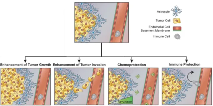
In sum, these studies attribute to astrocytes putative roles in the pathophysiology of primary brain tumors and brain metastases(Figure 1.5). However, the majority of these conclusions were obtained from in vitro studies, with obvious limitations. Taking in consideration the different cell types present in the brain and the interactions between them in the context of brain tumors, these studies fail to simulate the microenvironment complexity around the tumor. Considering that astrocytes communicate with brain cancer cells, it is crucial to understand the impact that these cells may have on glioma, and how the interaction with the surrounding cells in the tumor microenvironment in vivo can modulate disease progression.
Figure 1.5 - Putative roles of astrocytes in cancer progression - Astrocytes may modulate cancer progression through the enhancement of tumor growth and invasion, chemoprotection, and immune escape. Several secreted products by astrocytes can enhance cancer progression by acting directly with neoplastic cells, or by interfering with the role of other non-neoplastic cells (e.g. microglia, macrophages, lymphocytes). Astrocytes can also modulate the extracellular matrix (ECM) conformation, presenting a direct implication in the invasion of cancer cells (Adapted from (Placone et al., 2016)).
- 20 -
1.4
The dnSNARE model for study of astrocyte modulation of microenvironment
In order to assess the role of astrocytic vesicular release in GBM modulation in vivo, it was necessary to select a model with a specific impairment on this capacity. Several models in which astrocytes are modulated by genetic modifications, have been developed to understand the relevance of astrocytes in normal and diseased brain (Oliveira et al., 2015). Among them, the models in which the astrocytes are not able to release substances by exocytosis, such as the dnSNARE, Glast-iBot and GFAP-TeNT, appear as good candidates for the study of astrocytes influence in GBM. The dnSNARE model was selected, since the dnSNARE expression in astrocytes was showed by independent studies to impact the release of gliotransmitters in cell culture conditions and in vivo dnSNARE mice (Zhang et al., 2004, Pascual et al., 2005, Sultan et al., 2015) Moreover, being an inducible model, it prevents developmental effects that could mask potential alterations in the brain microenvironment. The dnSNARE model has been selected for studies in different disorders, among which stand out the studies regarding sleep deprivation and the subsequent effect on cognitive/emotional processes (Halassa et al., 2009, Florian et al., 2011). The release of ATP by astrocytes presented an active role for sleep homeostasis. Moreover, studies with this transgenic model suggested that astrocytes were able to modulate epileptogenesis and
pathophysiological consequences of epilepsy through pathways involving N-methyl-D-aspartate (NMDA)
receptors (Clasadonte et al., 2013). The targeting of astrocytes signaling, mediated by the vesicular release, have been suggested to have a potential benefit for the outcome of stroke in human patients by limiting the spread of damage (Hines and Haydon, 2013). In conclusion, this model was validated by different laboratories and is highly suitable for the purpose of this project.
- 21 -
1.5
Research goals
GBM are the most common and lethal tumors of the CNS, the median survival for the patients is approximately 15 months, with the present treatment. The current treatments available that target almost indiscriminately neoplastic and non-neoplastic cells have been insufficient against GBM. The role and importance of the glioblastoma microenvironment has been emphaticized in recent years, and future therapies may use the microenvironment as an additional target. Astrocytes constitute the majority of glial cells in the brain, and have been described to have an active role in the pathophysiology of GBM. Although the mechanisms are still unclear, in vitro studies, have revealed the importance of paracrine regulation of glioblastoma cells by astrocytes. In this work, it is hypothesized that the secreted products released in vesicles by astrocytes can be responsible for an influence in glioblastoma growth and invasion. To clarify this hypothesis, the dnSNARE mouse model that displays with an impairment in astrocytic vesicular release was studied. In this model, astrocytes have an impaired capacity to modulate the ECM via astrocytic vesicular release. Therefore, in vitro and in vivo complementary approaches were employed to assess whether the astrocytic modulation of the ECM via exocytosis influences GBM. We aimed to evaluate:
1) The effect of conditioned medium secreted by glial cultures derived from WT and dnSNARE mice in viability and migration capacity of a glioma cell line;
2) The effect of astrocytic vesicular release in tumor growth and mice survival, using an in vivo syngeneic orthotopic intracranial GBM model;
- 23 -
- 25 -
2 Materials and Methods
2.1
Cell lines and culture conditions
The mouse glioma 261 cell line (GL261), a kindly donation of Prof. Conceição Pedroso Lima from Center for Neuroscience and Cell Biology, was used in this project. In 1939, Seligman and Shear, obtained a carcinogenic induced mouse glioma model (GL261) through intracranial implantation of methylcholanthrene pellets in the brain mice, and since then this glioma model has been used in the study of GBM (Seligman et al., 1939, Newcomb and Zagzag, 2009).Cells were cultured in Dulbecco´s Modified Eagle Medium (DMEM; Gibco®, USA) supplemented with 10% Fetal Bovine Serum (FBS; Biochrom, UK) and 1% Penicillin-Streptomycin (Invitrogen, USA), which will be designated as complete DMEM from now on. Cells were maintained in a humidified atmosphere at 37ºC and 5% (v/v) CO2, and passaged to new flasks at sub-confluent levels. To perform in
vitro assays, when 80% confluence was reached, GL261 cells were washed with PBS and detached with trypsin at 37ºC for 5 minutes. Trypsin was inactivated using complete DMEM (twice the trypsin volume), collected and centrifuged at 900 rpm for 5 minutes. Cells were resuspended in complete DMEM, and using a 1:1 dilution of trypan blue dye and cell suspension, cells were counted using a Neubauer chamber. Cell density was calculated accordingly with the different assays performed.
2.2
The dnSNARE mouse model
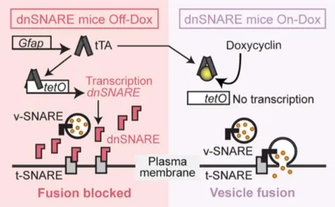
Experiments were conducted in mice expressing a transgenic dominant-negative domain of vesicular SNARE (dnSNARE) and their respective Wild-Type (WT) littermates were used as controls. Animals were obtained using crossing two transgenic mouse lines: GFAP-tTA, in which the expression of tetracycline transactivator (tTA) is mediated by the GFAP promoter; tetO.dnSNARE, in which the dominant-negative domain of vesicular SNARE (Synaptobrevin II/ VAMP2), the reporter enhanced green fluorescent protein (EGFP) and lacZ domain are coexpressed under the control of the tetO promoter. The dnSNARE mice present a “Tet-Off” tetracycline transcriptional activation system where in the absence of doxycycline (dox) the tTA protein binds to tetO operator, triggering the transgene expression, and blocking the vesicle fusion (v-SNARE) with plasma membrane domain (t-SNARE; Figure 2.1). The impairment in SNARE
- 26 -
complex assembly driven by the GFAP promoter results in blockade of exocytosis specifically in GFAP+
cells (Pascual et al., 2005, Fujita et al., 2014, Sultan et al., 2015).
To prevent expression of dnSNARE during mice development, doxycycline (Sigma-Aldrich) was administered in the drinking water (100 µg/ml), and removed 4 weeks before the experiments. Animals were kept in facilities with 12 h light/dark cycle with ad libitum access to food and water.
Figure 2.1. dnSNARE model – Schematic outlining of the “Tet-Off” system used in the transgenic model to impair exocytosis specifically in astrocytes (Fujita et al., 2014).
2.3
Mouse genotyping
Mice were ear tagged using a scissor, and a tail sample was collected for genotyping proposes. In ice, 300 µl of Cell Lysis (Citomed, Portugal) and 1.5 µl of proteinase K (200 mg/mL; Citomed, Portugal) were added to each sample, followed by a spin down to collect the supernatant. The samples were then left overnight to allow the tissue dissociation. In the next day, 100 µl of Protein Precipitation solution (Citomed, Portugal) were added to the cell lysis and after a quick vortex for homogenization, the samples were centrifuged at 14000 rpm during 5 minutes. To induce DNA precipitation, 300 µl of Isopropanol (100 %) were added to the samples, followed by a centrifugation (14000 rpm; 5min). The supernatant was carefully discarded and 300 µl of Ethanol 70 % were added to the pellet. One more centrifugation was done (14000 rpm; 1 min), the supernatant removed and the pellet was left to dry at
- 27 -
room temperature for 60 minutes. Finally, miliQ water was added to the samples and left to incubate at 65ºC for 1 h.
The genotyping was carried out by the polymerase chain reaction (PCR) technique with two pairs of primers, tTA and tetO, that were used in separated PCR mixtures to identify the transgenic mice. Additionally, the constitutive gene HSF-1 was used for control effects. The PCRs were performed in a thermocycler (Mastercycler®, Eppendorf, USA), and the amplified PCR products were separated on a 1.2% agarose gel prepared in Tris-Acetate-EDTA (TAE) running buffer that was boil, before the addition of the green safe (2%). DNA size marker and the samples were loaded in the gel, and electrophoresis at 150V run for 1h. Gel pictures were taken using a transilluminator (Alpha Innotech Corporation, Bio-Rad). The primer sequences and PCR conditions, used for genotyping are present in Table 1.
Table 1 - PCR conditions for genotype identification
Primer sequences for PCR
Primer Sequences
HSF-1 KO1 5´ - TCT CCT GTC CTG TGT GCC TAG C – 3´ HSF-1 KO2 5´- CAG GTC AAC TGC CTA CAC AGA CC – 3´ tTa forward 5´ - ACT CAG CGC TGT GGG GCA TT – 3´ tTa reverse 5´ - GGC TGT ACG CGG ACC CAC TT – 3´ TSL forward 5´- TGG ATA AAG AAG CTC ATT AAT TGT CA – 3 TSL reverse 5´- GCG GAT CCA GCA ATG ATA AGA – 3´
Reaction mix components (10 µl/reaction)
Mix/sample (µl) for tTa Mix/sample (µl) for tetO Buffer (NH4) SO4 10X 1 Buffer (NH4) SO4 10X 1
MgCl2 (25mM) 1.2 MgCl2 (25mM) 1.2 DMSO 99.9% 0.24 DMSO 99.9% 0.24 dNTPs (10mM) 0.24 dNTPs (10mM) 0.24 Primer tTa forward 0.4 Primer TSL forward 0.6 Primer tTa reverse 0.4 Primer TSL reverse 0.6 Primer HSF-1 KO2 0.3 Primer HSF-1 KO2 0.3 Primer HSF-1 KO2 0.3 Primer HSF-1 KO2 0.3 Taq DNA Polymerase 0.4 Taq DNA Polymerase 0.4 H20 miliQ 5.52 H20 miliQ 5.12
- 28 -
Amplification program (40 cycles)
Step Temperature (ºC) Duration (sec) Initial Denaturation 94 300
Denaturation 94 60
Annealing 61.6 60
Extension 72 60
Final extension 72 600
2.4
Primary culture of glial cells
Primary cultures of glial cells were obtained using a modified protocol of Schildge and colleagues (Schildge et al., 2013). Mice between 5 and 7 days’ age (P5-P7) previously genotyped were sacrificed by decapitation and the brain was removed and placed in a cold Hank´s Balanced Salt Solution (HBSS), to maintain pH and osmotic balance of the tissue. Using a magnifier, the olfactory bulbs and brainstem were removed and the hemispheres were separated and open. The hippocampus was carefully removed from both hemispheres, obtaining two cortexes that were used to follow the procedure. Meninges surrounding the cortical tissue were cautiously removed, to avoid contamination by meningeal cells and fibroblasts, and two “clean” cortex were obtained from each pup. Then, the samples from each animal were cut in several pieces and a quick spindown was done to pellet cortex tissue pieces. The supernatant was removed and 1 mL of dissociation medium (2.5 % trypsin; 87.5 % HBSS; 10 % DNase) was added per animal, followed by a 30 minutes’ incubation of the tissue in water bath at 37ºC. Next, a STOP solution (40% FBS and 60% HBSS) was added to inactivate trypsin, followed by a centrifugation at 800 rpm for 2 minutes. After centrifugation the supernatant was discarded and 1.5 mL of complete DMEM was added per sample. The samples were then vigorously resuspended with a pipette until no brain tissue was visible at human eye. One more centrifugation was done and the medium was changed, followed by a new resuspension. At last, with a Neubauer chamber a sample of cell suspension plus Trypan Blue was added (1:1) for quantification effects. For each T25 cell culture flask, 7 x 10⁵ cells were placed, and the medium(complete DMEM) was replaced after the first 48 hours, followed by a medium renovation each 3 days. The primary cells were left to grow for 18-20 days in an atmosphere at 37ºC supplement with 5% (v/v) CO2, the time necessary to reach an 80-90 % confluent state.
- 29 -
To obtain conditioned medium (CM) of glial cultures, cells in culture for 18-20 days were washed twice with PBS followed by another wash with DMEM without FBS. A volume of 3 mL of DMEM (1% Pen-Strep) per T25 was added, and glial cultures were placed at an incubator at 37ºC for 24 hours. After this time, CM derived from WT glial cultures (CM WT) and from dnSNARE glial cultures (CM dnSNARE), were collected and filtered (0.2 µm), followed by a “snap freeze” in liquid nitrogen for 5 minutes. Samples were stored at 80ºC negative, until needed.
2.5
Viability assays
2.5.1 Trypan blue assay Trypan blue assay is a well described test use to determine the number of viable cells present in a cell suspension. The principle is based that viable cells have an intact cell membrane and are able to exclude the trypan blue dye, where dead cells do not, presenting thus a blue cytoplasm (Strober, 2015). GL261 cells were plated in 12-well cell plates at a density of 2 x 10⁴ cells per well and allowed to adhereand grow in a complete DMEM for 48 hours. After this period, cells were washed twice with PBS, and conditioned medium (CM WT or CM dnSNARE) were added to the cells. For control effects, GL261 were also grown in to DMEM with 0 % FBS and DMEM with 10 % FBS. After 48 hours of CM exposure, cells were washed with 500 µl of PBS, followed with 200 µl of trypsin during 5 minutes at 37ºC. Trypsin was inactivated with 200 µl of complete DMEM, and 20 µl of cell suspension were collected to eppendorfs to which was added 20 µl of trypan blue solution. From this mix, 10 µl were placed in a Neubauer Chamber for viable cells counting effects. The results represent the mean of at least three independent experiments, each one in duplicate, and are normalized for the control group (0 % FBS).
2.5.2 MTT assay MTT (3-(4, 5, - dimethylthiazol-2-yl)-2, 5-diphenyltetrazolium bromide) tetrazolium reduction assay is a well stablished technique to analyze cell viability and metabolic cell activity. Viable cells are able to cleavage the tetrazolium salt MTT into formazan (blue/purple colored product), by the mitochondrial enzyme succinate-dehydrogenase. The quantity of formazan produced, absorbance recorded at 570 nm, is proportional to the number of metabolic active cells present in the sample (Slater et al., 1963, Denizot and Lang, 1986). GL261 cells were plated in 24-well cell plates at a density of