Connexins in the early development of the African clawed frog
Xenopus
laevis
(Amphibia): The role of the connexin43 carboxyl terminal tail in the
establishment of the dorso-ventral axis
Jaime Cofre
1and Eliana Abdelhay
21
Laboratório de Embriologia Molecular, Universidade Federal de Santa Catarina, Florianópolis,
SC, Brazil.
2Laboratório de Biologia Molecular Maury Miranda, Instituto de Biofísica Carlos Chagas Filho,
Universidade Federal do Rio de Janeiro, Rio de Janeiro, RJ, Brazil.
Abstract
Connexins are a family of related proteins identified in vertebrate forming gap junctions, which mediate cell-to-cell communication in early embryos, with an important role in establishing embryonic asymmetry and ‘communication compartments’. Byin situ hybridization, immunocytochemistry, reverse transcriptase PCR (RT-PCR) and western blotting we show that a Cx43-like molecule is present in oocytes and embryos of the African clawed frogXenopus laevis, with specific localization in the animal-vegetal axis. This specific distribution is suggestive for an important role for this protein in the establishment of the dorso-ventral axis. Antisense RNA and antibodies directed against rat carboxyl terminal tail of the Cx43 (CT-Cx43) and injected in 1-cell stage Xenopus embryos, induced pronounced al-terations in nervous system development, with a severe ventralization phenotype. Coherently, the overexpression of CT-Cx43 produced a dorsalization of the embryos. In antisense treated embryos, the expression of theβ-catenin gene is eliminated from the Nieuwkoop center, the pattern expression of theChordin, Xnot and Xbra is modified, with no effect in expression of theGoosecoid gene. In CT-Cx43 mRNA treated embryos the pattern of expression of the
β-catenin, Chordin, Goosecoid, Xnot and engrailed-2 genes is modified. The expression ofβ-catenin is increased in the Nieuwkoop center, the expression pattern ofChordin and Goosecoid is expanded to the posterior neural plate andengrailed-2 presents ectopic expression in the ventral region. Taken together our data suggest a role for CT-Cx43 as a maternal determinant with a critical function in the formation of the dorso-ventral axis inXenopus laevis. The Cx43 may be one of the earliest markers of the dorso-ventral axis in these embryos and could possibly be acting through regionalization of factors responsible for the establishment of this axis.
Key words:connexins, carboxyl terminal tail, dorso-ventral axis, early development in amphibians,Xenopus laevis
Received: December 14, 2005; Accepted: November 3, 2006.
Introduction
During the past several years, mouse genetic ap-proaches (Lo, 1999) and dominant negative techniques in frogs of the genus Xenopus (Paulet al., 1995) have demon-strated that cell-cell communication via gap junction chan-nels has an important role in establishing embryonic asymmetry during early development (Levin and Mercola, 1998; Levin and Mercola, 1999) and maintenance of appro-priate organ function during adult life (Lo, 1996). The es-sential role of gap junctions for normal development in Xenopus was suggested by experiments in which disrup-tion of gap juncdisrup-tional communicadisrup-tion using an
anti-connexin antibody injected into a dorsal blastomere led to the loss of anterior-dorsal structures (Warneret al., 1984). Additionally, experiments using the microinjection of dominant negative and wild type connexin mRNAs clearly showed that dorso-ventral differences in gap junctional communication within the early embryo are needed in left-right patterning (Levin and Mercola, 1998; Levin and Mer-cola, 1999).
Six Xenopus connexins have been identified to date. The Cx30 which appears at the gastrula and tailbud stages (Gimlichet al., 1988, 1990), the Cx41 which is not detected until adulthood (Bruzzoneet al., 1996) and four maternal connexins: Cx43, Cx43.4, Cx31 (Landesmanet al., 2003) and Cx38 (Gimlichet al., 1988, 1990). Unlike Cx38, ex-pression of Cx31, Cx43 and Cx43.4 continue zygotically (Landesmanet al., 2003)
The establishment of the dorso-ventral axis is a criti-cal initial step in pattern formation but how polarity origi-nates and is maintained is not well understood. Several studies have suggested that in Xenopus embryos the earli-est step in dorsal axis formation involves the rotation of cortical cytoplasm in the fertilized egg, and this rotation is postulated to localize cytoplasmic dorsal determinants in the future dorsal side (Yugeet al., 1990). This process in-duces dorsal expression of regulatory genes, which in turn establish the Spemann organizer at the start of gastrulation (Kageura, 1997). The Wnt, Vg1 and Noggin factors have dorsal determining activity, but do not display polarized dorso-ventral expression or activity during early develop-ment in Xenopus. Thus, attention has turned to the cyto-plasmic components of these pathways. The principal downstream signaling target of Wnt-1 is aβ-catenin (Lara-bellet al., 1997) and its overexpression is sufficient to in-duce a complete secondary axis (Guger and Gumbiner, 1995; Sokolet al., 1995).
Xenopus embryos exhibit a polarity in gap junctional permeability at the 32-cell stage, as dorsal blastomeres transfer the dye Lucifer Yellow more frequently than do ventral blastomeres (Guthrie, 1984). The gap junctional permeability is increased between ventral animal pole cells in Xenopus embryos treated with dorsalizing agents like lithium chloride (Nagajskiet al., 1989), Wnt-1 and Wnt-8 (Olsonet al., 1991). The dorso-ventral pattern and its rela-tion to cell coupling was further reinforced by studies with ultraviolet light, in which a ventro-posterior phenotype was observed, with a concomitant decrease in dye transfer on the dorsal side of the embryo (Nagajskiet al., 1989). Thus, ventralization seems to correlate with reduced dye transfer activity, while increased dye transfer correlates with dorsa-lization. These studies support the idea that gap junction channels may be involved in coordinating dorso-ventral polarity in the early embryo, although no direct experimen-tal evidence has yet been presented in favor of such a model.
In recent years, some evidence for the participation of connexins hemi-channels in cellular physiology has ap-peared, contributing in diverse and different processes as apoptosis, cellular proliferation, tumor growth, calcium wave propagation and ephaptic neuronal communication (Goodenough and Paul, 2003). These evidence indicates that connexins have independent functions besides cell-cell communication and also in many cases these functions are independent of the cellular membrane. Segretain et al. (2003) showed that the proliferation of tumor lines is asso-ciated with intracellular sequestration of endosomal Cx43 and, therefore, not inserted in the cellular membrane. More-over Zhanget al(2003) demonstrated that the injection of the Cx43 carboxyl tail alone is enough to produce a tumor suppressor effect. However, the role of cellular communi-cation in this process was completely excluded by the ex-periments of Olbina and Eckhart (2003), in which a mutant
of Cx43 for the second extracellular region was created. Immunofluorescence detected the mutant protein in the cy-toplasm but not in the plasmatic membrane, however, the effect of cellular growth suppression still was observed. At present, it cannot be affirmed that all connexin functions are related to communication channels and connexin func-tions may not be restricted to embryonic stages where intercellular communication has already been established. With this hypothesis in mind we searched for the expres-sion of a Cx43 homologue in the African clawed frog Xenopus laevis,this connexin being the first and more ubiq-uitous expressed connexin found during mouse develop-ment (Davies et al., 1996). We also used sense and antisense RNA microinjection to investigate the role of Cx43 during the early development ofX. laevis.
Material and Methods
Biological material and embryo experiments
Specimens of the African clawed frogXenopus laevis (Daudin, 1802) (Amphibia, Anura, Pipidae) were main-tained in aquaria at 16 °C on a diet of purina dog chow. Egg induction and fertilization is described below.
Embryos were obtained from adult (2 years old) fe-maleX. laevisby hormone induced egg laying and fertil-izedin vitrousing standard methods (Heasmanet al., 1991) and staged according to Nieuwkoop and Faber (1967).
To assay for possible functions of the Cx43-like prod-uct in the early development of the nervous system we in-jected synthetic Cx43 antisense RNA (500 pg/10 nL) into embryos at the one or two-cell embryo stage. As a specific control for the antisense injection assays, embryos were in-jected with 1 ng of Cx38 antisense RNA or 1 ng of Cx32 antisense RNA. Also, other groups of 1 cell stage embryos were injected with mRNA in the vegetal region to demon-strate the presence of ectopic CT-Cx43 after injection.
Molecular biology
Forin vitroRNA synthesis the Cx43(subcloned in the PGEM 7z+ vector), CT-Cx43 (subcloned in the PcDNA3 vector), chordin, goosecoid, engrailed-2, Xnot andXbragenes were transcribed as described by Harland and Weintraub (1985). The RNA (mRNA or antisense probe) was extracted by a standard procedure, precipitated with lithium chloride 7.5 M and diluted in water treated with diethylpyrocarbonate (DEPC) for injection or in situ hybridization.
For the in situ hybridization of whole-mount embryos digoxigenin or fluorescein labeled Cx43, chordin, goose-coid, Xnot, and Xbra cDNAs antisense RNA probes were constructed (Harland and Weintraub, 1985) and embryos prepared, hybridized and stained by the method of Harland (Harland, 1991).
and 1-cell stage Xenopus embryos (n = 10) with TRizol (Gibco) and treated with Dnase I Rnase-free (Ambion) and used to synthesize cDNA. The RT-PCR was performed in a finale volume of 50µL at 94 °C for 30 s, 58 °C for 1 min, 72 °C for 1 min 30 s and repeated for 33 cycles and the products were resolved in 1% (w/v) agarose gel and stained with ethidium bromide. The RT-PCR was amplified using a set of specific primers for Xenopus Cx43 (α1) (forward 5’-ggaattcggtacatgtatgggtttagc, reverse 5’-ggggtaccaggtcg tggtctgctactag), xCx38 (forward 5’-gtggacagacgagcagtc ag, reverse 5’-actcaactttatgtttgcat), rat Cx26 (forward 5’-atgtacaatggcttcttcat, reverse 5’-gggacttcaaatggcggcat.) and mouse Cx50 (forward 5’-aaaggaccgtgaagctgagg, re-verse 5’-acttctctcccacttccggt).
Immunocytochemistry and Western blotting
The embryos were fixed in 4% (w/v) formaldehyde solution for 1 h and a polyclonal antibody for residues 346-360 of rat Cx43 (rCx43) or a polyclonal antibody for
β-catenin were added together with a secondary alkaline phosphatase-coupled anti-rabbit antibody and developed using Nitro-Blue Tetrazolium Chloride (NBT) and 5-Bro-mo-4-Chloro-3’-Indolyphosphate p-Toluidine Salt (BCIP) (Sigma).
To investigate the presence of connexins during the early development of Xenopus, protein extracts were pre-pared by direct lysis in SDS sample buffer from 2-cell stage control embryos (n = 10), embryos at the 2-cell stage treated with Cx43 antisense RNA (n = 10) and embryos in-jected with antisense RNA and rescued with CT-Cx43 mRNA (n = 10). Rat liver and heart were used as a positive control. All protein extracts were analyzed by Western blotting.
The specific distribution of Cx43 expression in oocytes was determined by dissecting oocytes (n = 10) into the pigmented animal (An) and vegetal (Veg) hemisphere which were independently analyzed by Western blotting.
For Western blotting proteins were separated using 12.5% SDS-PAGE and transferred to Hybond-C extra (Amersham) membranes. Membranes were blocked using 5% non-fat dry milk in phosphate buffered saline Tween-20 (PBST) and incubated for 1 h with the polyclonal body rCx43 (Zymed), a polyclonal rCx32 anti-body or a monoclonal anti-actin antianti-body (Sigma) as probes and 1 h with the appropriate alkaline-phosphatase-coupled secondary antibody (Sigma) and developed with a standard protocol provided by the manufacturer.
Note that some specific methodological details are given in the legends to the accompanying figures.
Results
Expression pattern of Cx43-like protein
To investigate the presence of connexins during the early development of Xenopus, we first screened protein
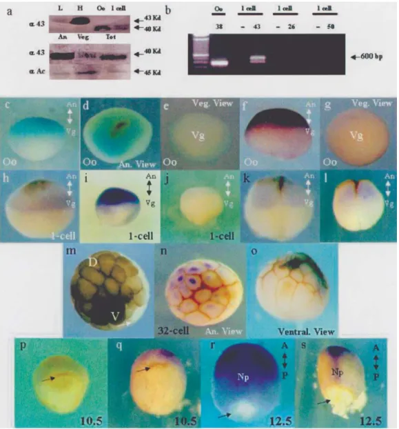
extracts from oocytes and 1-cell stage embryos using West-ern blotting. We detected a 40 KDa Cx43-like protein the oocytes and 1-cell stage embryos (Figure 1a, upper panel). The expression of this Cx43-like mRNA was detected by RT-PCR in the 1 cell stage embryos (Figure 1b).
The specific distribution of Cx43-like expression in the pigmented animal hemisphere and the vegetal hemi-sphere of dissected oocytes as detected by Western blotting is shown in Figure 1a. Surprisingly, we detected differen-tial expression of a Cx43-like protein was in the animal and vegetal regions of the oocytes (Figure 1a, middle panel), the Cx43-like protein being more abundant in the animal hemisphere. In this experiment much more vegetal hemi-sphere protein was loaded on the gel (Figure 1a, lower panel, actin control). These analyses were done with West-ern blots, because Xenopus embryos are opaque and only a very thin surface of the embryos can be stained with anti-bodies and also because whole-mount staining is not quan-titative. However, the specific distribution of this Cx43-like molecule in Xenopus oocytes and embryos was also confirmed by whole mountin situ hybridization and immunocytochemistry, the weakly-staining brown streaks visible in Figure 1 being the residual animal pole cortex pigments which we conserved in order to selectively iden-tify the animal hemisphere and the dorsal regions of the em-bryos. The earliest detectable expression of Cx43-like mRNA (Figure 1c-e) and protein (Figure 1f-g) in oocytes occurred before fecundation only in the animal hemisphere, confirming our Western blot results. This expression pat-tern was maintained throughout the 1-cell stage embryo (Figure 1h, i). At this stage the competition assay with the rCx43 antigenic peptide blocked recognition by the polyclonal antibody used (Figure 1j). Subsequently, with the first division of the embryo, the Cx43-like message and protein appeared localized in the animal hemisphere (Fig-ure 1k, l) but at the later 8 to 64-cell stages mRNA and pro-tein expression were confined to one domain of the animal blastomeres (Figure 1m, n, o), principally in less pigmented blastomeres in the presumptive dorsal region of the very early Xenopus embryos (Nieuwkoop and Faber, 1967), and was conserved in a specific dorsal pattern until the gastrula (Figure 1p, q) and neurula stage embryo (Figure 1r, s). These results are in line with the polarity found in 32-cell stage gap junctional permeability (Guthrie, 1984). Taken together these results suggest that a Cx43-like protein con-stitutes one of the first dorsal region markers appearing in early Xenopus embryos.
Phenotypic effects of CT-Cx43 sense and antisense RNA
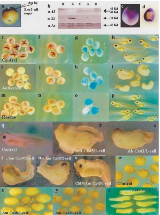
the expression of the Cx43-like protein was specifically re-duced at the injection site (Figure 2, compare c with d) and was dramatically reduced in the whole animal region in the 2-4 cell stage (Figure 2, compare e with i) and partially re-duced in the 32-cell stage embryos (Figure 2, compare f with j) as assayed by immunocytochemistry using a poly-clonal antibody for rat Cx43. Similar results were obtained by Western blot analysis, demonstrating a total reduction of Cx43-like expression using the Cx43 antisense RNA ap-proach (Figure 2b, compare columns C and A). These ex-periments were done as a control to demonstrate that Cx43 antisense RNA could eliminate the Cx43 expression de-tected by our antibodies. We recovered expression of the
The phenotypic analysis of embryos injected with Cx43 antisense RNA into the animal region, showed the ab-sence of eyes, a deformed head shape and severe ventra-lization (Figure 2, compare h with l and q with r), this phenotype being present in 62% (n = 50) of embryos in-jected with Cx43 antisense RNA. The same phenotype was also observed after injection of rCx43 polyclonal anti-body (Figure 2s), with 55% (n = 20) of embryos injected with anti-rCx43 polyclonal antibody displaying this pheno-type. This ventralization was rescued by injection of Cx43 mRNA after the antisense RNA injection (Figure 2p). When one blastomere was injected in a 2-cell stage embryo, the injected side showed ventralization (Figure 2t) while the other side of the same embryo appeared almost normal (Figure 2u), with 42% (n = 19) of injected embryos display-ing this phenotype. However, injection of a plasmid carry-ing Cx43 antisense RNA under the control of the CMV promoter showed little effect on the embryos, which were absolutely normal with respect to the absence of eyes, head, cement gland and ventralization (Figure 2v), indicating that the Cx43 connexin participates in dorso-ventral axis forma-tion at a very early stage during development. As a specific control for antisense injection assays, Xenopus embryos were injected with 1 ng of Cx38 antisense RNA (Figure 2x, z), a maternal connexin, or 1 ng of Cx32 antisense RNA (Figure 1y, z’), which were unable to produce dorso-ventral axis modifications in the neurula (Figure 2, Compare x and y with w) and tadpole stage (Figure 2, compare z and z’ with q).
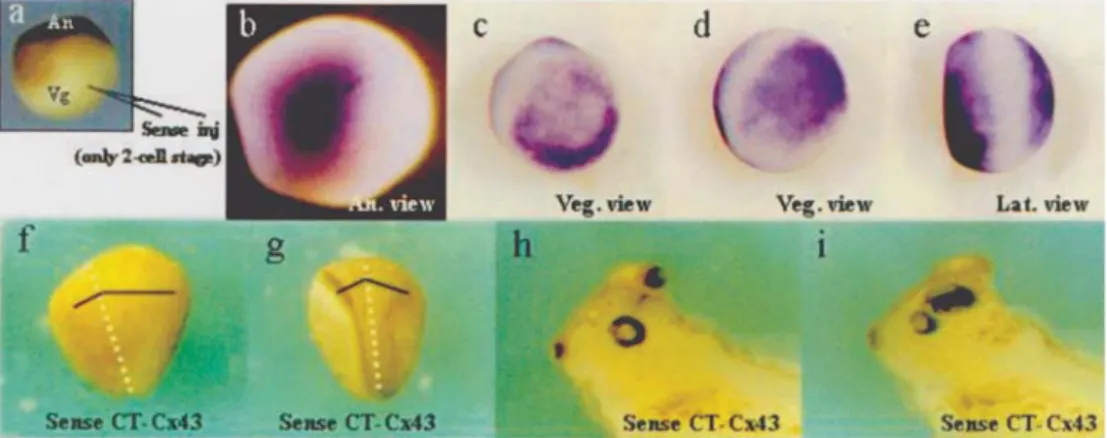
The role Cx43 has in dorsal-ventral axis establish-ment was further investigated by injecting CT-Cx43 sense mRNA into the vegetal hemisphere of 2-cell stage embryos (Figure 3a). We detected an expansion of the anterior neu-ral plate in the injected side in stages 12.5 (Figure 3f) and 13.5 (Figure 3g) and at the 38/39 stage the embryos showed
an additional eye on the injected side (Figure 3, compare h with i). This effect was observed in only 13% of sense in-jected embryos (n = 40). Also, other groups of 1 cell stage embryos were injected with mRNA in the vegetal region to demonstrate the presence of CT-Cx43 located ectopically after the injection of the mRNA (Figure 3, compare c-e with b).
Molecular effects of antisense and sense CT-Cx43
To define the molecular effects of Cx43-like ablation, we analyzed the expression of different dorsal markers, includingβ-catenin, in embryos injected with antisense Cx43 mRNA during the 1-cell stage. A strong reduction of bothchordin(Figure 4, compare a with b) andxnot (Fig-ure 4, compare j with k) expression was observed in early gastrula (stage 12.5) and neurula embryos (stage 10.5), re-spectively. The expression ofgoosecoidwas increased in 10 stage embryos or unaltered in stage 12.5 embryos (Figure 4, compare e with f and g with h). Sincegoosecoid displays cell-autonomous activation, this suggest that cell-autonomous events also may play a role in the defini-tion of the dorso-ventral axis in Xenopus (Lemaire and Gurdon, 1994). In addition,xbra showed increased ex-pression with expanded domains into the animal region at the early gastrula stage (10.5 stage) (Figure 4, compare m with n). The expansion of the mesodermal plate could be responsible for the restriction of neural territories in the presumptive ectoderm and consequently in the loss of dorso-anterior structures. This phenomenon probably re-flects the fact that the early patterning of the vertebrate mesoderm occurs by first establishing broad domains of gene expression which are subsequently refined by intergenic interaction (Ghysen and Dambly-Chaudière, 1988; Stern, 1954). Consistent with these results we
tected a strong reduction inβ-catenin expression in the 1-cell stage embryos injected with Cx43 antisense mRNA during the same stage (Figure 4).
To investigate the molecular effects of CT-Cx43 overexpression, we analyzed the expression ofchordin, goosecoid, xnot,engrailed-2andβ-cateninin 1-cell stage embryos which had had their vegetal region injected with CT-Cx43 sense mRNA. We detected expanded chordin andgoosecoidexpression at the posterior neural plate of 12.5 stage embryos (Figure 4c, i). When one blastomere of a 2-cell stage embryo was injected with the Cx43
antisense mRNAchordinexpression was reduced only in the injected side (Figure 4d).The expression ofxnotwas increased with expanded domains of the lateral lines be-fore gastrulation in a 10.5 stage embryo. We also detected ectopic expression ofengrailed-2(En-2) in the ventral re-gion of tadpole stage embryos (Figure 4p-q, this pheno-type being present in 57% of injected embryos, n = 69) when compared with normal En-2 expression (Figure 4o, s), this being a sign of embryo dorsalization. Some em-bryos displayed complete inversion of engrailed-2 ex-pression without dorsal exex-pression and showing a
complete neuralization of the ventral region (Figure 4r, this phenotype being present in 10% of injected embryos, n = 69). These results are consistent with the assumption that that the function of the Cx43-like protein is to dorsalize embryos and suggests that neural plate expan-sion could be caused by alterations in the expresexpan-sion of dorsal genes before or during neurulation, reinforcing the importance of connexins in the establishment and defini-tion of neural territories in the early development ofX. laevis. Coherently, the overexpression of CT-Cx43 pro-duced a clear increment ofβ-catenin concentration in the Nieuwkoop center (Figure 4, compare v with t). Our anal-yses of β-catenin expression in embryos injected with sense or antisense CT-Cx43 mRNA showed that CT-Cx43 modified the earliest molecular marker which acts by ini-tiating the cascade of events that induces the establish-ment of the dorso-ventral axis. The observed effect on
β-catenin expression is also coherent with the observed phenotypic effect in embryos treated with CT-Cx43 mRNA. However it is not clear how the asymmetric distri-bution of connexins relates to the establishment of differ-ences in the dorso-ventral axis.
Discussion
Heterologous characterization of Cx43-like protein
Connexin proteins have a well-established role in cell-cell communication (Kumar and Gilula, 1996). Along with the expansion of the list of connexin-associating pro-teins, reports have appeared that suggest that in addition to their channel function connexins might have other roles such as transcriptional and cytoskeletal regulation and that these functions are dependent of the specific interaction of CT-Cx43 and cellular proteins (Giepmans, 2004). The Xenopus Cx43 has been cloned by Gimlichet al.(1990), with this clone presented 87% of amino acids identities and 89% of nucleotides identities between Rat and Xenopus Cx43. Several works have been published using rat Cx43 as a tool to study the role of the gap junction in the early devel-opment of Xenopus. For example, (Paulet al., 1995) used a dominant negative approach with rat Cx43 mutated to block the activity of cell-cell communication in embryos, while (Levin and Mercola, 1998) described the importance of connexin in the establishment of left-right asymmetry using rat Cx43 as a tool. We applied the method of Levin and Mercola to detect a Cx43 homologue in the very early development of Xenopus and used antisense or ectopic sense mRNA injection assays and phenotypic or molecular analysis of the resulting embryos to propose a role for Cx43 in the induction of the dorso-ventral axis. The high identity between Rat and Xenopus Cx43 makes our antisense RNA an appropriate probe for detection and injection in Xenopus embryos. Moreover the primers used for our RT-PCR ex-periments were generic primers that we made taking full
advantage of the similarity between Cx43 in different spe-cies.
The present ‘connexin system’ nomenclature for gap junction subunits is primarily based on the molecular weight of proteins deduced from cDNA sequences (Beyer et al., 1987), but, however, very closely related proteins from different species can have different predicted molecu-lar weights. For instance, the mammalian Cx32 is 32 kD whereas the Xenopus homologue is 30 kD (Gimlichet al., 1988). Although he Cx43 protein is highly homologous be-tween Rat and Xenopus (Gimlich et al., 1990) Xenopus Cx43 can be found in several isoforms depending on the tis-sue analyzed. For example Gimlichet al., (1990) detected several variable-sized Cx43 transcripts from the ovary and characterized another Cx43 from Xenopus heart that was very abundant but smaller than the first cloned Xenopus Cx43. These observations are supported by our findings outlined in the present paper where we describe the detec-tion of a 40kD Cx43-like protein in the very early develop-ment of Xenopus.
Specificity of the probes and experiments
We consider that in order to demonstrate the specific-ity of any antisense RNA effect four criteria must be ful-filled. One criterion is to establish that other related and unrelated antisense RNAs have no such effects at compara-ble or higher doses. The Cx38 connexin is a unique mater-nal connexin representative of theα-connexin family (as is Cx43) while Cx32 is the principal representative of the
β-connexin family, both these connexins being used by us in doses twice as large as that used for Cx43 but did not pro-duced any phenotypic effect in the embryos (Figures 2x-z). A second criterion is to establish, by Western blot analysis and other techniques, that the antisense RNA is specifically decreasing the steady-state level of the protein under study while having no effect on other proteins used as controls. In our study we found reduced expression of a Xenopus Cx43-like protein by western blotting (Figure 2b) and immunocytochemistry (Figure 2d, i) while the expression of the actin positive control remained unaltered. A third cri-terion is to show that the phenotypic effects of appropriate antisense mRNA can be rescued by co-expression of wild type mRNA, as illustrated in Figure 2p. A fourth criterion is to demonstrate that antisense mRNA effects on connexin expression can be rescued by co-expression of wild type mRNA. All these criteria were fulfilled in our experiments, which thus demonstrate the specificity of the effect of ap-propriate antisense RNA against Cx43.
The antisense approach
studies has been suggested as a cause for the variability of reported results, and Morpholine oligo complementary Heasman techniques aimed at stabilizing the injected RNA have been proposed (Heasman, 2002; Heasman et al., 2000). In that regard we would like to point out that we did not use Morpholines and we presume that our antisense has at least 300-400 base pairs, rendering it more prone to rapid degradation. Secondly, and possibly more importantly, we have shown that the antisense injections decreased the ex-pression of the target protein, at least transiently (2 b, d, i, j). We appear to have found the correct time window for de-creasing expression of the Cx-like molecule, this being one of the major findings of our study. We believe that we were dealing with this Cx-like molecule at a stage when it was not acting as an intercellular channel and the transient de-crease of its expression during this critical time period dur-ing development produced the effects described in this paper (Figure 2l, r). In that regard, we feel that the main criticism directed at the use of antisense strategies (Heasman, 2002) does not affect our work. In fact, we think that if the expression of the protein is decreased for longer periods the effects reported in the literature for connexins will manifest themselves,i.e.delamination of cells in gas-trulation and a high percent of non-viable embryos (Paulet al., 1995). In our opinion, the host-transfer technique is un-suitable for achieving selective reduction in the 1 to 2 cell stage. We have also shown that antibody injection produces the same effect as antisense techniques (Figure 2s), sense RNA producing exactly the opposite effects to the anti-sense treatment (Figure 3). Our data also shows that antisense injections work, at least during the developmental period when it appears that that the target molecule plays a fundamental role in establishing the dorso-ventral pattern-ing of the embryo.
Connexins and their interactions
The binding of α-β-catenins to the zonula occlu-dens-1 (ZO-1) protein is required for Cx43 transport to the plasma membrane during the assembly of gap junctions (Wuet al., 2003) and the ZO-1 ‘scaffold’ protein might re-cruit signaling proteins into Cx43-based gap junctions (Giepmans, 2004). Recent studies have shown the forma-tion of an α-β-catenin- (ZO-1) -Cx43 complex in rat cardiomyocytes and epithelial cells. The first structural characterization of a connexin domain integrated into a multimolecular complex was recently published by (Sorgenet al., 2004). It is known that c-Src can disrupt the connexin43 (Cx43)-zonula occludens-1 (ZO-1) interaction and lead to down-regulation of gap junction intercellular communication, the ZO-1 and c-Src binding sites corre-sponding to widely separated Cx43 domains (Sorgenet al., 2004). The c-Src tyrosine kinase phosphorylates CT-Cx43 and is involved in G protein-mediated inhibition of Cx43 gap junctional communication (Giepmans, 2004).
(Moyer and Ehrlich, 2003) have shown that in normal fibroblasts Cx43 and β-catenin were evenly distributed throughout the cell, but in cells treated with hyaluronan these proteins were co-localized to the cell surface. Further-more, (Collares-Buzato et al., 2001) have shown that connexin 43 andβ-catenin are co-localized in neonatal pan-creatic islets cells treatedin vitrowith prolactin and consid-ered this interaction to be an important event in thein vitro maturation process of neonatal pancreatic islets cells. (Aiet al., 2000) characterized the Wnt family of secreted poly-peptides as regulators of Cx43 expression and gap junction channel function in mouse intact hearts and dissociated mouse myocytes and have shown that in response to Wnt signaling Cx43 co-localized with β-catenin in the junc-tional membrane and coherently in a transgenic cardio-myopathic mouse model exhibiting ventricular arrhythmias and gap junctional remodeling,β-catenin and Cx43 expres-sion being downregulated in conjunction.
These studies by other workers support our experi-ments involving mRNA overexpression and the injection of antisense in which we observed the simultaneous in-crease or reduction ofβ- catenin and Cx43 (Figure 4t, u, v). Despite lack of evidence of a direct interaction between connexins andβ-catenin to date, the physical interactions found so far and a possible function in gap junctional chan-nel-independent regulation of gene expression by Cx43 se-questering transcriptional regulators are noteworthy and relevant to developmental biology.
On the other hand, (Giepmanset al., 2001) identified the interaction between CT-Cx43 and alpha/beta-tubulin dimers (microtubules) which extend with their distal ends to Cx43-based gap junctions, suggesting that Cx43 gap junctions may play a novel role in regulating microtubule stability in contacted cells. Other observations support the premise that direct association of the cytoskeleton with gap junctions plays a significant role in the regulation of Cx43 expression and distribution through integrin-mediated sig-nal transduction pathways (Guoet al., 2003). It may be that the channel-independent effects of connexins, which have now been reported by many groups, might be explained by dysfunction or function of connexin-tail interacting pro-teins (Goodenough and Paul, 2003).
Overexpression approach
under-stand the complex network of interactions and regulation in the cytoplasm of the cells. In C6 glioma cells transfected with Cx43 it has been possible to characterize several genes, including those coding for a number of secreted fac-tors which may play a role in modulating the tumor pheno-type of the cells, helping to better understand the functional studies carried out with these cells (Naus et al., 2000). However, on the other hand, there are no publications de-scribing the effect of CT-Cx43 overexpression on the cellu-lar genes that can affect the interpretations of our data.
Final Considerations
During the research described in this paper we de-tected both mRNA and protein at the 1 or 2-cell stage of development when cell-cell communication is not yet es-tablished. The connexin signal detected suggests a coherent pattern with a maternal determinant, mainly based on the asymmetrical distribution of the connexin protein in the an-imal-vegetal axis of oocytes and embryos. Our functional studies of blockade and overexpression of the CT-Cx43 factor show that this can modify the establishment of the dorso-ventral axis, suggesting an important role for CT-Cx43 in the early formation of this axis. (Nascone and Mercola, 1997) have pointed our that dorso-ventral asym-metry is important in establishing the left-right patterning of the embryo and connexins are known to be involved in the genesis of this patterning (Levin and Mercola, 1998). The dorso-ventral axis is established during the initial events of embryonic development, before left-right axis formation. Since the critical events involved in dorso-ventral axis formation occurs between fecundation and the first cleavage of the embryo, strategies to ablate Cx expres-sion in 4-8 cell stage embryos cannot interfere with dorso-ventral patterning because they affect only left-right axis formation.
The carboxy-terminal (COOH) domains of connexins are located in the intracellular space and have important roles in regulation of cell-cell communication (Bruzzoneet al., 1996). Changes in Cx43-mediated gap junctional com-munication by growth factors (Hossainet al., 1999), onco-genes (Kanemitsuet al., 1997; Looet al., 1995) and activa-tors of protein kinases (Kanemitsuet al., 1998; Kanemitsu et al., 1997; Kimet al., 1999; Warn-Crameret al., 1996) have been shown to require an intact COOH domain. It is known that Cx43 is also a substrate for phosphorylation by protein kinase C (PKC), Mitogen-Activated Protein Kinase (MAPK) and Src protein kinase (v-Src), in which the SH3 (Src Homology-3) domain of v-Src binds to the COOH do-main of Cx43 (Hossainet al., 1999). This opens the possi-bility that the carboxyl domain of Cx43, through associa-tion with SH2 (Src homology-2) and SH3-containing proteins, could direct these proteins to separate compart-ments within the cytoplasm. The posterior cleavage of the embryo would then help to establish and generate the dorso-ventral axis. Based on these results, we propose that
a Cx43-like protein molecule acts as a maternal protein dur-ing the very early development in Xenopus and has a major role in the formation of the dorso-ventral axis.
Acknowledgments
We wish to thank Dr. A.C. Campos de Carvalho and M.O. Masuda for the opportunity to start this work as well for the critical review of this manuscript; Dr. E.L. Hertzberg for providing the anti-Cx43 antibodies; Dr. E. De Robertis for goosecoid andchordin; Dr. K. Cho for brachyuryand Dr. D. Kimmelman forXnotcDNA clones. We also thank Dr. E. Kalapotakis for the original design of Cx primers. We are grateful to D. Avanzi for technical as-sistance. This work was supported by grants from the Bra-zilian agency FAPERJ.
References
Ai Z, Fischer A, Spray DC, Brown AMC and Fishman GI (2000) Wnt-1 regulation of connexin43 in cardiac myocytes. J Clin Invest 105:161-171.
Beyer EC, Paul DL and Goodenough DA (1987) Connexin43: A protein from rat heart homologous to a gap junction protein from liver. J Cell Biol 105:2621-2629.
Bruzzone R, White TW and Goodenough DA (1996) The cellular internet: On-line with connexins. BioEssays 18:709-718. Collares-Buzato CB, Leite AR and Boschero AC (2001)
Modula-tion of gap and adherens juncModula-tional proteins in cultured neo-natal pancreatic islets. Pancreas 23:177-185.
Davies TC, Barr KJ, Jones DH, Zhu D and Kidder GM (1996) Multiple members of the connexin gene family participate in preimplantation development of the mouse. Developmental Genetics 18:234-243.
Ghysen A and Dambly-Chaudière C (1988) From DNA to form: The achaete-scute complex. Genes and Development 2:495-501.
Giepmans BN, Verlaan I and Moolenaar WH (2001) Connexin-43 interactions with ZO-1 and alpha- and beta-tubulin. Cell Commun Adhes 8:213-217.
Giepmans BNG (2004) Gap junctions and connexin-interacting proteins. Cardiovascular Research 62:233.
Gimlich RL, Kumar NM and Gilula NB (1988) Sequence and de-velopmental expression of mRNA coding for a gap junction protein in Xenopus. J Cell Biol 107:1065-1073.
Gimlich RL, Kumar NM and Gilula NB (1990) Differential regu-lation of the levels of three gap junction mRNAs in Xenopus embryos. J Cell Biol 110:597-605.
Goodenough DA and Paul DL (2003) Beyond the gap: Functions of unpaired connexon channels. Nature Reviews Molecular Cell Biology 4:285-295.
Guger KA and Gumbiner BM (1995) [beta]-catenin has Wnt-like activity and mimics the nieuwkoop signaling center in Xenopus dorsal-ventral patterning. Developmental Biology 172:115-125.
Guo Y, Martinez-Williams C and Rannels DE (2003) Gap junc-tion-microtubule associations in rat alveolar epithelial cells. Am J Physiol Lung Cell Mol Physiol 285:L1213-1221. Guthrie SC (1984) Patterns of junctional communication in the
Harland R and Weintraub H (1985) Translation of mRNA injected into Xenopus oocytes is specifically inhibited by antisense RNA. J Cell Biol 101:1094-1099.
Harland RM (1991)In situhybridization: An improved whole-mount method for Xenopus embryos. Methods Cell Biol 36:685-695.
Heasman J (2002) Morpholino oligos: Making sense of antisense? Developmental Biology 243:209.
Heasman J, Holwill S and Wylie C (1991) Fertilization of cultured Xenopus oocytes and use in studies of maternally inherited molecules. Methods Cell Biol 36:45-60.
Heasman J, Kofron M and Wylie C (2000) [beta]-catenin signal-ing activity dissected in the early Xenopus embryo: A novel antisense approach. Developmental Biology 222:124. Hossain MZ, Jagdale AB, Ao P, Kazlauskas A and Boynton AL
(1999) Disruption of gap junctional communication by the platelet-derived growth factor is mediated via multiple sig-naling pathways. J Biol Chem 274:10489-10496.
Kageura H (1997) Activation of dorsal development by contact between the cortical dorsal determinant and the equatorial core cytoplasm in eggs of Xenopus laevis. Development 124:1543-1551.
Kanemitsu MY, Jiang W and Eckhart W (1998) Cdc2-mediated phosphorylation of the gap junction protein, connexin 43, during mitosis. Cell Growth Differ 9:13-21.
Kanemitsu MY, Loo LWM, Simon S, Lau AF and Eckhart W (1997) Tyrosine phosphorylation of connexin 43 by v-Src is mediated by SH2 and SH3 domain interactions. J Biol Chem 272:22824-22831.
Kim DY, Kam Y, Koo SK and Joe CO (1999) Gating connexin 43 channels reconstituted in lipid vesicles by mitogen-activated protein kinase phosphorylation. J Biol Chem 274:5581-5587.
Kumar NM and Gilula NB (1996) The gap junction communica-tion channel. Cell 84:381-388.
Landesman Y, Postma FR, Goodenough DA and Paul DL (2003) Multiple connexins contribute to intercellular communica-tion in the Xenopus embryo. J Cell Sci 116:29-38.
Larabell CA, Torres M, Rowning BA, Yost C, Miller JR, Wu M, Kimelman D and Moon RT (1997) Establishment of the dorso-ventral axis in Xenopus embryos is presaged by early asymmetries in beta-catenin that are modulated by the Wnt signaling pathway. J. Cell Biol 136:1123-1136.
Lemaire P and Gurdon JB (1994) A role for cytoplasmic determi-nants in mesoderm patterning: Cell-autonomous activation of the goosecoid and Xwnt-8 genes along the dorsoventral axis of early Xenopus embryos. Development 120:1191-1199.
Levin M and Mercola M (1998) Gap junctions are involved in the early generation of left-right asymmetry. Developmental Bi-ology 203:90-105.
Levin M and Mercola M (1999) Gap junction-mediated transfer of left-right patterning signals in the early chick blastoderm is upstream of Shh asymmetry in the node. Development 126:4703-4714.
Lo CW (1996) The role of gap junction membrane channels in de-velopment. Journal Bioenergetic and Biomembrane 28:337-383.
Lo CW (1999) Genes, gene knockouts, and mutations in the anal-ysis of gap junctions. Developmental Genetics 24:1-4. Loo LWM, Berestecky JM, Kanemitsu MY and Lau AF (1995)
pp60 Src-mediated phosphorylation of connexin 43, a gap junction protein. J Biol Chem 270:12751-12761.
Moyer KE and Ehrlich HP (2003) Modulation of human fibroblast gap junction intercellular communication by hyaluronan. Journal of Cellular Physiology 196:165-170.
Nagajski DJ, Guthrie S, Ford CC and Warner AE (1989) The cor-relation between patterns of dye transfer through gap junc-tions and future developmental fate in Xenopus: The consequences of u.v. irradiation and lithium treatment. De-velopment 105:747-755.
Nascone N and Mercola M (1997) Organizer induction deter-mines left-right asymmetry in Xenopus. Developmental Bi-ology 189:68-78.
Naus CCG, Bond SL, Bechberger JF and Rushlow W (2000) Iden-tification of genes differentially expressed in C6 glioma cells transfected with connexin43. Brain Research Reviews 32:259.
Nieuwkoop P and Faber J (1967) Normal Table ofXenopus laevis
(Daudin). North-Holland Publishing Company, Amster-dam.
Olbina G and Eckhart W (2003) Mutations in the second extra-cellular region of connexin 43 prevent localization to the plasma membrane, but do not affect its ability to suppress cell growth. Mol Cancer Res 1:690-700.
Olson DJ, Christian JL and Moon RT (1991) Effect of Wnt-1 and related proteins on gap junctional communication in Xenopus embryos. Science 252:1173-1176.
Paul DL, Yu K, Bruzzone R, Gimlich RL and Goodenough DA (1995) Expression of a dominant negative inhibitor of inter-cellular communication in the early Xenopus embryo causes delamination and extrusion of cells. Development 121:371-381.
Segretain D, Decrouy X, Dompierre J, Escalier D, Rahman N, Fiorini C, Mograbi B, Siffroi J-P, Huhtaniemi I, Fenichel P and Pointis G (2003) Sequestration of connexin43 in the early endosomes: An early event of Leydig cell tumor pro-gression. Molecular Carcinogenesis 38:179-187.
Sokol SY, Klingensmith J, Perrimon N and Itoh K (1995) Dorsalizing and neuralizing properties of Xdsh, a maternally expressed Xenopus homolog of dishevelled. Development 121:1637-1647.
Sorgen PL, Duffy HS, Sahoo P, Coombs W, Delmar M and Spray DC (2004) Structural changes in the carboxyl terminus of the gap junction protein connexin 43 indicates signaling be-tween binding domains for c-Src and Zonula occludens-1. J Biol Chem 279:54695-54701.
Stern C (1954) Two or three bristles. Am Sci 42:213-247. Warn-Cramer BJ, Lampe PD, Kurata WE, Kanemitsu MY, Loo
LWM, Eckhart W and Lau AF (1996) Characterization of the mitogen-activated protein kinase phosphorylation sites on the connexin-43 gap junction protein. J Biol Chem 271:3779-3786.
Warner AE, Guthrie SC and Gilula NB (1984) Antibodies to gap-junctional protein selectively disrupt junctional com-munication in the early amphibian embryo. Nature 311:127-131.
Wu JC, Tsai RY and Chung TH (2003) Role of catenins in the de-velopment of gap junctions in rat cardiomyocytes. Journal of Cellular Biochemistry 88:823-835.
Yuge M, Kobayakawa Y, Fujisue M and Yamana K (1990) A cy-toplasmic determinant for dorsal axis formation in an early embryo ofXenopus laevis. Development 110:1051-1056. Zhang Y-W, Kaneda M and Morita I (2003) The gap
junction-independent tumor-suppressing effect of connexin 43. J Biol Chem 278:44852-44856.