481
Copyright ©2003 by Sociedade Brasileira de Pediatria
REVIEW ARTICLE
Abstract
Objective: To review recent data concerning osteoporosis and osteopenia in childhood and adolescence, focusing on diagnosis, prevention and treatment.
Sources of data: Literature review of Medline and Lilacs databases (1992 to 2002).
Summary of the findings: Childhood osteoporosis is defined and classified. Imaging and laboratory diagnostic techniques are emphasized, as well as prevention and drug treatment.
Conclusions: Pediatricians should identify the risk factors for osteoporosis and guide patients in terms of its prevention and treatment.
J Pediatr (Rio J). 2003;79(6):481-8: Osteoporosis, children, adolescents.
Osteoporosis in childhood and adolescence
Lúcia M.A. Campos,1 Bernadete L. Liphaus,2 Clóvis A.A. Silva,3 Rosa M.R. Pereira4
1. MSc. Assistant physician, Rheumatology Unit, Instituto da Criança do Hospital das Clínicas, School of Medicine, Universidade de São Paulo, USP, São Paulo, SP, Brazil.
2. MSc. Assistant physician, Rheumatology Unit, Instituto da Criança do Hospital das Clínicas, School of Medicine, Universidade de São Paulo, USP, São Paulo, SP, Brazil.
3. PhD. Chief of the Rheumatology Unit, Instituto da Criança do Hospital das Clínicas, School of Medicine, Universidade de São Paulo, USP, São Paulo, SP, Brazil.
4. PhD. Assistant professor, School of Medicine, Universidade de São Paulo. Chief of the Laboratory of Metabolic Bone Diseases, Department of Rheumatology, School of Medicine, Universidade de São Paulo, USP, São Paulo, SP, Brazil.
Manuscript received Jan 24 2003, accepted for publication Mar 26 2003.
Introduction
Osteoporosis is a significant health problem all over the world. From the age of 50 onwards, 30% of women and 13% of men may suffer some type of fracture.1 It is estimated that the incidence of fractures will quadruple over the next 50 years as a result of increased
life-expectancy.2 Osteopenia and osteoporosis are no longer exclusively the concern of adults and older people, since the bone mineral density of these age groups is dependent upon the peak bone mass acquired by the end of the second decade of life.3 The pediatrician has the responsibility of guaranteeing the conditions necessary for children and adolescents to develop the best possible quality of bone mass, avoiding fractures in adult life.
Bone metabolism
Bone tissue is made up of cells (osteoblasts and osteoclasts), minerals (calcium and phosphorous) and the organic matrix (collagen proteins and other proteins). Osteoblasts synthesize and mineralize the protein matrix with hydroxyapatite crystals, while osteoclasts promote bone reabsorption, thus maintaining constant tissue remodeling. The parathyroid hormone (PTH), 25(OH) vitamin D and 1,25(OH)2 vitamin D are the primary regulators of calcium homeostasis. There are two types of bone: trabecular and cortical. Trabecular bone is primarily found in the spine, skull, pelvis, and in the ultradistal radius, while cortical bone predominates in the long bones, the femoral neck and distal radius. Trabecular bone metabolizes more, making it more susceptible to changes in bone mass.3,6
During childhood bone formation exceeds reabsorption and remodeling is intense. There are two periods during which growth is accelerated: during the first two years of life and during adolescence (between 11 and 14 years for girls and 13 and 17 years for boys).7
Factors that interfere with bone formation can be divided into two groups: intrinsic and extrinsic factors. The former include hereditary factors (responsible for around 80% of final peak bone mass), race, sex and hormonal factors (growth hormone, insulin-dependant growth factor I, estrogen and testosterone), while extrinsic factors are related to nutritional elements, mechanical factors, habits, the existence of chronic diseases and the use of medications.6
Among risk factors for reduced peak bone mass, are the female sex, the Caucasian race, late puberty, low nutrient ingestion (calcium, vitamins, calories), smoking, excessive alcohol consumption, low weight for age and little physical activity.6 Chronic diseases, and in many cases the therapies used to treat them, can interfere with and worsen many of these elements, as we shall see later.
Clinical status
During childhood, both osteopenia and osteoporosis tend to be asymptomatic. In order to identify affected patients a minutely detailed investigation of risk factors is necessary. The primary sign of osteoporosis is the occurrence of fractures after light traumas or during daily activity. The fractures may occur in any location, with the most frequent areas being: spine (44%), proximal femur (20%) and forearm (14%).8 Clinical status varies with the area affected. There may be acute pain around the area of the fracture, where there will also be muscle spasms. On physical examination, height and weight development should be assessed and attention paid to the possibility of musculoskeletal abnormalities such as dorsal hyperkyphosis or physical signs of chronic diseases or conditions associated with osteoporosis.
Diagnosis
The World Health Organization defined a normal bone mineral density for adults as being between zero and + 1 standard deviation (SD) in relation to average values observed in healthy young adults (t-score). For children these values must be adjusted for age and sex ( z-score). Osteopenia is defined as being when bone mineral density is between -1 and -2.5 SD and osteoporosis as when it is below -2.5 SD.4 Golding et al.9 demonstrated that a reduction of one SD in total body bone density doubles the risk of fractures in girls.
Indications that bone mineral density (BMD) should be investigated are: estrogen deficiency, hypogonadism, a suspicion of osteopenia due to x-ray findings, primary asymptomatic hyperparathyroidism, chronic diseases and therapy with corticosteroids.5
Imaging methods
The method employed for measuring bone mineral density (BMD) in children is dual energy X-ray absorption (DEXA).6,10 Dual energy X-Ray absorption measures BMD both in the axial and the appendicular skeleton and is therefore capable of evaluating both trabecular and cortical bone. The DEXA method is considered the method of choice for measuring bone mass because it is fast, precise and causes low levels of radiation exposure. Bone densitometry detects bone mass losses of less than 5%, while the x-ray system detects losses of 30 to 50%. The interpretation of DEXA in children is a challenge because of the changes in bone size and geometry that occur during their growth and development. Correct interpretation of the data must take into account skeletal maturity, pubertal development, ethnic origins, weight and height of the patient.
Bone mass is recorded in terms of bone mineral content (BMC, in grams) and bone mineral density (BMD, in g/cm2), and both can be influenced by the size of the bones. Although BMD is adjusted to the bone area scanned, this does not correct differences in bone thickness. Thus, the true density is overestimated in large bones and underestimated in small ones. In order to avoid this problem a number of different mathematical models have been developed to estimate the volume of bone (g/cm3).11,12 In order to correct the bone mineral content of the whole body for bone size, Molgaard et al. have suggested taking the height and bone are of the individual into account.13 Despite the lack of consensus on the best method for adjusting for bone size, skeletal age and pubertal development should be considered when interpreting pediatric densitometric studies.
but the patient is subjected to a high dose of radiation, which can be minimized by using peripheral quantitative computerized tomography (pCTQ). Quantitative ultrasound is often used to assess the BMD of calcaneus and phalanges. It is an easily performed examination, of low cost and does not involve radiation. During childhood, however, the bone macrostructure of the areas being evaluated is constantly changing, which compromises the sensitivity of this examination.3,5,8
Biochemical markers of bone remodeling
The biochemical markers of bone remodeling can be divided into markers of formation and or reabsorption. These markers can be tested for in blood and urine. The results are difficult to interpret, especially in children and adolescents, since they reflect the growth and remodeling, which is intense at these ages. Average values and inter-individual variation are many times greater in children than in adults. Biochemical markers reach their maximum values at the start of adolescence (Tanner stage II), diminishing after this phase, despite continued gains in both size and mineral density of the bones. The wide variation in normal values and the need to adjust for stage of pubertal development limit the value that markers have in defining normal or abnormal bone remodeling. Furthermore, these are expensive tests, of low specificity and sensitivity and are influenced by diet, circadian cycle and renal function.14,15 Markers of bone formation include bone-specific alkaline phosphatase (BALP), an enzyme produced only by osteoblasts and essential to bone mineralization. Osteocalcine (OC) is a small collagen protein with uncertain function, synthesized by osteoblasts to be incorporated into the bone matrix. A fraction of recently liberated OC is liberated into circulation and can be measured by radioimmunoassay. Osteocalcine has been show to follow a circadian rhythm and to reflect bone formation. The carboxyl-terminal and amino-terminal propeptides of type I procollagen are liberated by the type I collagen molecule before incorporation into the matrix collagen fibrils. They can be measured in serum by immunoassay, but they may also reflect collagen metabolism in other locations, such as the skin. The non-collagen proteins produced by osteoblasts (OC and BALP) are the most sensitive and specific bone formation markers.16,17
Useful bone reabsorption markers are generally those that are products of collagen degradation. The type I collagen cross linked N-telopeptide (NTx) and cross linked C-telopeptide (CTx) are products of type I collagen degradation and can be measured by immunoassay in the urine and nowadays in serum as well. Pyridinoline and deoxypyridinoline (DPD) are cross linked covalents found in type I collagen, liberated during bone reabsorption, metabolized and found in urine either freely existing or bonded to peptides. They are more sensitive markers of bone reabsorption than hydroxyproline (the classical urinary
reabsorption marker). Tartrate resistant acidic phosphate (TRAP) is an enzyme liberated by osteoclasts, but also derived from erythrocytes. Its usefulness is limited because it is not stable in serum, even when frozen. Currently, the best markers for bone reabsorption definition are DPD and NTx or CTx.16,17
Classification
In childhood, osteoporosis is generally secondary to chronic diseases, and primary osteoporosis is a very rare entity. The main causes of childhood osteoporosis are classified in Table 1.
Table 1 - Classification of the causes of osteoporosis8,18,19
Primary osteoporosis
– Osteogenesis imperfecta – Idiopathic juvenile osteoporosis
Secondary osteoporosis Diseases of the digestive system
– Hepatobiliary disease
– Intestinal inflammatory disease
Nutritional diseases
– Malabsorption – Malnutrition
Neoplasic diseases
– Leukemias – Lymphomas – Neuroblastoma
Renal diseases
– Renal insufficiency – Tubular renal acidosis – Idiopathic hypercalciuria
Diseases of the connective tissue
– Juvenile rheumatoid arthritis – Systemic lupus erythematous – Juvenile dermatomyositis
Lung diseases
– Asthma – Cystic fibrosis
Endocrine diseases
– Hypopituitarism – Cushing syndrome – Hypertireoidism – Hypogonadism
Neuropsychiatric diseases
– Anorexia nervosa – Cerebral palsy – Paraplegia
Drugs
Primary osteoporosis
Osteogenesis imperfecta (OI) is a genetic disease which affects connective tissues, characterized by weak bones that fracture easily. It may be dominant or recessive autosomal, depending upon the location of the mutation that affects collagen chains (types I, II, III and IV). These types relate to extreme variations in severity and one patient may present less than ten fractures while another may present hundreds over the course of a life. Clinical characteristics vary depending upon disease type, but generally include bone fragility, bluish sclera, otosclerosis and dental abnormalities. Skin is excessively fine and there are healing abnormalities and joints are hypermobile.3,19
Idiopathic juvenile osteoporosis (IJO) is a rare disease with onset at the start of puberty (between 8 and 14 years of age), primarily affecting males. Its etiology is unknown, with negative calcium balance being detected in some patients. Idiopathic juvenile osteoporosis diagnosis is difficult since symptoms are non-specific. The principal clinical characteristics are: fractures of the long bones and vertebrae: consolidation of fractures with reduced bone density and frequently improves with skeletal maturity. Although the majority of cases occur in prepubescent children, there have been descriptions of pre-school cases. Manifestations vary with the degree of osteoporosis. Arthralgia is common, particularly of the knees and ankles. There may be lumbar pain, with or without vertebral fractures due to microtraumas. In the most serious cases lower limb involvement leads to difficulties walking. These patients may develop permanent bone deformities, resulting from consecutive fractures of long bone metaphyses. Prognosis is usually favorable.3,19
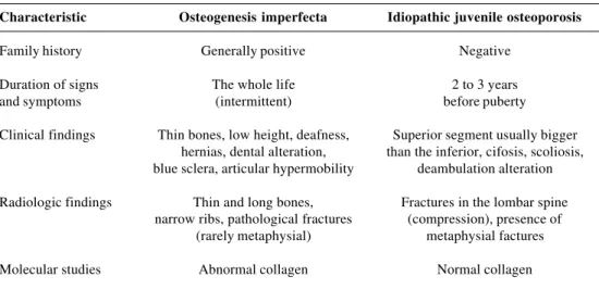
The main differences observed between OI and IJO are to be found in Table 2.
Secondary osteoporosis
The chronic diseases listed in Table 1 and the therapies used in the treatment of many of them encourage the development of osteoporosis, as described below.
Osteoporosis due to chronic diseases, such as, for example, juvenile rheumatoid arthritis, is multifactorial. It may be the result of the actions of the disease itself, of immobility due to arthritis, of myositis, of insufficient exposure to sunlight, of nutritional factors such as caloric-proteic malnutrition, of low calcium ingestion or of hormonal factors and/or drugs used to treat the disease, Inflammatory mediators which are highly active in many of chronic pathologies, perform an important role in bone reabsorption Interleukin 1 and tumor necrosis factor stimulate osteoblasts to produce cytokines which activate osteoclasts, while interleukin-6 promotes osteoclast precursor cell differentiation.20-22 Inflammatory activity also provokes reduced levels of osteocalcine, of insulin-dependent growth factors (IGF-1) and their transportation proteins. These interleukins also exacerbate catabolism and induce anorexia, reducing the ingestion of nutrients that are important to bone formation, such as calcium and vitamin D.8
Diseases which interfere with the absorption of nutrients, as is observed in intestinal inflammatory disease, cystic fibrosis, hepatobiliary disease and anorexia nervosa, lead to reduced body mass and loss of muscle mass, contributing to the development of osteoporosis. Diseases that interfere in the conversion of vitamin D into its active forms, such as chronic liver and kidney diseases, lead to secondary diminished bone formation.8
The reduction in physical activity and prolonged immobility that result from conditions such as juvenile rheumatoid arthritis, juvenile dermatomyositis or chronic neuropathies, result in reduced mechanical tension on the bones and so diminished stimulation to their formation.8
Table 2 - Differential diagnosis between osteogenesis imperfecta and idiopathic juvenile osteoporosis
Characteristic Osteogenesis imperfecta Idiopathic juvenile osteoporosis
Family history Generally positive Negative
Duration of signs The whole life 2 to 3 years
and symptoms (intermittent) before puberty
Clinical findings Thin bones, low height, deafness, Superior segment usually bigger hernias, dental alteration, than the inferior, cifosis, scoliosis, blue sclera, articular hypermobility deambulation alteration Radiologic findings Thin and long bones, Fractures in the lombar spine
narrow ribs, pathological fractures (compression), presence of (rarely metaphysial) metaphysial factures
During adolescence, the increase in bone density occurs primarily in the spine, probably in response to the activity of sexual hormones (estradiol and testosterone) in the trabecular component of the bones. Conditions that result in pubertal retardation in adolescents of both sexes, such as chronic inflammatory diseases, hypogonadism, anorexia nervosa or amenorrhea induced by exercise, can also be highlighted as causes of osteoporosis.23,24
A number of different medications can be considered to induce osteoporosis, such as corticosteroids, methotrexate, heparin, anticoagulants, phenobarbital, phenytoin and carbamazepine. Among these, corticosteroids stand out as they act directly on the osteoblasts reducing bone formation. They also act to reduce intestinal calcium absorption and increase its renal excretion, provoking secondary hyperparathyroidism, which results in increased bone reabsorption. In adults, the use of doses above 5 to 7.5 mg/day for a period of more than three to six months is considered to induce osteoporosis. The deleterious effect of corticosteroids on bone mass is most intense during the first six months of use.8,25-27
Preventative treatment
In the case of patients suffering from chronic diseases it is important that all of the risk factors present in each case are identified and treated or attenuated in the best manner possible.
Dietary guidance
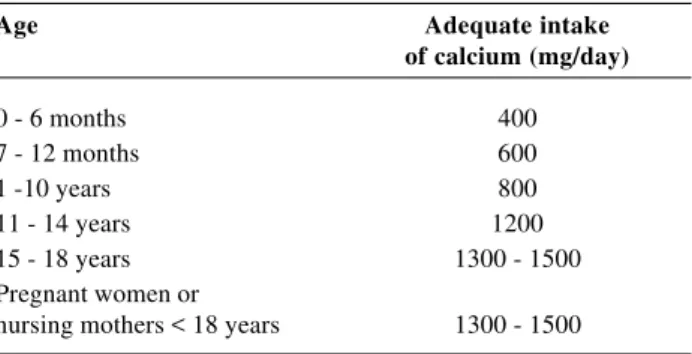
Diet should be rich in foods with a high level of calcium and avoid protein and phosphates (present in red meat, cereals and soy). Also recommended is restricted sodium ingestion since when it is ingested in high quantities it increases renal calcium excretion.28 Daily calcium ingestion varies with the age of the individual (Table 3). Calcium can be found in a number of different animal and vegetable foodstuffs. Milk and its derivatives contain the greatest proportion of bio-available calcium, although other sources can also be used (Table 4).
In cases where corticosteroids will be prescribed chronically, the preventative introduction of calcium and vitamin D supplements should be planned.26 The source of these vitamins in the diet may be animal or vegetable and the requirement varies from 400 to 800 units.28
Exposure to sunlight
Sufficient exposure to solar ultraviolet rays is necessary for adequate vitamin D production from its precursor, 17-deidrocolesterol, present in fat and skin. Sunlight should fall directly on the skin. Wearing clothes and exposure to sun from behind glass reduce the efficiency of epidermal vitamin D synthesis.29
Modification of habits
Adolescents should receive guidance with respect of the negative effects of alcohol consumption, coffee and smoking on bone metabolism and consequentially on peak bone mass.28
Physical activities
Physical activity, and especially exercise against the force of gravity, should be stimulated. Activities such as walking, running and weight training, have a greater effect on the bones than activities which do not involve load, such as cycling and swimming. Physical activity should be undertaken in a regular manner (three to four times a week, for a minimum of thirty minutes). Female adolescents who exercise with great intensity can develop amenorrhea, compromising bone mass gain. Bones are more susceptible
Table 3 - Daily recommended intake of calcium according to age group29
Age Adequate intake
of calcium (mg/day)
0 - 6 months 400
7 - 12 months 600
1 -10 years 800
11 - 14 years 1200
15 - 18 years 1300 - 1500
Pregnant women or
nursing mothers < 18 years 1300 - 1500
Table 4 - Calcium sources30
Food Calcium mg
Natural yogurt (1 cup) 415
Fruit yogurt (1 cup) 345
Low-fat milk (1 cup) 302
Whole milk (1 cup) 291
Mozzarella cheese (30 g) 183
Milk ice-cream (1 cup) 176
Pudim (1/2 cup) 146
Can of sardines 371
Roasted fish 117
Cooked spinach (1/2 cup) 122
Cooked broccoli (1 cup) 94
Beet cooked with leaves (1/2 cup) 82
Beans (1 cup) 81
Egg (unit) 28
Lasagna (portion) 460
Spaghetti (1 cup) 14
Hamburger with cheese (unit) 135
to losses in bone mass due to inactivity than they are capable of gaining it by increased physical activity. A loss of one percent of bone mass, which can occur after a week of restricted activity, can take a year to be recovered by increased physical activity.28,29
Pharmacological treatment
Initial treatment for osteoporosis should be performed with calcium and vitamin D supplements. Calcium can be supplemented using a variety of different calcium salts. The most recommendable is calcium carbonate, which offers the greatest quantity of elemental calcium (40%). Calcium citrate makes 21% of elemental calcium available, lactate 13% and glutamate 9%. Replacement is recommended at a rate of 500 mg to 1 g per day, and should be ingested with meals to aid absorption. Some patients may present dyspepsia, nausea and constipation, as side effects.8,26,29
Vitamin D can be supplemented using multivitamins or in association with calcium salts, since it is not commercially available in isolation in our country.29 The recommended dose varies from 400 to 800 units per day. Active forms of Vitamin D (calcitriol and alphacalcidiol) can also be used in cases where there is reduced hepatic or renal metabolism. It this form the dose is 0.5 to 1 mcg per day, associated or not with calcium.28
Thiazide diuretics inhibit renal calcium excretion in the proximal tubule and can be used with patients with hypercalciuria. The recommended dose is 25 mg/day.29
Drugs that stimulate bone formation or reabsorption can also be used, as described below.
Drugs that stimulate bone formation
Fluorine (sodium fluoride or monofluorophosphate) is a potent trabecular bone formation stimulator. However, the effective dose is very close to the toxic dose and there is controversy over the quality of the bone formed as a result, since bone may be formed with an overly high fluoride content, which limits its use. There are also reports of osteomalacia associated with its use. In order to minimize these effects, it should be administered in association with calcium and vitamin D. Other side effects are: nausea, vomiting, epigastralgia, diarrhea, melena and arthralgia. The recommended dose is 0.5 to 1 mg/kg/day, although further trials are necessary to confirm its efficacy for osteoporosis treatment. In Brazil it is not available in isolation but it can be used in multivitamin form.28,29
Parathormone (PTH) is continually secreted by the parathyroid. Its action causes a catabolic reaction in the skeleton, as exemplified in the severe primary hyperparathyroidism model. However, if parathormone is administered in low doses, in an intermittent manner a significant anabolic property is observed, particularly in
trabecular bone. The recommended dose for adults is 20 µg of subcutaneous PTH daily. Clinical trials have demonstrated increased BMD in the spine and hips and a reduced risk of vertebral and non- vertebral fractures when compared with a placebo.31 There are not yet any reports of its use with children or adolescents.
Drugs that reduce bone reabsorption
Bisphosphonates act directly on the osteoclasts, reducing their number and level of activity and act indirectly on the osteoblasts, increasing bone formation. They can be used with children with osteoporosis secondary to chronic diseases, such as juvenile rheumatoid arthritis, or in cases of chronic corticosteroid use, apparently without suppressing bone remodeling and without adverse effects on linear growth. The use of second generation bisphosphonates (pamidronate) has been shown to be effective for the treatment of osteoporosis in children, however, its intravenous form restricts its use. Alendronate (third generation bisphosphonate) is the most often used. It should be given 30 to 60 minutes before breakfast, with the patient sitting, since its principle side effects are gastroesophageal reflux and esophagitis and its absorption is reduced in the presence of food. The dose is 5 to 10 mg per day.7,8,32-34 Calcitonin acts to inhibit osteoclasts and also has a central analgesic effect. It is indicated for acute pain secondary to vertebral fractures. Calcitonin should be used in association with calcium.35 Commercially, calcitonin comes from a number of different species, such as from humans, salmon, pigs and mares. Calcitonin may be injectable (intramuscular or subcutaneous) or in the form of a nasal spray. The nasal spray, which allows calcitonin to pass through the nasal mucosa results in fewer adverse effects. The recommended doses can be found in Table 5. It has not been standardized for use with pediatric patients.28
Follow-up
Calcitonin Route Daily dose Treatment duration
Salmon IM or SC 100 - 200 IU 15 days/month Humane IM or SC 50 - 100 mg 15 days/month Salmon Nasal spray 200 - 400 IU 15 days/month Eel Nasal spray 80 - 160 IU 15 days/month Table 5 - Calcitonin administration28
IM = intramuscular; SC = subcutaneous.
The identification of osteoporosis risk factors is the responsibility of pediatricians as is the guidance of their patients in relation to its treatment and monitoring, thus guaranteeing a healthy peak bone mass during adolescence, which will without doubt contribute to better quality of life during adult life, since adult osteoporosis is inversely proportional to peak bone mass acquired during childhood.
References
1. NIH Consensus Statement, n. 111. Osteoporosis prevention, diagnosis and therapy. 2000;17:1-36.
2. Rigss BL, Melton LJ. The worldwide problem of osteoporosis: insight afforded by epidemiology. Bone. 1995;17(5 Suppl): S505-11.
3. Van der sluis IM, Muinck Keizer-Schrama SMPF. Osteoporosis in childhood: bone density in children in health and disease. J Pediatr Endocrinol Metab. 2001;14:817-32.
4. The WHO Study Group. Assessment of fracture risk and its application to screening for postmenopausal osteoporosis. Switzerland: World Health Organization; 1994.
5. Zerbini CAF. Osteoporose: uma revisão. Jovem Médico. 1998;2:89-94.
6. Cassidy JT. Osteopenia and osteoporosis in children. Clin Exp Rheumatol. 1999;17:245-50.
7. McDonagh JE. Osteoporosis in juvenile idiopathic arthritis. Curr Opin Rheumatol. 2001;13:399-404.
8. Kiss MHB. Osteoporose. In: Setian N, editor. Endocrinologia Pediátrica: Aspectos Físicos e Metabólicos do Recém Nascido ao Adolescente. 2nd ed. São Paulo: Savier; 2002. p. 354-62. 9. Golding A, Jones IE, Taylor RW, Manning PJ, Williams SM.
More broken bones: a four-year double cohort study of young girls with and without distal forearm fractures. J Bone Miner Res. 2000;15:2011-18.
10. Pereira RMR, Corrente JE, Chahade WH, Yoshinari NH. Evaluation by dual X-ray absorptiometry (DXA) of bone mineral density in children with juvenile chronic arthritis. Clin Exp Rheumatol. 1998;16:495-501.
11. Lu PW, Cowell CT, Lloyd-Jones AS, Briody JN, Howman-Giles R. Volumetric bone mineral density in normal subjects, aged 5-27 years. J Clin Endocrinol Metab. 1998;83:1420-5-27. 12. Kroger H, Kotaniemi A, Vaino P, Alhava E. Bone densitometry
of the spine and femur in children by dual-energy x-ray absorptiometry. Bone Mineral. 1992;17:75-85.
13. Molgaard C, Thomsen BL, Michaelsen KF. Influence of weight, age and puberty on bone size and bone mineral content in healthy children and adolescents. Acta Paediatr. 1998;87:494-9.
14. Pereira RMR, Falco V, Corrente JE, Chahade WH, Yoshinari NH. Abnormalities in the biochemical markers of bone turnover in children with juvenile chronic arthritis. Clin Exp Rheumatol. 1999;17:251-5.
15. Mora S, Pitukcheewanont P, Kaufman FR, Nelson JC, Gilsanz V. Biochemical markers of bone turnover and the volume and the density of bone children at different stages of sexual development. J Bone Miner Res. 1999;14:1664-71.
16. Swaminathan R. Biochemical markers of bone turnover. Clin Chim Acta. 2001;313:95-105.
17. Guarnero P, Delmas PD. Biochemical markers of bone turnover in osteoporosis. In: Marcus R, Feldman D, Kelsey J, editors. Osteoporosis. 2nd ed. San Diego: Academic Press; 2001. p. 459-77.
18. Saggese G, Baroncelli GI, Bertelloni S. Osteoporosis in children and adolescents: diagnosis, risk factors and prevention. J Pediatr Endocrinol Metab. 2001;14:833-59.
19. Pereira RMR. Osteoporose juvenil. Acta Fisiátrica. 1997;2: S135-8.
20. Horowitz, MC. Cytokines and estrogen in bone: anti-porotic effects. Science. 1993;260:626-7.
21. Pereira RMR. Osteoporose e doenças reumáticas juvenis. In: Oliveira SKF, Azevedo ECL, editors. Reumatologia Pediátrica. 2nd ed. Rio de Janeiro: Revinter; 2001. p. 409-13.
22. Castro TC, Terreri MT, Szejnfeld VL, Castro CH, Fisberg M, Gabay M, et al. Bone mineral density in juvenile systemic lupus erythematosus. Braz J Med Biol Res. 2002;35:1159-63. 23. Rubin K, Schirduan V, Gendreau P, Sarfarazi M, Mendola R,
Dalsky G. Predictors of axial and peripheral bone mineral density in healthy children and adolescents, with special attention to the role of puberty. J Pediatr. 1993;123:863-70.
24. Moretto PA. Contribuição ao estudo da osteoporose em crianças e adolescentes portadores de lúpus eritematoso sistêmico [dissertation]. São Paulo: Faculdade de Medicina da Universidade de São Paulo; 1996.
25. Eastell R, Reid DM, Compston J, Cooper C, Fogelman I, Francis RM, et al. A UK Consensus group on management of glucocorticoid-induced osteoporosis: an update. J Int Med. 1998;244:271-92.
26. Boulos P, Adachi JD. Guidelines for the prevention and therapy of glucocorticoid-induced osteoporosis. Clin Exp Rheumatol. 2000;18:S79-86.
27. Recommendations for the prevention and treatment of glucocorticoid-induced osteoporosis: 2001 update. American College of Rheumatology Ad Hoc Committee on Glucocoritcoid-Induced Osteoporosis. Arthritis Rheum. 2001;44:1496-503. 28. Szejnfeld VL. Atualização terapêutica em osteoporose. Rheuma.
1995;2:4-7.
29. Pinto Neto AM, Soares A, Urbanetz AA, Souza ACA, Ferrari AEM, Amaral B, et al. Consenso brasileiro de osteoporose 2002. Rev Bras Reumatol. 2002;42:343-54.
30. Nutritive Value of Foods. Home and Garden Bulletin. US Department of Agriculture. Washington, DC: US Government Printing Office; 1988.
31. Neer RM, Arnaud CD, Zanchetta JR, Prince R, Gaich GA, Reginster JY, et al. Effect of parathyroid hormone on fractures and bone mineral density in postmenopausal women with osteoporosis. N Eng J Med. 2001;344(19):1434-41.
32. Brumsen C, Hamdy NAT, Papapoulos SE. Long-term effects of bisphosphonates on the growing skeleton. Studies of young patients with severe osteoporosis. Medicine. 1997;76:266-83. 33. Roux C, Dougados M. Bisphosphonates in the prevention and
treatment of glucocorticoid-induced osteoporosis. Clin Exp Rheumatol. 2000;18:S49-52.
Corresponding author: Lúcia Maria de Arruda Campos Rua Joaquim Antunes, 135/701 CEP 05415-010 – São Paulo, SP, Brazil
Tel.: +55 (11) 3063.2678 – Fax: +55 (11) 3081.7373 E-mail: [email protected]