Reconstruction of alveolar cleft with allogenous
bone graft: Clinical considerations
Omar Gabriel da Silva Filho1, Terumi Okada Ozawa2, Cláudia Bachega3, Marco Aurélio Bachega4
Introduction:Secondary bone grafting consists in a routine procedure on the treatment of patients with alveolar cleft. Usually, it is performed by the end of the mixed dentition, when the permanent canine is erupting, with autogenous cancellous bone from the iliac crest.
Objective: The present article discusses the alternative of autogenous bone grafting with allogeneic bone, obtained from human bone bank, illustrating the result with the presentation of a clinical case of left unilateral alveolar cleft.
Keywords:Orthodontics. Cleft palate. Bone transplant.
How to cite this article: Silva Filho OG, Ozawa TO, Bachega C, Bachega MA. Reconstruction of alveolar cleft with allogenous bone graft: Clinical consider-ations. Dental Press J Orthod. 2013 Nov-Dec;18(6):138-47.
Submitted: November 23, 2009 - Revised and accepted: October 18, 2010
» The authors report no commercial, proprietary or financial interest in the products or companies described in this article.
» Patients displayed in this article previously approved the use of their facial and in-traoral photographs.
Contact address: Omar Gabriel da Silva Filho
Rua Silvio Marchione, 3-20, Bauru/SP - CEP: 17.043-230 E-mail: [email protected]
1 Orthodontist, Hospital of Rehabilitation of Craniofacial Anomalies
(HRAC-USP), Bauru.
2 Orthodontist and Director of the Dentistry Department, HRAC-USP, Bauru. 3 Periodontist, Professor of the Specialization Course in Periodontics, University
of Marília (UNIMAR).
4 Orthodontist, Professor of the Specialization Course in Periodontics, University
of Marília (UNIMAR).
Introdução:o enxerto ósseo secundário consiste em um procedimento rotineiro no tratamento de pacientes com fissura alveolar. Via de regra, é realizado no final da dentadura mista, na época de erupção do canino permanente, com osso medular autógeno retirado da crista ilíaca.
Objetivo:o presente artigo discorre sobre a alternativa de enxerto ósseo autógeno realizado com osso alógeno, obtido de banco de ossos humanos, ilustrando o resultado com a apresentação de um caso clínico de fissura alveolar unilateral do lado esquerdo.
GENERAL CONSIDERATIONS ON ALVEOLAR
CLEFT AND SECONDARY BONE GRAFTING
The clet lip and alveolar ridge (pre-incisive fora-men clets) and the complete clet lip and palate
(trans-incisive foramen clets)19 demand surgeries, at strategic
time, for reconstitution of the morphological defect. The primary plastic surgeries, cheiloplasty and palato-plasty, reconstruct the clet lip and palate in sot tissue during childhood, from 3 and 12 months of age, re-spectively. The alveolar bone disruption, exactly at the height of the upper lateral incisor, persists ater primary plastic surgeries, maintaining the discontinuity of the alveolar arch and upper arch. The normalization of the alveolar bone defect, persistent ater primary plastic surgeries, is achieved with marrow-cancellous bone
autogenous grating from the iliac crest.1,2 This
proce-dure is performed during the mixed dentition, more precisely near the period of eruption of the permanent canine adjacent to the clet, between 9 and 12 years of age — therefore, it is designated as secondary bone
grating.20 Thus, the secondary alveolar bone grating
represents fundamental part in the process of rehabili-tation of patients with clet involving the alveolar ridge. Figure 1 illustrates the facial and occlusal condition of the patient subjected to primary plastic surgeries and to secondary bone grating, within the conventional rehabilitation protocol. The advantages of secondary bone grating are related to the elimination of alveolar defect without negative interference on the maxillary
growth potential.18 The grated bone becomes alveolar
bone (Fig 2), continuing the alveolar ridge and pro-viding support to the nasal base. The newly formed
alveolar bone allows spontaneous7,12 or forced21 tooth
movement, and ofers better periodontal conditions
for the teeth adjacent to the clet.1,2,25 Eliminating the
alveolar bone defect, it is possible to inish the reha-bilitation procedure without prosthesis in the area of the involved lateral incisor, and with the continuous alveolar arch, without interruption (Figs 1 and 2). Ul-timately, the neoformed bone allows implant instal-lation with prosthetic purpose. The success index of secondary bone grating is clinically and
radiographi-cally25,28 measured, as exempliied on Figures 1 and 2.
From the radiographic point of view, the periapical ra-diographs of the grat area and the occlusal radiograph of the maxilla allows evaluating the level of neoformed bone in the clet region. However, the most accurate
bone diagnosis is performed with CT, for it allows a three-dimensional evaluation and the depth reading of
the neoformed bone.10 Several radiographic
research-es have proved the succresearch-ess of secondary bone
grat-ing,1,2,5,12,17,25,28,27 making it deinitive in the therapeutic
process of clets involving alveolar ridge.
CONSIDERATIONS ON GRAFTING WITH
AUTOGENOUS BONE
The bone to be grated in the alveolar clet region can be autogenous, allogeneic or synthetic. The autog-enous secondary alveolar bone grating technique is the most widely used on the rehabilitation of clet
pa-tients and was technically detailed by Boyne,3 in 1974,
and by Boyne and Sands,4 in 1976.
In a short period, about 3 months, the grafted autogenous bone totally reintegrates to the area, the radiographic distinction between the cleft limit and the neoformed bone is difficult. The osteoblasts sur-vive, functionally, in an autogenous graft, ensuring bone vitality.
The autogenous bone grat from the iliac crest has been the favorite by rehabilitators, worldwide, in-cluding at the Hospital of Rehabilitation of Craniofa-cial Anomalies of USP (HRAC-USP, in Bauru/SP). The iliac crest bone, enough to completely ill the al-veolar defect, is medullary, to favor and accelerate the transformation of the grated bone. Other donor sites, such as tibia,13 rib,29 skullcap,6 retromolar region16 and
mandibular symphysis,14 have been used. The iliac
bone is the most used because the iliac crest features a large amount of medullary bone, with greater amount
of osteoinductive cells.24 The disadvantages comprise
the morbidity resultant from obtainment of grat and the necessity of a medical professional to obtain the grat, oten an Orthopedist.
Although the height of the bony septum formed in the clet region is similar in the bones obtained from
the iliac crest and mandibular symphysis,22 the
man-dibular symphysis can only be used in narrow clets, which do not require large amount of tissue to be illed.
CONSIDERATIONS ON ALLOGENEIC BONE
OBTAINED FROM BONE BANK
Using allogeneic bone (allograts),8 eliminates the
Figure 1 - A) Right lateral facial photograph. B) Frontal facial photograph of patient with cleft involving the alveolar ridge on the right. The secondary bone grafting procedure continues the alveolar ridge, creating periodontal conditions favorable to the movement of teeth adjacent to the congenital bone defect.
C) Left lateral facial photograph. D) Facial photograph with zoom on the cleft region. E) Upper occlusal photograph: alveolar cleft (unilateral pre-incisive foramen), segmenting the alveolar ridge on the left upper lateral incisor region — beginning of mixed dentition. F) Lower occlusal photograph. G) Right lateral intraoral photograph. H) Frontal intraoral photograph. I) Left lateral intraoral photograph.
Figure 2 - A) The longitudinal radiographic evaluation by means of periapical radiographs of the cleft area shows the incorporation of the autogenous bone
grafted in the subjacent bone tissue. B) Permanent canine eruption in the graft area. C) Canine eruption of in the graft area. D) Orthodontic movement in the grafted area.
A
A D
G
B
B
E
H
C
C D
F
molecular composition exert chemical and mechanical properties required for diferentiation of parenchymal
cells and for the tissues physiological demand.15 Certain
molecules that compose the matrix act as regulators of the cell function – which, in the case of bone tissue, are the BMPs. These are secreted by osteoblasts in case
of bone deposition.15 Such characteristics, extracellular
matrix and presence of BMPs, are present in allograts. By the data collected in literature, there are no reports on reconstruction of alveolar clet with allogeneic bone. Therefore, the objective of the present article consists in presenting, through a clinical case, the reconstruction of the alveolar clet with allogeneic bone from bone bank. of a donor site and a medical professional to obtain the
bone to be grated. The allograts present tive and osteoinductive properties. The osteoconduc-tive properties are assigned to the mineralized collagen structure, forming a scafold that allows bone deposi-tion. The osteoinductive properties are assigned to the presence of BMPs (bone morphogenetic proteins), which are released by osteoclastic activity,
biostimula-tion to the grat incorporabiostimula-tion.15,39,31 With the
devel-opment of tissue engineering, fundamental principles were established for the regeneration of certain tissues.23 Among them, the use of extracellular matrix similar to the tissue to be regenerated, once its structure and
Figure 3 - A) Right lateral facial photograph.
B) Frontal facial photograph. C) Right lateral intraoral photograph. D) Frontal intraoral pho-tograph. E) Left lateral intraoral photograph.
F) Upper occlusal photograph. G) Lower occlu-sal photograph.
A
D
G B
E C
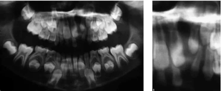
Figure 4 - A) The panoramic radiograph reveals the upper left alveolar bone defect with tooth agenesis # 22. In addition, the presence of a pre-canine tooth adjacent to the alveolar cleft is found. Such tooth is common in clefts that completely disrupt the alveolar ridge. B) The periapical radiograph of the upper anterior region reveals, more clearly, the alveolar bone defect on the upper left, along with tooth agenesis #22 and presence of pre-canine tooth adjacent to the alveolar cleft.
A B
CLINICAL CASE DESCRIPTION
The patient, with unilateral complete clet lip and alveolar ridge (pre-incisive foramen clets), on the let-side, underwent primary cheiloplasty during child-hood and initiated the orthodontic follow-up at the beginning of the mixed dentition (Fig 3), with alveolar bone defect typical of a unilateral complete alveolar ridge clet (Fig 4). The treatment protocol involved the control of the occlusion development, assisting permanent teeth eruption and pre-canine behavior in
Figure 5 - A) Right lateral intraoral photograph.
B) Frontal intraoral photograph. C) Left lateral in-traoral photograph. Control of permanent teeth eruption, in the mixed dentition, with persistent alveolar bone defect. D) Upper occlusal photo-graph. E) Lower occlusal photograph.
A
D
B
E
C
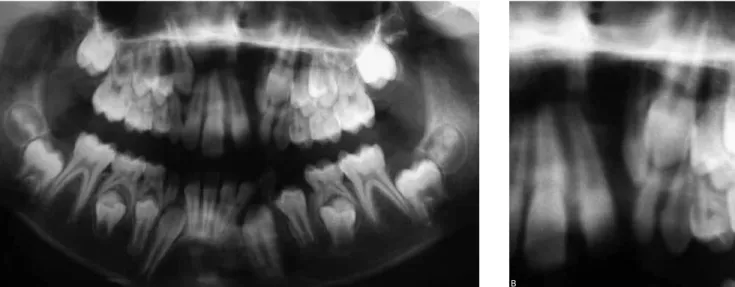
Figure 6 - A) Radiographic image compatible to the alveolar bone defect in complete alveolar cleft. B) Detailed, the alveolar bone defect in complete alveolar cleft is observed. The pre-canine and canine teeth, in the cleft area, present favorable eruption path towards the bone defect.
A B
Figure 8 - A) Occlusal radiograph before secondary bone grafting. B) Occlusal radiograph after bone grafting. The comparison of occlusal radiographs, before
and after bone grafting, reveals the structural difference on the alveolar ridge, assigned to the bone grafting.
A B
Figure 7 - A) Panoramic radiograph shows the procedure of secondary bone grafting performed using allogeneic bone, obtained from human bone bank,
to restore the alveolar ridge and allow the eruption of canine and pre-canine by bone grafting. B) This section, obtained from the panoramic radiograph, illustrates the grafted region.
Figure 10 - A) Panoramic radiograph by the end of the orthodontic treatment. The periodontal condition illustrated on the image is very favorable. B) This image demonstrates more clearly the favorable periodontal condition.
A B
Figure 9 - A) Right lateral intraoral photograph.
B) Frontal intraoral photograph. C) Left lateral intraoral photograph. By the end of the orth-odontic treatment, the left pre-canine substi-tutes the lateral incisor. There is evidence and space enough for the increase of the pre-canine crown. D) Upper occlusal photograph. E) Lower occlusal photograph.
A
D
B
E
C
Figure 11 - In this radiographic image, the pre-canine tooth is observed replacing the corresponding
DISCUSSION
When choosing the grafting material to recon-struct the alveolar processes, the decision of estab-lishing a donor site on the receptor itself can give the procedure a dimension of greater amplitude, once the post-surgical morbidity becomes more signifi-cant, contributing decisively on the patient recovery. This factor was shown to be relevant on the decision for the use of alternatives to autogenous sources.
The main characteristic assigned to allogeneic bone consists in its versatility, with osteoconduc-tion and osteoinducosteoconduc-tion potential assigned to its macroscopic characteristics. Both potentials must be considered when planning to use it or when the results are evaluated. Why this double potential of osteoconduction and osteoinduction? Which factors contribute to this?
Despite the absence of osteogenic phase, the pro-cess of incorporation of allogeneic graft is similar to of autogenous graft. The graft is placed in a dead area,
filled with blood clot. The dead area is hypoxic (PO2
of 5 to 10 mmHg) and acidotic (pH 4 to 6), containing platelets, leukocytes, red cells and fibrin, in a com-plex structure. Associated with the graft, sectioned capillaries were observed, with clot and exposed en-dothelial cells. Externally to the periosteal closure,
the tissue is normoxic (PO2 of 45 to 55 mmHg) and
presents physiological pH (pH of 7.42). The release of growth factors (PDGF, TGF-b and IGF), from the platelet degranulation, exert primordial functions on the regenerative process. It is highlighted the angio-genic activity of capillary sprouts in the graft inte-rior, by the induction of mitosis in endothelial cells, differentiation and proliferation of fibroblasts and pre-osteoblasts in functional osteoblasts, secretion of bone matrix by osteoblasts and of collagen matrix by fibroblasts, providing support to the capillary growth. In autogenous grafts, the capillaries appear in the graft on the third day and the complete penetration is visu-alized between the 14th and 17th day.
In autogenous grafts, the formation of bone phase I is originated from endosteal osteoblasts that de-limit the trabecular bone surfaces. This bone from phase I is immature and disorganized, with absence of Haversian systems and little structural integrity. It develops four weeks after grafting. The end of the graft revascularization eliminates the small tension of
oxygen necessary to keep the macrophages activity. They leave the area since they are no longer neces-sary for the graft maintenance. The phase I bone will, mandatorily, be subjected to a process of resorption/ apposition, which will lead to formation of a mature bone, with lamellar architecture and Harvesian sys-tems, denominated phase II bone. With a developed periosteum and endosteum, it is a self-sustaining bone, with complete structural integrity.
The maturation of the regenerated bone involves the IGF and the bone morphogenetic proteins. These are insoluble acidic proteins, released by osteoclastic resorption on the process of normal bone remodeling, which occurs 0.7% a day in normal bone, and can oc-cur as fast as 5 to 8% a day in a grat in maturation.
The release of BMP and IGF links the bone resorp-tion to neoformaresorp-tion, acting in adjacent pluripotent cells and pre-osteoblasts, inducing its proliferation and diferentiation in functional osteoblasts, which secrete, actively, the bone matrix. This way, the grating cycle develops from a cell transplantation, which is placed in a complex biomechanical environment, to a mature and functional bone, which is self-sustaining by means of a
normal cycle of resorption/remodeling.9,15
The BMPs present in the osteoconductive alloge-neic matrix are glycoproteins classified into 15, being 2, 4 and 7 those of greater influence on the osteo-inductive process, since they effectively act on the differentiation of pluripotent mesenchymal cells in osteogenic precursors. When different samples of al-logeneic bone tissue, from different bone banks, were subjected to immunohistochemical analysis, it was observed that the best clinical results (bone forma-tion) were associated with presence of BMPs 2 and 7. Therefore, the osteoinductive potential is variable from bank to bank and, in the same bank, from donor
to donor.11 Another physiological principle of
por-tion. The medullary is much more easily vascularized and incorporated; however, more easily resorbed, in relation to the cortical. The particulate grafts, how-ever, are very easily vascularized and, consequently, resorbed. One of the factors for choice of graft is the type of defect that will be reconstructed. The influ-ence of the allograft processing must also be consid-ered, since it will affect its osteoregenerative poten-tial. The method for allograft processing must ensure the sterilization, preservation of biological
character-istics and reduction of antigenicity.26 The bone that
was used here undergoes a process of acid treatment with sterile HCl associated with a solution of Ca at 5%, water pumping for 12 hours, lyophilization and storage at 80°C, which ensures its availability for five years. Such procedure ensures its sterilization and the preservation of osteoinductive and osteoconductive
biological properties. However, the careful selection of donor is fundamental to prevent transmission of infectious diseases. The bone allografts are an alterna-tive to reconstruction of alveolar clefts. On the one hand, the graft with structural (cortical/medullary) and biological characteristics (amount and type of BMPs present) and, on the other hand, the receptor, with its inherent osteoclastic activity, are the variable that will affect the clinical results.
CONCLUSION
1. Abyholm FE, Bergland O, Semb G. Secondary bone grafting of alveolar clefts: a surgical orthodontic treatment enabling a non-prosthodontic rehabilitation in cleft lip and palate patients. Scand J Plast Reconstr Surg. 1981;15(2):127-40.
2. Bergland O, Semb G, Abyholm FE. Elimination of the residual alveolar cleft by secondary bone grafting and subsequent orthodontic treatment. Cleft Palate J. 1986;23(3):175-205.
3. Boyne PJ. Use of marrow-cancellous bone grafts in maxillary alveolar and palatal clefts. J Dent Res. 1974;53(4):821-4.
4. Boyne PJ, Sands NR. Combined orthodontics surgical management of residual palato-alveolar clefts defects. Am J Orthod. 1976;70(1):20-37. 5. Brattström V, McWilliam J. The inluence of bone grafting age on dental
abnormalities and alveolar bone height in patients with unilateral cleft lip and palate. Eur J Orthod. 1989;11(4):351-8.
6. Denny AD, Talisman R, Bonawitz SC. Secondary alveolar bone grafting using milled cranial bone graft: a retrospective study of a consecutive series of 100 patients. Cleft Palate Craniofac J. 1999;36(2):144-53. 7. El Deeb M, Messer LB, Lehnert MW, Hebda TW, Waite DE. Canine
eruption into grafted bone in maxillary alveolar cleft defects. Cleft Palate J. 1982;19(1):9-16.
8. Feoilof ET, Garcia JR. Técnicas de obtenção, processamento, armazenamento e utilização de homoenxertos ósseos: Protocolo do Banco de Ossos da Escola Paulista de Medicina. Rev Bras Ortop. 1996;31(11):895-903.
9. Marx RE, Garg AK. Bone structure, metabolism, and physiology: its impact on Dental Implantology. Implant Dent. 1998;7(4):267-76. 10. Hamada Y, Kondoh T, Noguchi K, Iino M, Isono H, Ishii H, et al.
Application of limited cone beam computed tomography to clinical assessment of alveolar bone grafting: a preliminary report. Cleft Palate Craniofac J. 2005;42(2):128-37.
11. Han B, Tang B, Nimni ME. Quantitative and sensitive in vitro assay for osteoinductive activity of demineralized bone matrix. J Orthop Res. 2003;21(4):648-54.
12. Hinrichs JE, el-Deeb ME, Waite DE, Bevis RR, Bandt CL. Periodontal evaluation of canines erupted through grafted alveolar cleft defects. J Oral Maxillofac Surg. 1984;42(11):717-21.
13. Kalaaji A, Lilja J, Elander A, Friede H. Tibia as a donor site for alveolar bone grafting in patients with clefts lip and palate: long-term experience. Scand J Plast Reconstr Surg Hand Surg. 2001;35(1):35-42.
14. Koole R. Ectomesenchymal mandibular symphysis bone graft: an improvement in alveolar cleft grafting? Cleft Palate Craniofac J. 1994;31(3):217-23.
15. Marx RE, Kline SN, Johnson RP, Malinin TI, Matthews JG 2nd, Gambill V. The use of freeze-dried allogeneic bone in oral and maxillofacial surgery. J Oral Surg. 1981;39(4):264-74.
16. Natsume N, Kawai T. Lower jaw bone graft of the alveolar cleft region in a cleft lip and palate patient using the third-molar extraction procedure. Plast Reconstr Surg. 1994;93(6):1309-10.
REFERENCES
17. Nightingale C, Witherow H, Reid FD, Edler R. Comparative reproducibility of three methods of radiographic assessment of alveolar bone grafting. Eur J Orthod. 2003;25(1):35-41.
18. Semb G. Efect of alveolar bone grafting on maxillary growth in unilateral cleft lip and palate patients. Cleft Palate J. 1988;25(3):288-95.
19. Silva Filho OG, Ferrari Junior FM, Rocha DL, Freitas JAS. Classiicação das issuras lábio-palatais. Breve histórico, considerações clínicas e sugestão de modiicação. Rev Bras Cir. 1992;82(2):59-65.
20. Silva Filho OG, Ozawa TO, Carvalho RM. Enxerto ósseo secundário. In: Trindade IEK, Silva Filho OG. Fissuras labiopalatinas: uma abordagem interdisciplinar. 1ª ed. São Paulo: Ed. Santos; 2007. p. 239-60. 21. Silva Filho OG, Teles SG, Ozawa TO, Filho LC. Secondary bone graft
and eruption of the permanent canine in patients with alveolar clefts: literature review and case report. Angle Orthod. 2000;70(2):174-8. 22. Sindet-Pedersen S, Enemark H. Reconstruction of alveolar clefts
with mandibular or iliac crest bone-graft: a comparative study. J Oral Maxillofac Surg. 1990;48(6):554-8.
23. Spector M. Basic principles of tissue engineering. In: Lynch SE, Genco RJ, Marx RE. Tissue engineering: applications in maxillofacial surgery and Periodontics. 2th ed. Illinois: Quintessense; 1999. p. 3-16.
24. Steinberg B, Chiego DJ Jr, Huizinga PJ, Wozney JM, Wikesjö UM. Efect of human bone morphogenetic protein 2 implant on tooth eruption in an experimental design. J Craniofac Surg. 1999;10(4):338-41.
25. Trindade IK, Mazzottini R, Silva Filho OG, Trindade IE, Deboni MC. Long-term radiographic assessment of secondary alveolar bone grafting outcomes in patients with alveolar clefts. Oral Surg Oral Med Oral Pathol Oral Radiol Endod. 2005;100(3):271-7.
26. Vastel L, Meunier A, Siney H, Sedel L, Courpied JP. Efect of diferent sterilization processing methods on the mechanical properties of human cancellous bone allografts. Biomaterials. 2004;25(11):2105-10.
27. Williams A, Semb G, Bearn D, Shaw W, Sandy J. Prediction of outcome of secondary alveolar bone grafting in children born with unilateral cleft lip and palate. Eur J Orthod. 2003;25(2):205-11.
28. Witherow H, Cox S, Jones E, Carr R, Waterhouse N. A new scale to assess radiographic success of secondary alveolar bone grafts. Cleft Palate Craniofac J. 2002;39(3):255-60.
29. Witsenburg B, Peter H, Freihofer M. Autogenous rib graft for reconstruction of alveolar bone defects in cleft patients. Long-term follow-up results. J Craniomaxillofac Surg. 1990 Feb;18(2):55-62. 30. Wong ME. Allogenic bone and bone healing. J Oral Maxillofac Surg.
2005;63(8 Suppl):14.