Structural, Optical and Thermal Characterizations of PVA/MAA:EA Polyblend Films
Received: November 04, 2017; Revised: March 19, 2018; Accepted: June 05, 2018
Films of polyvinyl alcohol (PVA), Methacrylic Acid - Ethyl Acrylate (MAA:EA) copolymer and their blends PVA:MAA:EA of composition 80:20, 60:40, 50:50, 40:60, 20:80 (wt %) were prepared by using the solution cast technique. The prepared films were investigated by structural, optical and thermal studies. X-ray diffraction (XRD) scans revealed the semicrystalline nature of the blends for lower concentrations of PVA up to 60 wt % and the amorphous nature for higher ones. Fourier transform infra-red spectroscopy (FTIR) of blend samples indicates that there is a compatibility between PVA and MAA:EA copolymers through the formation of hydrogen-bonding between their polar groups. SEM image of polymer blend suggested the presence structural reorganization of polymer chains. UV-Visible spectral analysis revealed that the intensity of the shoulder around 271 nm decreases with increasing MAA:EA content. In DSC analysis, a single glass transition temperature for each blend was observed, which supports the existence of compatibility of such systems. From the observed results, 50:50 (wt %) PVA/MAA:EA is found to be the optimum blending ratio.
Keywords: PVA/MAA:EA film, XRD, FTIR, SEM, TGA, DSC, optical and thermal parameters.
*e-mail: [email protected]
1. Introduction
Polyvinyl alcohol (PVA) is a polymer with carbon chain backbone attached with hydroxyl groups. These OH groups can be a source of hydrogen bonding and hence assist in the formation of polymer blends. PVA is non-toxic, water-soluble synthetic polymer, which is widely used in the polymer blends due to its good physical and chemical properties, excellent film forming characteristics, emulsifying capability, non-carcinogenic, biodegradable and biocompatible qualities1.
These unique characteristics enable it for its applicability in pharmaceutical fields, drug coating agents, material for surgical structures and cosmetic industries2.
Nowadays, the polymer electrolytes are of immense interest because of their potential applications in the field of thin film formation, interfacial contacts, desirable sizes etc. The development of miniature electronic gadgets like mobile phones and laptops has been remarkable in the last decade to a strong need for high energy batteries. However, those polymer electrolytes are unable to satisfy certain properties such as good mechanical strength, high ionic conductivity, long - term phase stability, tensile strength, and good abrasion resistance. In order to overcome these drawbacks, copolymers (or blending of polymers) have been considered for enhancing the properties of polymer electrolytes. Polymer blend can be more useful because of its easier preparation method and its ease to control the properties of polymer electrolytes by changing the composition of the blended polymer. These polymer blends have gained an ever - increasing importance as an excellent way to develop, without chemical synthesis, new materials with improved
properties. From the scientific point of view, polymer blends can enlighten structural properties relationships helping us to understand the underlying physics of polymer interactions. Due to the importance in the field of polymer applications, special emphasis has been given to those blends in which the polymeric components are believed to be mixed at the molecular level mixtures3.
Recently, some of the polymer blend electrolytes such as ( PVA - PANI )4, ( PVA - PAN ), (PVP - PVA)5,6, (PMVEMA
- PVA) , (PVA - PEI), (PVA - PVdF), (PEO - PVA)7, (PVA -
PAA)8, (PVA - PEG)9, (PEO - PMMA)10, (PVC - PS), (Chitosan
- PVA)2, (PVA - NyC), (PEMA - PVdF), (PVA - NaAlg) etc.
have been studied extensively by various techniques. These polymer electrolytes satisfy most of the properties required in the field of battery applications.
Methacrylic Acid - Ethyl Acrylate (MAA:EA) copolymer has been occupied the center stage amongst the copolymers because of its good environmental stability, easier processability and transparency. MAA:EA copolymer is having a substantial charge capacity and dopant dependent electrical and optical properties11. Both PVA and MAA:EA possesses film forming
property. Accordingly, PVA/ MAA:EA film was thought to be a good candidate to enhance the cost to performance ratio on commercial products and of significant practical applications12.
However, polyvinyl alcohol (PVA) - Methacrylic Acid-Ethyl Acrylate (MAA:EA) polymer blend electrolyte has yet to be studied. In the present work, structural, optical and thermal properties of PVA/MAA:EA blends in the range of composition 80:20, 60:40, 50:50, 40:60, 20:80 wt% were investigated.
aDepartment of Physics, Vikrama Simhapuri University PG Centre, Kavali, 524201, India Tellamekala Siddaiaha, Pravakar Ojhaa, Neeruganti Obularajugari Gopal Velikanti Ramesh Kumara,
2. Materials and Methods
MAA:EAcopolymer (1:1) dispersion 30 percent is a dispersion in water of a copolymer of Methacrylic Acid and Ethyl Acrylate having a mean relative molecular mass of about 250000 (supplied by Merck Millipore India Ltd.,) and had high purity. Polymer films of PVA/MAA:EA blend in different concentrations were prepared at room temperature by solution casting method. The desired concentrations of PVA/ MAA:EA solution (80:20, 60:40, 50:50, 40:60, 20:80wt %) were prepared by using distilled water. The solution was stirred magnetically for 10-12 hours to get a homogeneous mixture and then cast onto plastic dishes. The solution was slowly evaporated at room temperature to obtain free-standing pure and blended films at the bottom of the dishes.
In order to investigate the nature of the copolymer films, differential scanning calorimetry (DSC) measurement was carried out by a SEIKO calorimeter (DSC - 220) with a continuous heat rate of 10ºC/min under a nitrogen atmosphere from 40 to 600ºC. The thermogravimetric analysis (TGA) was done using SEIKO thermal analysis system (TGA - 20) in the presence of nitrogen flow from 40 to 600ºC, at the heating rate of 10ºC/min. X-ray diffraction measurements were carried out using a Siemens D5000 diffractometer with CuKα radiation (λ= 1.5406 Å). The films were scanned at 2θ angles between 10 and 40º with a step size of 0.02º. FTIR spectra of these films were recorded using a Perkin-Elmer FTIR spectrometer. The morphology of the polymer films was characterized by the Hitachi (TM - 3000 and H - 8100) electron microscope with scanning attachment. The spectra were taken over a wavenumber range 500 - 4000 cm-1. UV-Vis
absorption spectra of the prepared samples were recorded in the range 200 - 800 nm at room temperature using JASCO UV-VIS-NIR spectrophotometer (model - V.700). From the optical absorption data, the optical constants such as band edge, optical band gap (both direct and indirect) were determined.
3. Results & Discussion
3.1. Fourier transform infra-red spectroscopy
(FTIR)
Polymer structure and their chemical species could be investigated by using FTIR spectroscopy. Infrared spectra of these materials vary according to their composition and may be able to show the occurrence of complexation and interaction between various constituents. The mutual interaction between them generates change in their vibrational modes of the atoms or molecules in the material which introduces change in physical and chemical properties of the constituents of the complex.
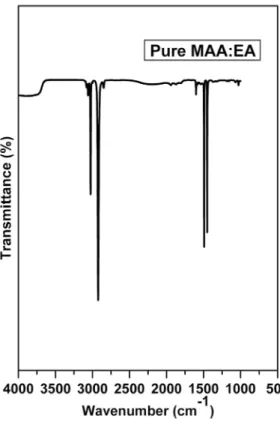
The FTIR spectrum for pure MAA:EA copolymer is shown in the Figure 1. The spectrum exhibits bands characteristics
of stretching, bending vibrations of O - H, C - H, C = O, CH3 - O and CH2 + α - CH3 groups. The observed FTIR
band positions and their assignments are presented in Table 1. In this film, a strong band observed at 1452 cm-1 has
been assigned to bending mode vibration corresponding to CH3 group, while another strong band observed at 1492
cm-1 has been attributed to C - H bending of CH 2
13. The
FTIR absorption bands appeared as weak bands in 1601 cm-1 range are attributed to carbonyl stretching of the acid
and ester units14. A broad and very strong band observed at
2923 cm-1, arising from C - H stretching frequency shows
the presence of methylene group11. Asymmetric C - H
stretching of methyl groups shows a peak at 3026 cm-1 and
C - H stretching of the methylene group gives two peaks at 3059 cm-1 and 3082 cm-1.
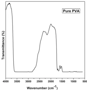
As shown in Figure 2, the FTIR spectrum of pure PVA exhibits several bands characteristic of stretching and bending vibrations of O - H, C - H, C = C and C - O groups. A broad and very strong band observed at 2813 - 3601 cm-1, which
arises from O - H stretching frequency is an indicator of the presence of hydroxyl groups. Another band observed at 2905 cm-1 indicates an asymmetric stretching mode of theCH
2
group. The band corresponding to CH2 symmetric stretching
vibration occurs at about 2168 cm-1. Absorbance at 1720
cm-1 corresponds to an acetyl C = O group, which could be
explained on the basis of intermolecular hydrogen bonding with the adjacent OH group. The shape of the carbonyl bands ( C = O ) for blend samples is different from that of pure
homopolymers, indicating a change in the balance of true and associated carbonyl groups in the blends. The increase in the multiplicity of hydroxyl band (OH) with increasing MAA:EA content in the blend samples may reflect possible interactions through hydrogen bonding of hydroxyl bread on the backbone of PVA and MAA:EA. Similar behavior was observed by Attia and Abd El - Kader 12 on PVA/ 2HEC
polyblend films. The increased intensity of bands indicates that greater amount of MAA:EA have complexed with the oxygen and hydrogen atoms. The effect of blending on the modes of vibration is observed interms of increase in the intensity of bands and appearance of new bands, which results from the formation of crosslinks between the MAA:EA and oxygen atoms of carbonyl groups. This indicates the increase in the basicity of C = O groups with an increase in the MAA:EA concentration. Absorption band observed at 1711cm-1 is due to C = O of carbonyl group. A moderate
absorption peak at 1658 cm-1 has been attributed to the C =
C stretching mode. Another band at 1566 cm-1 was attributed
to stretching vibration of C = C and a characteristic alcohol band at 1080 cm-1 was assigned to the stretching of C - O of
PVA, which is affected by hydrogen bonding along with C - H and O - H bending 15. A C - C stretching mode of absorption
is observed at 918 cm-1. The band observed at 1349 cm-1 has
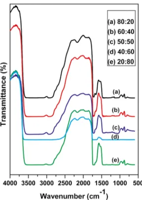
been attributed to combination frequencies of (CH + OH). The miscibility between the polymer and the copolymer (PVA / MAA:EA) blend is due to the hydrogen bonding between PVA hydroxyl groups and carbonyl groups of MAA:EA copolymer. The possibility of interactions between the polymer pair in the blend through specific polar groups is examined using FTIR analysis. FTIR spectra of PVA/
MAA:EA blends (80:20, 60:40, 50:50, 40:60, 20:80 wt %) are also shown in Figure 3(a - e). The FTIR spectra of all blended films exhibit bands, whose vibrational frequencies are found to be similar to several bands observed for pure PVA polymer film. IR features of PVA are more dominant even in 20:80 composition because PVA sample has dense molecular packing in the crystal and also stronger intermolecular hydrogen bonds comparing with MAA:EA, which are responsible for disappearance of functional groups of MAA:EA in the polymer blend. The FTIR spectrum shows shift in some of the band positions and change in the intensities of some other bands compared with pure films. O - H stretching frequency observed at 2813 - 3601 cm-1 for pure PVA shows a large
shift towards the low-frequency region in the blended films, which indicates the presence of a broad range of associated hydroxyls in blended films. The CH2 stretching frequency at
2905 cm-1 shifts towards higher frequency for all the blends
and the shift is found to be very high for 50:50 (wt %) blend i.e 2939cm-1. CH
2 stretching frequency observed at 2905cm -1
for pure PVA disappeared for 40:60 (Wt %) blend film. This change indicates the possibility of MAA:EA copolymer to be attached to methylene (CH2) and C - H in the side
chain of PVA molecule 16. The shift in the 2813 - 3601 cm-1
band corresponding to OH groups resulted from in inter/ intramolecular interactions in PVA/MAA:EA blends. The band at 2168 cm-1 is assigned to CH
2 symmetric stretching
vibrations. For the blend samples, the band shifts to lower frequency and the intensity of the band is decreased, which indicates the change in the chemical structure.
The appearance of a band at 1658 cm-1 (C = C) confirms
the semi-crystalline nature of the blends as reported by Ragab6. A weak peak around 1658 cm -1 is observed in the
present work, which confirms the semicrystalline nature
Table 1. FTIR peak assignment of pure and PVA/MAA:EA blend films.
Wavenumber
(cm-1) Band assignment
1452 CH3– O bending 1492 C – H bending of CH2
1601 C = O stretching
2923 C – H stretching of methylene group
3026 C – H stretching methyl group
3059 C – H stretching methylene group
3082 C – H stretching methylene group
2813 – 3601 O – H stretching frequency
2905 Asymmetric stretching mode of CH2group
2168 CH2symmetric stretching vibration 1711 C = O stretching
1658 C = C stretching
1566 C = C stretching vibration
1349 CH + OH combination frequencies
1080 C – O stretching
918 C – C stretching
polymer blend films. This occurs as the elimination of water proceeds isolating C = C in acyclic ring structure.
The small absorption band around 918 cm-1 was found to
be characteristic of the syndiotactic structure of the prepared films, Nagura et al.17 reported that the syndiotacticity of the
PVA samples causes dense molecular parking in the crystal and stronger intermolecular hydrogen bonds , which inturn are responsible for the disappearance of the molecular motion. This band was appeared in the spectra of pure PVA and other blend samples. These results manifested the conclusion of the specific interaction in PVA/MAA:EA blend matrix. In the case of PVA/MAA:EA, FTIR spectra shows shifts in some bands and change in the intensities of other bands comparing with the pure films which indicates the considerable coupling between the polymer and copolymer.
3.2. Differential scanning calorimetry (DSC)
Thermal characterization techniques are the convenient tools to determine the physical and chemical changes such as phase transitions, glass transition temperature (Tg) and melting temperature (Tm). A temperature range of 0 - 250ºC at a heating rate of 10ºC/min was utilized under nitrogen atmosphere. The DSC patterns in Figure (4) show endothermic peaks of glass transition temperature (Tg) and melting temperature (Tm). Tg of polymer blends is a typical characteristic which is used in studying the miscibility and interaction between polymers and Tm is mostly used in
investigating the crystallization of polymers. In Figure 4, one can see that all blended films have only one Tg indicating
that the blends are homogeneous. Theoretically, the glass transition temperature of miscible polymer blends could be calculated by using Fox equation18.
(1)
In which W1, W2 and are weight fractions and the glass transition temperatures of component 1 and 2 respectively. The observed transition temperature Tg(o) and theoretical value Tg(th) of the blends are presented in Table 2.
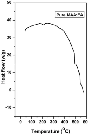
For pure PVA, Tg observed is 96.3ºC 19 and for copolymer
(MAA:EA) shown in the Figure 5, it is 103ºC 11. The small
traces at about 41ºC for the MAA:EA sample could be due to a small amount of moisture. The Tg for blend varies from 90 to 103.6ºC with the blend ratio. All the observed transition temperatures show variation compared to their theoretical values calculated by equation (1). This deviation from the theoretical value of the glass transition temperature is also observed in an earlier report 20 in which they attributed this
behavior to deviation with respect to free volume linearity in the blends. Changes in the free volume are related to specific interactions between polymer chains, increasing specific interactions lead to the formation of a more compact molecular network and diminish the free volume of molar
Figure 3. FTIR curves of PVA/MAA:EA blend samples (a) 80:20
(b) 60:40 (c) 50:50 (d) 40:60 (e) 20:80
Figure 4. DSC curves of (a) Pure PVA and PVA/MAA:EA blend
sample (b) 80:20 (c) 60:40 (d) 40:60 (e) 20:80 (f) 50:50
T
T
W
T
W
1
g blend g g
1 2
1 2
=
+
chains. In this system, the mobility of the chains decreases due to the presence of hydrogen bonds.
An endothermic peak was observed at around 222ºC for pure PVA and MAA:EA copolymer shows it at 210ºC, which corresponds to the melting temperature of PVA and MAA:EA respectively. A slight shift of Tm towards lower temperature has been observed for blended samples, which reveals the transition from semi-crystalline to an amorphous phase. The lowering of Tm for blend films is quite common and has been related to decreasing spherulite sizes21 and their
surface free energy. As a result of more flexible amorphous environment getting trapped in or adjacent to the crystalline matrix, the suppressed crystalline portion of the MAA:EA copolymer melts probably at lower temperatures. This clearly indicates the suppression of crystallites and there by increase in the amorphous content in the polymer. This leads to favoring in transport and thus to enhance the ionic conductivity of the electrolyte22.
3.3. Thermogravimetric analysis (TGA)
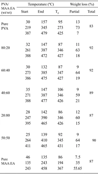
Figure (6), shows dTG thermograms of pure films and PVA/MAA:EA blends with a heating rate of 10ºC/min in the temperature ranging from room temperature to 800ºC. It is found that there are three temperature regions over which most of the weight loss occurs. These stages can be distinguished in the diagram of weight loss ( TG% ) during heating as well as more clearly in the diagram of derivative mass loss (DTA). The decomposition steps and percentage weight loss for individual polymers and their blends are represented in Table 3.
For pure PVA the first weight loss occurs between 30 - 157ºC with a weight loss of 13 %, which corresponds to the removal of water. The second weight loss occurs between 219 - 345ºC and corresponds to the thermal degradation of PVA intermolecular hydrogen bonding. The third degradation between 387 - 479ºC corresponds to the decomposition of PVA main backbone chain. The DTG curve of the pure MAA:EA copolymers shown in inset of Figure (6), reveals that there are three distinct steps of weight loss. The 4 - 5% weight loss below 170ºC is attributed to the loss of adsorbed water23. The second and third steps observed in
the temperature ranges 210 - 400ºC and above 400ºC are the regions of major weight loss and appeared to be due to the extensive degradation of the polymer backbone. The
Table 2. Comparison of observed transition temperatures Tg(o) and
theoretical Value Tg(th) of the blends.
PVA/MAA:EA Tg(th)ºC Tg(o)ºC
20:80 100.00 103.6
40:60 99.07 94.7
60:40 98.3 95.0
80:20 97.21 99.6
50:50 98.59 90.0
Figure 5. DSC curve of pure MAA:EA copolymer
Figure 6. DTG curves of pure and blended PVA/MAA:EA samples
(a) Pure PVA (b) 80:20 (c) 60:40 (d) 40:60 (e) 50:50 (f) 20:80 (g) Pure MAA:EA
The blends of PVA and MAA:EA also exhibit three weight loss stages as shown in Figure (6), followed by a final decomposition at 500ºC for all the membranes. The lower values of weight loss percentage in the first decomposition step are responsible for the volatilization of small molecules and or evaporation of residual absorbed water. The second weight loss may be due to the splitting of monomers and bond scission in the polymeric backbone and also the ionic interaction between the homopolymers. The major weight loss percentage in the third decomposition step is attributed to fragmentation of the macromolecular structure of both PVA polymer and MAA:EA copolymer 12. The blends thus
exhibit a relatively good thermal stability than pure films. Therefore, it can be concluded that the blend sample of 50:50 wt % has a more thermal stability and lower order than the other samples.
3.4. X - ray diffraction analysis (XRD)
The X-ray diffraction technique is a useful tool to determine the structure and crystallization of the polymer
matrices. Figure 7 shows the X - ray diffraction patterns of PVA and MAA:EA copolymers as well as their blend samples at room temperature in the scanning range 0º≤2θ≤40º. Pure PVA spectrum [Figure 7 (a)] shows small peaks at 2θ = 0.6º, 1.46º and relatively broad peak at 2θ = 19.52º diffused in the hallow amorphous region25. The appearance
of sharp reflections and diffuse scattering is characteristic of crystalline and amorphous phases of convectional semicrystalline polymers. Accordingly, the PVA/MAA:EA blends [Figure 7 (b-f)] also exhibited semicrystalline nature. From the spectra of PVA/MAA:EA blends, one distinct peak at 2θ = 19.52º corresponding to crystal, which is responsible for separation as it comprises of functional groups such as - CH2 - CH3 and - OH , has not undergone any significant
change after adding more amount of PVA. However, the spectrum of pure MAA:EA copolymer shown in the inset of Figure 7 exhibits a broad amorphous halo with a scattered intensity maximum corresponding to 2θ = 16.39º. The patterns revealed that there are no sharp diffraction lines and the broad background scattering of MAA:EA copolymer suggests the presence of amorphous nature.
XRD patterns of blend samples with concentrations 60, 40, 20 and 50 (wt %) PVA (Figure 7) exhibit the characteristics of pure PVA, but varying intensity for the reflection peak.
Table 3.TGA and DTG data for individual Polymers and their
blends of PVA/ MAA:EA (wt/wt).
PVA/ MAA:EA (wt/wt)
Temperature (ºC) Weight loss (%)
Start End TP Partial Total
Pure PVA 30 219 387 157 345 479 95 273 425 13 73 7 83 80:20 32 261 388 147 387 472 87 346 427 11 63 18 92 60:40 30 273 386 132 385 475 87 347 427 9 64 19 92 40:60 35 271 388 147 387 477 106 346 426 9 59 21 89 20:80 28 247 395 142 390 465 86 346 426 12 60 15 87 50:50 25 264 411 139 410 465 92 345 431 9 64 17 90 Pure MAA:EA 46 135 243 135 243 458 86 194 367 7.5 35 55.65 87
Figure 7. XRD curves of pure and blended PVA/MAA:EA samples
Thus, one can say that the semicrystalline nature of PVA is decreased upon mixing with the relatively higher content of MAA:EA copolymer. For such blends, compatibility between the semicrystalline and amorphous components of copolymers is possible. Thus, it can be suggested that the crystalline forms in PVA do not prevent the compatibility between amorphous regions of the two polymers in the blend system26. The tendency of varying crystallinity in blend
sample implies a decrease of a number of hydrogen bonds that are formed between PVA and MAA:EA if present7.
It can be seen that the peaks at θ = 0.6º and 1.46º got slightly displaced in the complex 80 wt % PVA polymer blend film. The intensity of the peak at 2θ = 19.520 decreased
and peak width increased to 80 wt % of PVA film. The decrease in the intensities of 80 % complexed PVA blend films causes a decrease in the degree of crystallinity and a simultaneous increase in the amorphous nature of complexed films. Hence this amorphous nature of the film is responsible for greater ionic diffusivity which may results in high ionic conductivity, which can be obtained in amorphous polymers that have flexible backbone16. This observation confirms
that complexation has taken place in the amorphous phase.
3.5. Scanning electron microscopy (SEM) Studies
The Scanning electron microscope produces images of a sample by scanning it with a focused electron beam. The beam of electrons interacts with electrons in the sample, which produces various signals that can be detected and that contain the information about the sample’s surface morphology and composition. The beam of electrons generally scanned by the method of raster scan pattern, and the position of the beam is combined with detected signal to create an image. SEM is often used to study the compatibility between various components of the polymer electrolytes through the detection of phase separations and interfaces27,14. The
compatibility between the polymer blend and the inorganic dopants has great influence on the properties of thermal, mechanical and ionic conductivity of the polymer electrolytes.
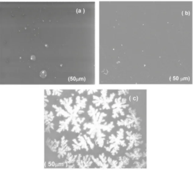
Figure 8 (a-c) shows the SEM images of Pure MAA:EA, Pure PVA and PVA/MAA:EA (50:50) blend.It can be seen from Figure 8 (a) that pure MAA:EA copolymer film shows smooth surface, suggesting that MAA and EA molecules may disperse in the soft-segment phase with little influence on the micro phase separation and mixing of the hard and soft segments28. SEM of pure PVA film exhibits no features
attributable to any crystalline morphology as shown in Figure 8 (b). So the semicrystallinity of PVA discussed earlier is likely to be submicroscopic in nature. SEM image of polymer blend electrolyte is shown in Figure 8 (c).The side branches are not well correlated along the length of the dendrites trunk. The growth of the dendritic-like shape, which represents the gathering of the branched aggregate clusters, became more clear leading to the formation of condensed
aggregated dendrites shape. This suggests the presence of structural reorganizations of polymer chains10.
3.6. UV - Visible absorption studies
The study of optical absorption, the absorption edge, in particular, has been proved to be the very useful in the elucidation of the optical properties and optical constants of crystalline and non - crystalline materials. Figure 9 shows the absorption spectra of PVA and MAA:EA copolymers and their blends 80:20, 60:40, 50:50, 40:60, and 20:80 (wt %) PVA/MAA:EA in the wavelength range 200 - 800 nm. The spectra of pure PVA and pure MAA:EA copolymer exhibited a shoulder like band at 274 and 221nm as shown in Figure 9 (a) and insert of Figure 9 respectively, which may be due to π→π* electronic transition (k - band) of
carbonyl groups29. All the blended samples contain only one
remarkable shoulder like a band with an irregular deviation in the position. The absorption of all blended samples increases as copolymer content increases. This is the evidence for the miscibility between copolymers. The UV - Visible spectra for the blended PVA/MAA:EA samples with the composition of 40:60, 50:50, and 20:80 (wt %) contained sharp intense bands at 213, 208 and 201 nm, beside a shoulder like band at about 276 nm.
The absorption coefficient ‘α' is related to absorbance ‘A’ by the equation
(2)
Where A is the absorbance and d is the thickness of the sample. When direct band gap exists, the absorption coefficient has the following dependence on the energy of the incident photon30
(3)
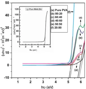
Where Eg is the band gap, C is a constant, υ is the frequency of light and h is plank’s constant. A plot of (αhυ)2
vs hυ is shown in Figure 10. The intercept on the energy axis on extrapolating the linear portion of the curve to zero absorption value may be taken as the value of the band gap. For both pure PVA and MAA:EA films, the direct band gap lies at 5.87 and 5.16 eV respectively. The direct band gap values for blended films vary from 5.77 eV to 5.05 e V and are presented in Table 4. For indirect transitions, which requires phonon assistance, the absorption coefficient has the following dependence on the photon energy11.
(4)
Where Ep is the phonon energy associated with transition and A and B are the constants depending on the band structure.
.
d
A
2 303
#
a
=
S X
h
C h
E
g1 2
a y
=
Q
y
-
V
h A h Eg Ep B h Eg Ep
2
Figure 8. SEM images of (a) Pure MAA:EA (b) Pure PVA (c) PVA/MAA:EA (50:50) blend
The indirect band gap values can be calculated by plotting (αhυ)1/2 vs (hυ) as shown in Figure 11. For both pure PVA
and MAA:EA films, the indirect band gap values lie at 5.68 and 4.82 eV respectively. The values of all blended samples vary from 5.28 to 4.39 eV (Table 4).
The position of absorption edge was obtained by extrapolating the linear portion of α vs hυ plot (Figure 12) to zero absorption value. For both PVA and MAA:EA films, the absorption edge lies at 5.94 and 5.04 eV respectively, and the absorption edge for the blends vary from 5.79 to 4.83 eV (Table 4)
From Table 4, it is evident that the values of direct and indirect band gap varies with increase in MAA:EA content. This may be reflecting the induced changes in the number of available final states according to blend compositions12.
Among the all blend samples, the sample with 50:50 (wt %) shows minimum optical band gap value (Table 4), this indicates that it shows more semiconducting nature than
remaining blend samples. Figure 9. Absorption spectra of pure and blended PVA/MAA:EA
Figure 10. (αhυ)2 vs hυ curves of pure and PVA/MAA:EA blend
samples (a) Pure PVA (b) 80:20 (c) 60:40 (d) 40:60 (e) 50:50 (f) 20:80 (g) Pure MAA:EA
Table 4. Values of absorption edge and optical band gap of pure
and PVA/MAA:EA blend samples.
PVA/ MAA:EA
(wt/wt)
Absorption edge (eV)
Optical bandgap energy (eV)
Direct Indirect
Pure PVA 5.94 5.87 5.68
80:20 5.79 5.77 5.28
60:40 5.62 5.74 5.21
50:50 4.83 5.05 4.39
40:60 4.89 5.16 4.47
20:80 5.04 5.12 4.68
Pure
MAA:EA 5.04 5.16 4.82
4. Conclusions
The existence of maximum absorbing edge and minimum in band tail for 50:50 (wt/wt) % PVA/MAA:EA blend sample indicate that it has more carrier concentration in the localized levels. On the basis of the observed data from the XRD, FTIR, DSC and TGA, it could be concluded that the formation of hydrogen bonding between PVA and MAA:EA is possible with in the investigated composition ranges. FTIR spectra of all blend samples shows the variation in the intensities of peaks with increasing the MAA:EA content in the polymer matrix , which indicate the considerable interaction between PVA and MAA:EA. XRD studies on the blend samples of 20, 40, 50 and 60 wt % PVA content revealed that the semi-crystalline structure of PVA is essentially sustained while
Figure 11. (αhυ)1/2 vs hυ curves of pure and PVA/MAA:EA blend
samples(a) Pure PVA (b) 80:20 (c) 60:40 (d) 50:50 (e) 40:60 (f) 20:80 (g) Pure MAA:EA
Figure 12. α vs hυ curves of pure and PVA/MAA:EA samples
(a) Pure PVA (b) 80:20 (c) 60:40 (d) 40:60 (e) 20:80 (f) 50:50 (g) Pure MAA:EA
(Tm). Out of all the blend films, PVA/MAA:EA with 50:50 (wt %) shows optimal properties.
5. References
1. Swaroop K, Francis S, Somashekarappa HM. Gamma irradiation synthesis of Ag/PVA hydrogels and its antibacterial activity.
Materials today: Proceedings. 2016;3(6):1792-1798. 2. Abraham A, Soloman PA, Rejini VO. Preparation of
Chitosan-Polyvinyl Alcohol Blends and Studies on Thermal and Mechanical Properties.Procedia Technology. 2016;24:741-748.
3. Muthuvinayagam M, Gopinathan C. Characterization of proton conducting polymer blend electrolytes based on PVdF-PVA. Polymer. 2015;68:122-130.
4. El-Sayed S, Abdel-Baset T, Elfadl AA, Hassen A. Effect of nanosilica on optical, electric modulus and AC conductivity of polyvinyl alcohol/polyaniline films. Physica B: Condensed Matter. 2015;464:17-27.
5. Shanthi B, Muruganand S. Structural, vibrational, thermal, and electrical properties of PVA/PVP biodegradable polymer.
International Journal of Scientific Engineering and Applied Science. 2015;1(8):348-351.
6. Ragab HM. Spectroscopic investigations and electrical properties of PVA/PVP blend filled with different concentrations of nickel chloride. Physica B: Condensed Matter. 2011;406(20):3759-3767.
7. Abdelghany AM, Meikhail MS, Elsheshtawy NA, Salah HY. Structural and Thermal Stabilization Correlation of PEO/ PVA-AgCl Polymer Composites. Middle East Journal of Applied Sciences. 2015;5(5):1-6.
8. Abbas AK, Naife RM, Rashid FL, Hashim A. Optical properties of (PVA-PAA-Ag) nanocomposites. International Journal of
Science and Research. 2015;4(1):2489-2492.
9. Rubee BH, AlHussien SA. Study of optical properties of (PVA-PEG-ZNO) nanocomposites. International Journal of
Science and Research. 2016;5(5):1794-1799.
10. Okerberg BC, Marand H, Douglas JF. Dentritic crystallization in thin films of PEO/PMMA blends: A comparison to crystallization in small molecule liquids. Polymer. 2008;49(2):579-587.
11. Kumar YM, Bhyagyasree K, Gopal NO, Ramu C, Nagabhushana H. Structural, Thermal and Optical Properties of Mn2+ Doped Methacrylic Acid - Ethyl Acrylate (MAA:EA) Copolymer Films.
Zeitschrift für Physikalische Chemie. 2016;231(5):1039-1055.
12. Attia G, El-kaderA MFH. Structural, Optical and Thermal Characterization of PVA/2HEC Polyblend Films. International Journal of Electrochemical Science. 2013;8(4):5672-5687.
13. Kumar KK, Ravi M, Pavani Y, Bhavani S, Sharma AK, NarasimhaRao VVR. Investigations on PEO/PVP/NaBr complexed polymer blend electrolytes for electrochemical cell applications. Journal of Membrane Science. 2014;454:200-211.
14. Abdelrazek EM, Abdelghany AM, Oraby AH, Asnag GM. Investigation of Mixed Filler Effect on Optical and Structural Properties of PEMA Films. International Journal of Engineering
& Technology. 2012;12(4):98-102.
15. Hemantha Kumar GN, Lakshmana Rao J, Gopal NO, Narasimhulu KV, Chakradhar RPS, VaradaRajulu A. Spectroscopic investigations of Mn2+ ions doped poly(vinyl)alcohol films.
Polymer. 2004;45(16):5407-5415.
16. Balaji Bhargav P, Madhu Mohan V,Sharma AK, Rao VVRN. Structural, Electrical and Optical Characterization of Pure and Doped Poly (Vinyl Alcohol) (PVA) Polymer Electrolyte Films.
International Journal of Polymeric Materials. 2007;56(6):579-591.
17. Nagura M, Mastuzawa S, Yamaura K, Ishikawa H. Tacticity Dependence of Molecular Motion in Crystal of Poly(vinyl alcohol). Polymer Journal. 1982;14:69-72.
18. Larrañaga M, Mondragon I, Riccardi CC. Miscibility and mechanical properties of an amine-cured epoxy resin blended with poly(ethylene oxide). Polymer International. 2007;56(3):426-433.
19. Chen N, Zhang J. The role of hydrogen-bonding interaction in poly(vinyl alcohol)/poly(acrylic acid) blending solutions and their films. Chinese Journal of Polymer Science. 2010;28(6):903-911.
20. Mano V, Ribeiro e Silva MES, Silva ME, Barbani N, Giusti P. Binary blends based on poly(N-isopropylacrylamide): Miscibility studies with PVA, PVP, and PAA. Journal of Applied Polymer
Science. 2004;92(2):743-748.
21. Bandara LRAK, Dissanayake MAKL, Mellander BE. Ionic conductivity of Plasticized(PEO)-LiCF3SO3 electrolytes.
Electrochimica Acta. 1998;43(10-11):1447-1451.
22. Mohan VM, Qiu W, Shen J, Chen W. Electrical properties of poly(vinyl alcohol) (PVA) based on LiFePO4 complex polymer electrolyte films. Journal of Polymer Research. 2010;17(1):143. 23. Farces S. Influence of gamma-ray irradiation on optical and
thermal degradation of poly (ethyl-methacrylate) (PEMA) polymer. Natural Science. 2012;4(7):499-507.
24. Zain NF, Zainal N, Mohamed NS. The influences of ionic liquid to the properties of poly(ethylmethacrylate) based electrolyte.
PhysicaScripta. 2015;90(1):015702.
25. Liu C, Xiao C, Liang H. Properties and structure of PVP-lignin "blend films". Journal of Applied Polymer Science. 2005;95(6):1405-1411.
26. Cheung YW, Guest MJ. A study of the blending of ethylene-styrene copolymers differing in the copolymer ethylene-styrene content: Miscibility considerations. Journal of Polymer Science Part B:
Polymer Physics. 2000;38(23):2976-2987.
27. Zhang S, Lee JY, Hong L. Visualization of particle distribution in composite polymer electrolytes systems. Journal of Power
Sources. 2004;126(1-2):125-133.
28. Madhav Kumar Y, Gopal NO, Ramu C, Babu S, Lakshmana Rao J, Nagabhushana H,et al. Structural, thermal and optical properties of Cu+2 doped methacrylic acid-ethyacrylate (MAA:EA) copolymer films. Bulletin of Materials Science. 2017;40(5):877-886.
29. Shehap AM. Thermal and Spectroscopic Studies of Polyvinyl Alcohol/Sodium Carboxy Methyl Cellulose Blends. Egypt
Journal of Solids. 2008;3(1):75-91.