R E S E A R C H
Open Access
Identification of two novel cytolysins from the
hydrozoan
Olindias sambaquiensis
(Cnidaria)
Vidal Haddad Junior
2, Fernando Zara
1, Sergio Marangoni
3, Daniela de Oliveira Toyama
4,
Alex Jardelino Felizardo de Souza
1, Simone Cristina Buzzo de Oliveira
1,2and Marcos Hikari Toyama
1,5*Abstract
Background:Although the hydrozoanOlindias sambaquiensisis the most common jellyfish associated with human envenomation in southeastern and southern Brazil, information about the composition of its venom is rare. Thus, the present study aimed to analyze pharmacological aspects ofO. sambaquiensisvenom as well as clinical manifestations observed in affected patients. Crude protein extracts were prepared from the tentacles of animals; peptides and proteins were sequenced and submitted to circular dichroism spectroscopy. Creatine kinase, cytotoxicity and hemolytic activity were evaluated by specific methods.
Results:We identified two novel cytolysins denominated oshem 1 and oshem 2 from the tentacles of this jellyfish. The cytolysins presented the amino acid sequences NEGKAKCGNTAGSKLTFKSADECTKTGQK (oshem 1) and
NNSKAKCGDLAGWSKLTFKSADECTKTGQKS (oshem 2) with respective molecular masses of 3.013 kDa and 3.375 kDa. Circular dichroism revealed that oshem 1 has random coils and smallα-helix conformation as main secondary
structure whereas oshem 2 presents mainly random coils as its main secondary structure probably due to the presence of W (13) in oshem 2. The hemolysis levels induced by oshem 1 and oshem 2 using a peptide concentration of 0.2 mg/mL were, respectively, 51.7 ± 6.5% and 32.9 ± 8.7% (n = 12 and p≤0.05). Oshem 1 and oshem 2 showed significant myonecrotic activity, evaluated by respective CK level measurements of 1890.4 ± 89 and 1212.5 ± 103 (n = 4 and p≤0.05). In addition, myonecrosis was also evaluated by cell survival, which was measured at 72.4 ± 8.6% and 83.5 ± 6.7% (n = 12 and p≤0.05), respectively. The structural analysis showed that both oshem 1 and oshem 2 should be classified as a small basic hemolytic peptide.
Conclusion:The amino acid sequences of two peptides were highly similar while the primary amino acid sequence analysis revealed W (22th) as the most important mutation. Finally oshem 1 and oshem 2 are the first cytolytic peptides isolated from theOlindias sambaquiensisand should probably represent a novel class of cytolytic peptides.
Keywords:Olindias sambaquiensis, Hemolytic, Myonecrosis, Cytotoxicity, Cytolysin, Cnidaria venom
Background
The phylum Cnidaria is subdivided into five classes. Some species are important in human medicine since they can cause severe envenomation by the injection of venom through specialized cells. In Brazil, these organisms are in-cluded in the Hydrozoa, Scyphozoa and Cubozoa classes [1,2]. The class Hydrozoa has two species frequently asso-ciated with human envenomation: the Portuguese
man-of-war (Physalia physalis) and Olindias sambaquiensis
(Figure 1), the latter being related to the majority of acci-dents in the most populated regions of Brazil [1,2]. Like the other cnidarians, they present stinging cells equipped with small organelles known as nematocysts that contain small threads ejected along with the venom when stimu-lated mechanically or chemically [3]. Furthermore, nema-tocysts are found throughout the body of the cnidarians and not just in their tentacles [4-6].
Olindias sambaquiensis(“relojinho” in the Portuguese language), is a small hydrozoan jellyfish that provokes mild accidents on the southeastern and southern Brazilian coast where it is commonly found in winter and autumn [1,2,7]. Despite the medical relevance and abundance of
* Correspondence:mhtjpn@yahoo.com
1São Paulo Experimental Coast Campus, São Paulo State University (UNESP –
Univ Estadual Paulista), São Vicente, São Paulo State, Brazil
5UNESP, Campus do Litoral Paulista, Unidade São Vicente, Praça Infante D.
Henrique, s/n, São Vicente, SP CEP 11330-900, Brasil
Full list of author information is available at the end of the article
this species in Brazil, there are not any studies characte-rizing its venom biochemically. The present work aimed to report the structural and biological characterization of hemolytic proteins from the nematocysts of Olindias sambaquiensis.
Methods
The jellyfish were collected off the São Vicente beach (latitude 23º57'47"– South, longitude 46º23'31"–West) and their tentacles were then subjected to three freeze-thaw cycles. After the last freeze-freeze-thaw cycle, the solution was centrifuged at 20,000 g for 60 minutes at 4°C. Crude protein extracts were prepared from the tentacles re-moved from the jellyfish body using forceps and im-mediately immersed in an ice-cold 0.1% trifluoroacetic acid (TFA). The supernatant was recovered and filtered through a 0.45-μm filter, followed by a second ultrafiltra-tion using a 0.22-μm filter.
The desired fraction was then lyophilized and the re-sulting peptides were subjected to hemolysis assay on freshly prepared human erythrocytes.
Protein sequencing was performed as previously de-scribed by Oliveira et al. [8]. Briefly, two milligrams of the purified protein was dissolved in 200μL of a 6 mol/L guanidine chloride solution (Merck, Germany) containing 0.4 mol/L of Tris–HCl and 2 mmol/L of EDTA (pH 8.15). Nitrogen was blown over the top of the protein solution for 15 minutes, after which the protein solution was re-duced with DTT (6 M, 200μL) and subjected to a second incubation under a nitrogen atmosphere for 90 minutes. After this incubation time, 80μL of iodoacetic acid was added to the solution (50 mM of cold iodoacetic and car-boxymethylated 14C-iodoacetic acid), followed by a third incubation under a nitrogen atmosphere after which the reaction tube was sealed. To remove excess reagent and
to purify the peptides, we used a preparative C5 reverse phase column.
Circular dichroism (CD) spectroscopy: the purified pep-tide was dissolved in 10 mM sodium phosphate pH 7.4 and the solution was adjusted to final protein concentra-tion of 10μM. After centrifugation at 4500 ×gfor five mi-nutes, samples were transferred to a 1 mm path-length quartz cuvette. Circular dichroism spectra in the wave-length range 185–300 nm were acquired in-house on a J720 spectropolarimeter (Jasco Corp., Japan) using a band-width of 1 nm and a response time of 1 s. Data collection was performed at room temperature. With a scanning speed of 100 nm/min, a total of nine scans was accumu-lated for each sample and all the spectra were corrected by subtraction of buffer blanks.
The release of creatine kinase (CK) from damaged muscle cells was followed by use of the Kit 47-UV (Sigma-Aldrich Co., USA) to measure the enzyme activity in mouse plasma. For this determination five groups of mice (18–22 g) were injected in the right gastrocnemius muscle with 50μL of 0.5 mg. mL–1
toxins (n = 4) while the con-trol group received PBS. For the CK determination in plasma, the animals were lightly anesthetized with halo-thane immediately before and three hours after toxin or PBS injection for blood collection according to the guide-lines for the care and use of laboratory animals [9].
Mice were bled from the tail three hours after injection and blood was collected into heparinized tubes, with one aliquot being used for CK determination. Plasma was sep-arated by centrifugation and stored at 4°C for subsequent determination of CK activity. The procedure for the meas-urement of CK activity was conducted following the in-structions of the kit (Kit 47-UV, Sigma Chemical Co., USA). Enzyme activity is reported as international units per liter (U/L), in which 1 U is the amount that catalyzes the transformation of 1 μmol of substrate at 25°C. The cytotoxicity assay was conducted according to Passero
et al. [10] with toxins (or PBS as control) incubated with cells for 60 minutes. Cytotoxicity of the toxins from the
Olindias sambaquiensis in J774 cells in culture was as-sayed using 3-(4,5-Dimethylthiazol-2-yl)-2,5-diphenyl tet-razolium bromide (MTT). Briefly, cells in 96-well plates (106 macrophage/well) in culture medium (RPMI 1640) for 48 hours at 35°C and 5% CO2 were incubated with various toxin concentrations ranging from 0.78 to 100.00 μg/well for 48 hours. The cells were then washed and incubated with MTT for four hours, followed by SDS with 10% HCl for the determination of formazan conver-sion, which was monitored by absorbance at 595 nm. This experiment was carried out in triplicate.
Hemolytic activity of the toxin was measured quan-titatively in terms of attenuance on human red blood cells (RBC) at room temperature using a Spectramax Microtiter® plate reader (Molecular Devices, USA). Human
blood, freshly collected with heparin, was centrifuged to re-move the buffy coat, and the erythrocytes obtained were washed three times in 0.85% saline and stored at 4ºC. Toxins at desired concentrations were added in the first well to erythrocyte buffer (140 mM NaCl, 10 mM Tris– HCl, pH 7.4), and then serially diluted two-fold. RBCs (100 μL; D630 = 0.5) in erythrocyte buffer were added to the toxins, and hemolysis was monitored by measuring attenuance at 630 nm for 20 minutes at room temperature. The final volume was 200μL per well. The hemolysis per-centage was determined at the end of the assay using the following equation of Malovrhet al. [11]:
Hemolysisð Þ% ðDmax−DobsÞ Dmax−Dmin 100
in which Dobs was the measured attenuance in the well after 20 minutes and Dmax is the maximal attenuance by distilled water and Dmin is the minimal attenuance by buffer used in the test. For this assay we firstly de-termined the hemolytic CI50 values for oshem 1 and oshem 2, which were 324μg and 198μg, respectively.
The animal utilization was approved by the Committee for Ethics in Animal Experimentation of the Institute of Biology, UNICAMP, certificate number 1320–1. We uti-lized Swiss mice supplied by CEMIB, the Multidisciplin-ary Center for Biological Research of UNICAMP.
Results and discussion
Accidents involving the hydrozoanOlindias sambaquien-sis are common in South America and may provoke
dermatitis, skin lesions, edema and pain.Olindias samba-quiensispoisoning is characterized as moderate to severe, which can lead to cardiovascular complications including cardiopulmonary arrest. Despite its clinical importance and the seriousness of symptoms observed, this is the first study of the biochemical characterization and isolation of toxic components fromOlindias sambaquiensis.
The protocol used for this work essentially followed the methods used for Bunodosoma caissarum [12] and
Phyllorhiza punctata[13]. The crude extract from Olin-dias sambaquiensis revealed the presence of at least 17 fractions, of which only one fraction, denominated oshem, showed a moderate hemolytic activity (Figure 2a), and was subjected to novel fractionation on C18 reverse chro-matography using non-linear gradient of buffer B (66.6% of acetonitrile in 0.1% TFA) at a constant flow rate of 1.0 mL/min. Under this condition, we purified two iso-forms from oshem that were designated oshem 1 and oshem 2 (Figure 2b).
Sea anemone toxins comprise mainly proteins and pep-tides that are cytolytic or neurotoxic with their potency
varying according to the structure and action site, and are efficient at targeting different animals, such as insects, crustaceans and vertebrates. Summarily, the cnidarians venom includes 3.5 to 6.5 kDa voltage-gated sodium (NaV) channels toxins and 3–5 kDa voltage-gated potas-sium (KV) channel toxins and ~20 kDa pore-forming toxins [14]. In the case of pore-forming toxins, there are three other less frequent classes of toxins that can be clas-sified as: group I, which consists of peptides from 5 to 8 kDa; group III with a molecular mass of 80 kDa; and the pore-forming toxins with phospholipase A2 activity and molecular mass around 20 to 40 kDa [15]. Toxins oshem 1 and oshem 2 contain hydrophilic low-molecular-weight peptides, and can be classified as group I toxins, which are peptides of 5 to 8 kDa, following the classification estab-lished by Anderluh and Macek [15].
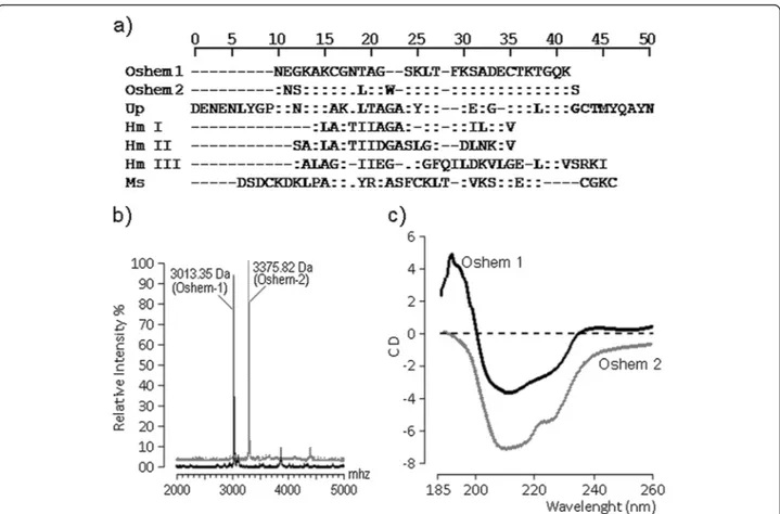
The N-terminal amino acid sequencing of oshem 1 and oshem 2 was determined after reduction. Both were subjected to automatic amino acid sequencing using a Procise® Protein Sequencer (Applied Biosystems, USA). The phenylthiohydantoin (PTH) derivatives of the amino
acids were identified by an Applied Biosystems model 450 microgradient PTH analyzer. (Figure 3a) and a 50% amino acid identity with Up (Urticina piscivora). The molecular masses of oshem 1 and 2 were measured ac-cording to the description of Toyama et al. [16], and found to be, respectively, 3.013 kDa and 3.375 kDa (Figure 3b). The circular dichroism showed that oshem 1 presents anα-helix structure not found in oshem 2, which revealed mainly the presence of random coil structures (Figure 3c) while the most important shifts observed were L (10th) and W (13th), which should be involved in modification of the secondary structure between oshem 1 and oshem 2. This structural replacement from oshem 1 to oshem 2 induced a significant modification in the secondary structure of these toxins. The struc-tural contents of oshem 1 comprise: percentage (%) by CD analysis of α-helix 33; β-sheet 48 and random coils 19; whereas the secondary structural contents of oshem 2 are: percentage (%) by CD analysis ofα-helix 04; β-sheet 68 and random coils 28.
Data collection was performed at room temperature, with a scanning speed of 100 nm.min−1
. Nine scans were accumulated for each sample whereas all spectra were corrected by subtraction of buffer blanks. The CD spec-tra are expressed as theta machine units in millidegrees.
The hemolytic activity levels of oshem fraction before its fractionation in oshem 1 and oshem 2 was evaluated at 12.4 ± 3.4% (n = 12 and p≤0.05), whereas the levels of oshem 1 and 2 were 51.7 ± 6.5% and 32.9 ± 8.7% (n = 12 and p≤0.05), respectively, and the hemolytic potency
of oshem 1 was two-fold less than that of α-hemolysin (Figure 4a). CK level after the injection of oshem was 759.50 ± 34 (n = 4, p≤0.05), whereas treatment with
oshem 1 or 2 increased CK levels by 1890.4 ± 89 and 1212.50 ± 103, respectively (n = 4, p≤0.05) (Figure 4b).
Cytotoxicity assay results were expressed as percentage of viable cells. Macrophage cell viability was moderately de-creased in the presence of whole oshem fraction before its fractionation in oshem 1 and oshem 2 was 92.4 ± 3.4% (n = 12, p≤0.05). For oshem 1 and 2 the values were 72.4 ± 8.6% and 83.5 ± 6.7% (n = 12 and p≤0.05),
res-pectively (Figure 4c). Cytolysins adopt a stable soluble structure, which undergoes a conformational change when brought into contact with a membrane, leading to an ac-tive, membrane-bound form that inserts spontaneously into the membrane [17-19]. The helix structure of oshem 1 appears crucial for exerting its full hemolytic activity, while the presence of W (22th) virtually destroyed its helix structure but did not abolish its hemolytic, myonecrotic or cytotoxic effect. The CD structure of oshem 2 showed the presence of random coil and beta-sheet structure, rais-ing the possibility that beta sheet may make an important structural contribution to the biological and pharmaco-logical activity of oshem 1 and 2.
The hemolysis percentage was calculated by the fol-lowing formula:
Hemolysis¼ðA540 in the peptide solution–A540 in PBSÞ
A540 in 1%Triton−X100–A540 in PBS
ð Þ 100%
Figure 4 shows the measurement of CK levels after three hours of toxin injection. The analysis of the figure revealed that oshem 1 and oshem 2 induced a significant increasing of CK levels in relation to oshem purified from the first chromatography. But this myonecrotic effect was lower if compared to α-hemolysin from Staphylococcus aureus.
Conclusions
Olindias sambaquiensis Muller, 1861 (Olindiidae) is a hydromedusae common in southern and southeastern
Figure 4Hemolytic effect of oshem, oshem1 and oshem 2 and alpha-hemolysin compared with alpha-hemolysin of
Brazilian coastal regions and Atlantic coast of Argentina and Uruguay [7]. The accident caused by this jellyfish has been described as presenting mild pain, round plaques and no systemic symptoms [1,2,20,21]. Our results suggest that the oshem fractions exert significant pharmacological and toxic effects that can play an important role in the clinical manifestation induced byO. sambaquiensis.
Ethics committee approval
The present study was approved by the Committee for Ethics in Animal Experimentation on the São Paulo Experimental Coast Campus of UNESP. The animal utili-zation was approved by the Committee for Ethics in Animal Experimentation of the Institute of Biology, UNICAMP, certificate number 1320–1. The utilized Swiss mice were supplied by CEMIB, the Multidisciplinary Center for Biological Research of UNICAMP.
Competing interests
The authors declare that there are no competing interests.
Authors’contributions
TLP and VHJ were responsible for the clinical and experimental study of the venom; FJZ, SM, DOT, AFS, VCGS and SCBO carried out the immunoassays and participated in the sequence alignments; MHT was the senior author of the manuscript. All authors read and approved the final manuscript.
Acknowledgments
The authors would like to thank FAPESP (Projeto Temático Biota, 2010/50188-8), CNPq and CAPES for their funding of this research.
Author details
1São Paulo Experimental Coast Campus, São Paulo State University (UNESP –
Univ Estadual Paulista), São Vicente, São Paulo State, Brazil.2Botucatu Medical
School, São Paulo State University (UNESP–Univ Estadual Paulista), Botucatu,
São Paulo State, Brazil.3Department of Biochemistry, Institute of Biology,
State University of Campinas (UNICAMP), Campinas, São Paulo State, Brazil.
4Center of Biological and Health Sciences, Mackenzie Presbyterian University,
São Paulo, São Paulo State, Brazil.5UNESP, Campus do Litoral Paulista,
Unidade São Vicente, Praça Infante D. Henrique, s/n, São Vicente, SP CEP 11330-900, Brasil.
Received: 21 August 2013 Accepted: 30 January 2014 Published: 25 March 2014
References
1. Haddad V Jr, da Silveira FL, Cardoso JL, Morandini AC:A report of 49 cases of cnidarian envenoming from southeastern Brazilian coastal waters.
Toxicon2002,40(10):1445–1450.
2. Haddad V Jr, Silveira FL, Migotto AE:Skin lesions in envenoming by cnidarians (Portuguese man-of-war and jellyfish): etiology and severity of accidents on the Brazilian Coast.Rev Inst Med Trop Sao Paulo2010,
52(1):43–46.
3. Watson GM, Hessinger DA:Evidence for calcium channels involved in regulating nematocyst discharge.Comp Biochem Physiol Comp Physiol 1994,107(3):473–481.
4. Alsen C:Biological significance of peptides fromAnemonia sulcata.Fed Proc1983,42(1):101–108.
5. Norton RS:Structure and structure-function relationships of sea anemone proteins that interact with the sodium channel.Toxicon 1991,29(9):1051–1084.
6. Honma T, Shiomi K:Peptide toxins in sea anemones: structural and functional aspects.Mar Biotechnol (NY)2006,8(1):1–10.
7. Júnior MN, Haddad MA:Variações morfológicas emOlindias sambaquiensis(Cnidaria, Hidrozoa, Limnomedusae) no Litoral de Guaratuba, Paraná Brasil.Rev Bras Zool2006,23(3):879–882.
8. de Oliveira DG, Toyama MH, Martins AM, Havt A, Nobre AC, Marangoni S, Câmara PR, Antunes E, de Nucci G, Beliam LO, Fonteles MC, Monteiro HS:
Structural and biological characterization of a crotapotin isoform isolated fromCrotalus durissus cascavellavenom.Toxicon2005,42(1):53–62. 9. Herling AW, Maas J, Seeger K:Guidelines for the care and use of
laboratory animals.InDrug Discovery and Evaluation: Pharmacological Assays.Edited by Vogel HG, Vogel WH. Germany: Springer Berlin Heidelberg; 1997:726–740.
10. Passero LF, Tomokane TY, Corbett CE, Laurenti MD, Toyama MH:
Comparative studies of the anti-leishmanial activity of three Crotalus durissus ssp. venoms.Parasitol Res2007,101(5):1365–1371.
11. Malovrh P, SepcićK, Turk T, Macek P:Characterization of hemolytic activity of 3-alkylpyridinium polymers from the marine spongeReniera sarai.
Comp Biochem Physiol C Pharmacol Toxicol Endocrinol1999,124(2):221–226. 12. Martins RD, Alves RS, Martins AM, Barbosa PS, Evangelista JS, Evangelista JJ, Ximenes RM, Toyama MH, Toyama DO, Souza AJ, Orts DJ, Marangoni S, de Menezes DB, Fonteles MC, Monteiro HS:Purification and characterization of the biological effects of phospholipase A(2) from sea anemone
Bunodosoma caissarum.Toxicon2009,54(4):413–420.
13. Carneiro RF, Nascimento NR, Costa PP, Gomes VM, de Souza AJ, de Oliveira SC, Dos Santos Diz Filho EB, Zara FJ, Fonteles MC, de Oliveira Toyama D, Toyama MH, Santos CF:The extract of the jellyfishPhyllorhiza punctata
promotes neurotoxic effects.J Appl Toxicol2011,31(8):720–729. 14. Frazão B, Vasconcelos V, Antunes A:Sea anemone (Cnidaria, Anthozoa,
Actiniaria) toxins: an overview.Mar Drugs2012,10(8):1812–1851. 15. Anderluh G, Macek P:Cytolytic peptide and protein toxins from sea
anemones (Anthozoa: Actiniaria).Toxicon2002,40(2):111–124. 16. Toyama MH, Toyama DO, Joazeiro PP, Carneiro EM, Beriam LO, Marangoni
LS, Boschero AC:Biological and structural characterization of a new PLA2 from theCrotalus durissus collilineatusvenom.Protein J2005,
24(2):103–112.
17. Anderluh G, Macek P:Dissecting the actinoporin pore-forming mechanism.
Structure2003,11(11):1312–1313.
18. Parker MW, Feil SC:Pore-forming protein toxins: from structure to function.Prog Biophys Mol Biol2005,88(1):91–142.
19. Kokelj F, Mianzan H, Avian M, Burnett JW:Dermatitis due toOlindias sambaquiensis: a case report.Cutis1993,51(5):339–342.
20. Mianzan HW, Fenner PJ, Cornelius PF, Ramírez FC:Vinegar as a disarming agent to prevent further discharge of the nematocysts of the stinging hydromedusaOlindias sambaquiensis.Cutis2001,68(1):45–48. 21. Mosovich JH, Young P:Picadura de medusaOlindias sambaquiensis:
análisis de 49 casos.Medicina (B Aires)2012,72(5):380–388. doi:10.1186/1678-9199-20-10
Cite this article as:Junioret al.:Identification of two novel cytolysins from the hydrozoanOlindias sambaquiensis(Cnidaria).Journal of Venomous Animals and Toxins including Tropical Diseases201420:10.
Submit your next manuscript to BioMed Central and take full advantage of:
• Convenient online submission
• Thorough peer review
• No space constraints or color figure charges
• Immediate publication on acceptance
• Inclusion in PubMed, CAS, Scopus and Google Scholar
• Research which is freely available for redistribution