RESUMO.- [Mapeamento dos sítios de latência e reativa-ção pelo herpesvírus bovino tipo 5 (BoHV-5) e por mu-tante deletado no gene da timidina quinase em ovinos.] Um recombinante do herpesvírus bovino tipo 5 com dele-ção no gene da timidina quinase (BoHV-5tkΔ) foi capaz de
estabelecer latência e reativar - embora ineficientemente - em modelo experimental em ovinos (Cadore et al. 2013). Como a reativação de alfaherpesvírus defectivos na TK em tecido neural é improvável, o presente estudo investigou os sítios de latência e reativação por esse recombinante em ovinos. Para isso, grupos de ovinos foram inoculados com a cepa de BoHV-5 parental (SV-507/99) ou com o recombi -nante BoHV-5tkΔ. Durante a infecção latente (dia 40 pós --infecção, pi) a distribuição do DNA do vírus recombinante no encéfalo de ovinos infectados experimentalmente foi similar ao do vírus parental (SV-507/99). O DNA de ambos os vírus foi detectado consistentemente por PCR nos gân -glios trigêmeos (TGs), frequentemente nas tonsilas farín
-Mapping the sites of latency and reactivation by bovine
herpesvirus 5 (BoHV-5) and a thymidine kinase-deleted
BoHV-5 in lambs
1Gustavo C. Cadore2, Gian Marcon2, Mario Celso Sperotto Brum3, Rudi Weiblen2
and Eduardo F. Flores2*
ABSTRACT.- Cadore G.C., Marcon G., Brum M.C.S., Weiblen R. & Flores E.F. 2013. Mapping the sites of latency and reactivation by bovine herpesvirus 5 (BoHV-5) and a thymi-dine kinase-deleted BoHV-5 in lambs. Pesquisa Veterinária Brasileira 33(12):1409-1415. Departamento de Medicina Veterinária Preventiva, Universidade Federal de Santa Maria, Camobi, Santa Maria, RS 97105-900, Brazil. E-mail: eduardofurtadoflores@gmail.com
A thymidine kinase (tk)-deleted bovine herpesvirus 5 (BoHV-5tkΔ) was previously shown to establish latent infection and reactivate - even poorly - in a sheep model (Cadore et al. 2013). As TK-negative alphaherpesviruses are unlike to reactivate in neural tissue, this study investigated the sites of latency and reactivation by this recombinant in lambs. For this, groups of lambs were inoculated intranasally with the parental BoHV-5 strain (SV-507/99) or with the recombinant BoHV-5tkΔ. During latent infection (40 days post-inoculation, pi), the distribution of recombinant virus DNA in neural and non-neural tissues was similar to that of the parental virus. Parental and recombinant virus DNA was consistently detected by PCR in trigeminal ganglia (TGs); frequently in palatine and pharyngeal tonsils and, less frequently in the retropharyngeal lymph nodes. In addition, latent DNA of both viruses was detected in several areas of the brain. After dexamethasone (Dx) administration (day 40pi), the recombinant virus was barely detected in nasal secretions contrasting with marked shedding of the parental virus. In tissues of lambs euthanized at day 3 post-Dx treatment (pDx), reverse-transcription-PCR (RT-PCR) for a late viral mRNA (glycoprotein D gene) dem -onstrated reactivation of parental virus in neural (TGs) and lymphoid tissues (tonsils, lymph node). In contrast, recombinant virus mRNA was detected only in lymphoid tissues. These results demonstrate that BoHV-5 and the recombinant BoHV-5tkΔ do establish latent infec -tion in neural and non-neural sites. Reactiva-tion of the recombinant BoHV-5tkΔ, however, appeared to occur only in non-neural sites. In anyway, the ability of a tk-deleted strain to reactivate latent infection deserves attention in the context of vaccine safety.
INDEX TERMS: Bovine herpesvirus, BoHV-5, thymidine kinase, recombinant, latency, sheep.
1 Received on July 9, 2013.
Accepted for publication on August 14, 2013.
2 Setor de Virologia, Departamento de Medicina Veterinária Preventiva
(DMVP), Centro de Ciências Rurais (CCR), Universidade Federal de Santa Maria (UFSM), Santa Maria, RS 97105-900, Brazil. *Corresponding author: eduardofurtadoflores@gmail.com
3 Faculdade de Veterinária, Universidade do Pampa (Unipampa), BR-472
geas e palatinas e, com menos frequência, nos linfonodos retrofaríngeos. Após administração de dexametasona (Dx), o vírus recombinante foi raramente detectado nas secre -ções nasais, contrastando com excreção abundante do ví-rus parental. RT-PCR para mRNA de um gene tardio (glico -proteína D) realizado em tecidos de animais eutanasiados 3 dias pós-Dx demonstrou reativação do vírus parental em tecido neural (TGs) e não-neural (tonsilas, linfonodo). Em contraste, a reativação do vírus recombinante ficou restrita ao tecido linfoide. Esses resultados demonstram que tan -to o BoHV-5 parental quan-to o recombinante estabelecem latência em sítios neurais e não-neurais. No entanto, o re -combinante BoHV-5tkΔ parece reativar apenas nos tecidos não-neurais (linfoide). De qualquer forma, a capacidade do recombinante reativar a infecção latente deve ser conside -rada no contexto de segurança vacinal.
TERMOS DE INDEXAÇÃO: Herpesvírus bovino, BoHV-5, timidina
quinase, recombinante, latência, ovinos.
INTRODUCTION
Bovine herpesvirus type 5 (BoHV-5) is the agent of non --suppurative, frequently fatal meningoencephalitis in cat -tle, a disease frequently described in Argentina (Perez et al. 2003) and Brazil (Salvador et al. 1998, Rissi et al. 2008). BoHV-5 is an enveloped DNA virus classified within the family Herpesviridae, subfamily Alphaherpesvirinae, genus Varicellovirus (Roizman et al. 1992). BoHV-5 is closely rela -ted to bovine herpesvirus 1 (BoHV-1), the agent of bovine infectious rhinotracheitis and vulvovaginitis/balanopos -thitis (Kahrs 2001). Like other alphaherpesviruses, BoHV-1 and BoHV-5 establish latent infections in sensory nerve ganglia and can be reactivated spontaneously or by corti -costeroid administration (Rock 1994, Vogel et al. 2003).
The BoHV-5 genome is a linear double-stranded DNA mo -lecule of approximately 138 kb in length and encodes at least 70 gene products (Delhon et al. 2003). Approximately half of viral-encoded products are believed to be non-essential (NE) for virus replication in tissue culture (Delhon et al. 2003). Deletions of individual NE genes – especially the gene enco -ding the enzyme thymidine kinase (TK) - have been used to produce attenuated BoHV-1 strains for potential use in vacci -nes (Kit et al. 1985, Chowdhury 1996, Kaashoek et al. 1996). Herpesvirus-encoded TK is involved in the metabolism of deoxyribonucleotides (dNTPs), an enzymatic activity that is necessary for viral DNA synthesis and genome replication in neurons (Tenser 1994). Deletion or inactivation of tk gene leads to deficient virus replication in neurons and reduced neurovirulence of alphaherpesviruses (Coen et al. 1989, Mengeling 1991, Tenser 1994, Ferrari et al. 2000). TK-dele -ted BoHV-1 mutants have been shown to be attenua-ted to different levels (Kit et al. 1985, Chowdhury 1996, Kaashoek et al. 1996), and a tk deletion BoHV-5 mutant was attenuated for rabbits (Silva et al. 2010) and calves (Santos et al. 2011).
Although herpesvirus-encoded TK is required for virus replication in neurons, it is not necessary for the establish -ment of latent infection (Tenser et al. 1979, Coen et al. 1989, Volz et al. 1992, Kaashoek et al. 1996, Chen et al. 2004). In contrast, TK activity is required for efficient reactivation in
neural tissue (Coen et al. 1989, Volz et al. 1992, Kaashoek et al. 1996, Ferrari et al. 1998, Chen et al. 2004). Thus, it is gene -rally accepted that TK-defective alphaherpesviruses do not reactivate - or reactivate poorly - from sensory nerve ganglia (Tenser et al. 1979, Coen et al. 1989, Chen et al. 2004).
In a recent study, a tk-deleted recombinant BoHV-5 strain (BoHV-5tk∆, Brum et al. 2010a) was demonstrated to establish latent infection in lambs, a proposed animal model. In addition, the recombinant was reactivated - even poorly - upon dexamethasone (Dx) administration, being shed in small amounts in nasal secretions. As truly TK-ne -gative alphaherpesviruses are unlikely to reactivate in neu -ral tissue, the present study aimed at investigating the sites of latency and reactivation by this recombinant in lambs.
MATERIALS AND METHODS
Experimental design
Lambs were inoculated intranasally (IN) with the parental vi
-rus (BoHV-5 SV-507/99, n=15) or with the recombinant (BoHV --5tk∆, n=15) and submitted to clinical, virological and serological monitoring during acute infection. At day 40 post-inoculation (pi), 9 animals of each group were euthanized for tissue collection. To
-tal DNA extracted from TGs was submitted to a nested-PCR for de
-tection of latent viral DNA. The remaining lambs (n=6 from each group) were submitted to Dx treatment; three of each group were euthanized at day 3pDx for tissue collection and the remaining were monitored for virus shedding and seroconversion.
Viruses and cells
The recombinant virus containing a deletion of the tk gene
(BoHV-5tk∆) was constructed out of a well characterized Brazi
-lian strain, BoHV-5 SV-507/99 (Brum et al. 2010a). The parental virus SV-507/99 was isolated from a cow with neurological di
-sease in southern Brazil and has been submitted to nucleotide sequencing of the entire genome (Delhon et al. 2003). All proce
-dures of virus multiplication, isolation and serological tests were performed in a MDBK-derived cell line named CRIB (ATCC-CRL 11883). Cells were maintained in minimum essential medium (MEM, Invitrogen, Brazil), supplemented with 10% fetal bovine serum (Nutricell, Brazil), 100 U/mL of penicillin and 100µg/mL of streptomycin (Nutricell, Brazil). The two viruses were used at passage # 6 in CRIB cells.
Animals, virus inoculation and monitoring
Thirty Pollwarth lambs of both genders, aging 4 to 6 months, were randomly allocated in two groups of 15 animals each and inoculated with either virus (parental or recombinant). Each ani
-mal received an inoculum of 2mL, divided in the two nostrils, con -taininga total dose of 106.7TCID
50/mL. After inoculation, animals
were monitored clinically on a daily basis and nasal swabs for virus isolation and quantification were collected up to day 15 pi; blood for serology was collected at days 0 and 40 pi. At that day, 9 ani
-mals of each group were euthanized for tissue collection. TGs were collected aseptically and stored at -80°C until use. The remaining lambs (n= 6 for each group) were then submitted to five daily intra
-muscular administrations of dexamethasone (Dx, 0.2 mg/kg/day; Decadronal®, Aché, Brazil). Three animals from each group were
euthanized for tissue collection at day 3pDx. The remaining lambs were monitored as described for acute infection. Swabs for virus isolation and quantification were collected up to day 15 post Dx tre
-mendations by the Brazilian Committee on Animal Experimenta
-tion (COBEA; law # 6.638 of May 8, 1979). The animal experiments were approved by the Institutional Ethics and Animal Welfare Committee (UFSM, approval # 96/2010 of January 18, 2011).
Sample processing
Viral isolation and quantification from nasal swabs were per
-formed in CRIB cells according to standard protocols (Diel et al. 2007). Virus titers in nasal secretions were expressed as log10T
-CID50/ml. Sera obtained at days 0 and 40 pi; and at day 15 pDx
were submitted to a standard virus neutralizing assay (VN) for neutralizing antibodies, testing two-fold dilutions of sera against 100-200 TCID50 of virus (Diel et al. 2007). Geometric mean titers
(GMT) of neutralizing antibodies of each group were calculated according to Thrusfield (2005).
Tissue collection, DNA extraction and nested-PCR
Upon necropsy (40dpi), the brain was removed and the areas were collected individually to avoid contamination. The following areas were collected separately and processed for PCR: olfactory bulbs, olfactory cortex, cerebral cortex (anterior, temporal, occipi
-tal), thalamus, midbrain, pons, bulb, medulla and trigeminal gan
-glia (TG). In addition, retropharyngeal lymph nodes and tonsils (pharyngeal and palatine) were collected. After cleaning, tissues were minced with a razor blade and submitted to total DNA extrac
-tion using phenol-chloroform protocol (Vogel et al. 2003). DNA extraction of nasal swabs was performed using DNAzol® Reagent
(Invitrogen, Carlsbad, CA, USA) according to the manufacturer’s protocol. After extraction, DNA was solubilized in Tris-EDTA (80 µL) and stored at -80 ºC until testing. DNA concentration was me
-asured by ultra violet light (UV) absorbance at 260 nm. Total DNA was submitted to a nested PCR using two set of primers of the glycoprotein B (gB) amplified according Diel et al. (2007). External primers, used in first reaction were – forward: 5’-CCAGTCCAGG
-CAACCGTCAC-3’ and reverse: 5’-CTCGAAAGCCGAGTACCTGCG-3’. The internal primers, used in second reaction were – forward: 5’-GTGGTGGCCTTTGACCGCGAC-3’ and reverse: 5’-GCTCCGGC
-GAGTAGCTGGTGTG-3’. The first PCR reaction amplifying a 444 bp DNA fragment and the second reaction results in a 294 bp ampli
-con. The PCR products were added with 3µL GelRed® (Biotium,
Inc., CA, USA) and analyzed under UV light after electrophoresis in an 1% agarose gel. Total DNA extracted from the brain of a con
-trol non-infected lamb, and from a calf with acute BoHV-5 infection was used as negative and positive controls, respectively.
For swabs, nasal secretions obtained from an uninfected lamb were tested in parallel as negative control. PCR for tk gene applied
on nasal swabs used the following primers: forward: 5’-GACG
-TCGTGACCCTCGTGTTTG-3’ and reverse: 5’-TAGGAAGGCGCACG
-TGTTCG-3’. The PCR amplifying a 285 pb DNA fragment and was carried out in a 25 µL volume containing 1X PCR buffer, 10mM dNTPs, 100ng of each primer, 2.5 units Taq polymerase, 1.5mM MgCl2, 10% DMSO, and 100ng DNA as template. The PCR condi
-tions consisted of initial denaturation at 94°C for 5 min followed by 35 cycles at 94°C for 45 s, 60°C for 30 s and 72°C for 45 s, and final extension of 10 min at 72°C. The PCR to gB gene was the same as described previously.
RNA extraction, cDNA synthesis and reverse transcription PCR (rt-PCR)
The TGs, tonsils and retropharyngeal lymph node collected at intervals after Dx treatment (day 3pDx) were minced with a razor blade and submitted to total RNA extraction using TRI
-zol® Reagent (Invitrogen, Carlsbad, CA, USA) according to the
manufacturer’s protocol and eluted in 30µL of buffer, and stored at -80°C. The DNA was removed by digestion with 1 U of Deoxyri
-bonuclease I, Amplification Grade (Invitrogen, Carlsbad, CA, USA) at room temperature for 15 min. The first-strand cDNA synthe
-sis from 8µL of total RNA, extracted from TGs, retropharyngeal lymph node and tonsils, was carried out using a SuperScript® III
First-Strand Synthesis SuperMix (Invitrogen, Carlsbad, CA, USA) using 100 µM oligo DT primers, according to the manufacturer’s protocol. The rt-PCR step conditions primer sets were 50 min at 50°C and 5 min at 85°C. The complete absence of contaminating DNA within these RNA samples was verified by conducting PCR amplification on sample aliquots that were not subjected to re -verse transcription.
Total RNA (1.5µL) extracted from TGs and lymphatic tissues were reverse transcribed with 100 U SuperScript® III First-Strand
Synthesis SuperMix (Invitrogen, Carlsbad, CA, USA) using 100µM oligo DT primers, according to the manufacturer’s protocol. The rt-PCR step conditions primer sets were 50 min at 50°C and 5 min at 85°C.
RESULTS
Virus replication during acute infection
Lambs inoculated with SV-507/99 shed virus in nasal secretions up to days 9 to 12 pi, with a mean period of ex -cretion of 11.1 days (Table 1). Lambs inoculated with the recombinant BoHV-5tk∆ shed virus for a shorter period of time (p<0.05), up to days 6 to 12 (average of 8.5 days). Vi -rus shedding peaked at day 1 pi (parental vi-rus) and 2 pi (recombinant), and the highest titers were observed at day 4 pi for SV-507/99 (105.9TCID50/ml) and 2 pi for BoHV-5tk∆ (105.1 TCID50/ml) (not shown). Virus titers were also signi -ficantly higher in secretions of lambs inoculated with the parental virus, noticeably at day 1 pi and between days 4 at 9 pi (p<0.01). Most lambs inoculated with the parental and recombinant strains developed neutralizing antibodies at
Table 1. Virus shedding in nasal secretions during acute infection and virus neutralizing titers at day 40 post inoculation (pi) in lambs inoculated with parental bovine herpesvirus type 5 (BoHV-5) SV-507/99 and recombinant
(BoHV-5tkΔ)
Group Virus shedding in nasal secretions Virus neutralizing antibody
Animal # Day post inoculation (pi) Day pi
2 4 6 8 10-14 0 40
SV-507/99 3 5.6a 4.0 2.1 0.9 -b < 2 4
4 4.0 3.5 4.1 1.0 - < 2 2 5 3.9 3.0 3.3 1.0 - < 2 2
7 4.0 3.0 2.5 1.8 - < 2 8
8 4.5 1.3 3.3 1.1 - < 2 2
9 4.6 4.0 4.1 1.0 - < 2 8
10 3.9 3.1 4.3 1.5 - < 2 4 11 3.1 4.1 4.0 1.5 - < 2 4 12 4.1 0.9 3.0 1.1 - < 2 4 BoHV-5tk∆ 1 4.1 2.1 +c 1.5 - < 2 2 2 4.0 0.9 3.1 0.9 - < 2 2
6 3.0 1.8 + 1.0 - < 2 4
13 4.5 2.0 3.0 0.9 - < 2 4 14 4.7 2.0 2.7 1.0 - < 2 4
16 3.9 2.3 2.0 1.0 - < 2 2
17 3.3 1.9 2.5 - - < 2 4
18 4.0 2.6 2.0 - - < 2 2
19 5.1 1.9 1.0 1.3 - < 2 4
a Positive sample; viral titers expressed as log
10TCID50/mL.
b Negative
sample after inoculation in cell culture. c Positive sample after second or third passage in cell culture (titer <100.9TCID
day 40 pi (Table 1). The geometric mean titer (GMT) of the parental group (2.36) was higher than the GMT of the group BoHV-5tk∆ (1.90). A few lambs inoculated with either virus presented a mild, transient serous nasal secretion between days 2 to 6 pi. No systemic signs were recorded. These re -sults demonstrate that the recombinant BoHV-5tk∆ repli -cated to moderate titers in the nasal mucosa of lambs.
Distribution of latent viral DNA at day 40pi
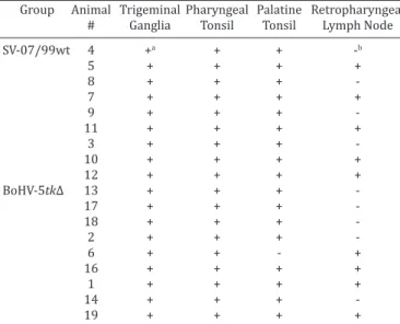
Nested-PCR examination of total DNA extracted from tis -sues of lambs euthanized at day 40pi revealed the presence of viral DNA in the TGs, pharyngeal and palatine tonsils of vir -tually all inoculated animals, regardless the group (Table 2).
Retropharyngeal lymph nodes were also frequently
positive in both groups. No infectious virus was detected upon inoculation and three passages of tissue homogena -tes into CRIB cells, confirming the status of latent infection. These results confirm our previous findings (Cadore et al. 2013) that the recombinant BoHV-5tk∆ retains its ability to establish latent infection in the TGs of inoculated lambs. In addition, both parental and recombinant viruses were sho -wn to establish latency in regional lymphoid tissue.
In addition, both parental and recombinant viral DNA was detected by PCR in several sections of the brain (not shown). No major differences in frequency and distribution were evident between the groups, indicating that the re -combinant retained its ability to invade and to replicate, to a certain extent, in the brain.
Virus shedding upon Dx administration
Following Dx administration - starting at day 40pi -, all three lambs inoculated with parental strain and kept for monitoring shed infectious virus in nasal secretions (mean duration = 5 days [4-6]) (Table 3). Shedding was first de -tected at day 3 and lasted up to day 9 pDx in one lamb. The peak in virus titers was observed at days 5 and 6 pDx, with titers reaching 104.1TCID50/mL. In contrast, nasal secretions from BoHV-5tk∆-inoculated lambs were negative for infec -tivity upon virus inoculation in cell cultures (Table 3). Swa -bs of lam-bs inoculated with the recombinant virus were positive for viral DNA by PCR (Table 3). Four (66.6%) and three (50%) lambs of the parental and recombinant virus group, respectively, showed a four-fold increase in VN titers after Dx treatment. Taken together, these results confirm our previous findings (Cadore et al. 2013) and demonstra -te that the recombinant BoHV-5tk∆ was able to reactivate following Dx treatment.
Monitoring virus reactivation in TGs and lymphoid tis-sues
In order to investigate the sites/origin of reactivated vi -rus, a search for viral mRNA by RT-PCR was performed in Table 2. Detection of viral DNA by PCR in neural and
non-neural tissues of lambs inoculated with the parental bovine herpesvirus type 5 (BoHV-5) SV-507/99 or with the
recombinant (BoHV-5tk∆), at day 40 post-inoculation
Group Animal Trigeminal Pharyngeal Palatine Retropharyngeal
# Ganglia Tonsil Tonsil Lymph Node
SV-07/99wt 4 +a + + -b
5 + + + +
8 + + +
7 + + + +
9 + + +
11 + + + +
3 + + +
10 + + + +
12 + + + +
BoHV-5tk∆ 13 + + +
17 + + +
18 + + +
2 + + +
6 + + - +
16 + + + +
1 + + + +
14 + + +
19 + + + +
a Positive sample by nested-PCR. b Negative sample by nested-PCR.
Table 3. Virus shedding in nasal secretions and virus neutralizing antibodies after dexamethasone (Dx) treatment in lambs inoculated with parental bovine herpesvirus
type 5 (BoHV-5) SV-507/99 and recombinant (BoHV-5tk∆)
Group Virus shedding in nasal secretions Virus neutralizing antibody
Animal # Day after dexamethasone administration (pDx) Day pDx
0-2 3 4 5 6 7 0 3 7
SV-507/99wt 7 -a †- 8 16
9 - †- 8 16
11 - †- 4 16
3 - - - 2.1b 1.7 - 4 32
10 - - - 1.3 1.5 1.3 4 8
12 - +c 2.1 1.9 1.3 - 4 16
BoHV-5tk∆ 2 - †(+)d 2 4
6 - †- 4 4
16 - †- < 2 8
1 - - (+) (+) - (+) 2 16
14 - - - (+) (+) - 4 32
19 - - (+) (+) (+) - 4 8
a Negative sample. b Positive sample. Viral titers expressed as log
10 TCID50/mL.
c Positive sample after second or third passage in cell culture(titer <100.9TCID
50/mL).
d Samples negative by virus isolation –
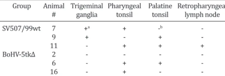
TGs and tonsils/lymph nodes of lambs submitted to eutha -nasia at day 3 after Dx treatment. Total RNA was submitted to a RT-PCR for mRNA of a late viral gene. As presented in Table 4, evidence of viral gene expression (and reactiva -tion) was demonstrated in TGs of the parental virus group (2/3). No viral gene expression was detected in TGs of lam -bs inoculated with the recombinant virus. Evidence of re -activation was also detected in pharyngeal tonsils of both groups (2/3), palatine tonsils of parental (2/3) and recom -binant (1/3) and retropharyngeal lymph node of parental virus group (1/3). These results indicate that reactivation of parental virus occurred in neural (TG) and non-neural tissues (tonsils, lymph node). Reactivation of the recombi -nant virus was not detected in TGs but seemed to occur in the tonsils, as demonstrated by detection of gB mRNA.
mined to date. Nevertheless, low levels of Dx-induced reac -tivation have been also demonstrated for this recombinant in calves (Santos et al. 2011) and lambs (Cadore et al. 2013). The absolute need of alphaherpesvirus-encoded TK activity for virus reactivation is controversial. A number of studies have shown that truly TK negative HSV mutants derived from laboratory strains do not reactivate in nerve ganglia (Tenser et al. 1987, Coen et al. 1989, Tenser et al. 1996, Chen et al. 2004), yet some mutants derived from clinical isolates might reactivate inefficiently (Horsburgh et al. 1998, Griffi -ths et al. 2003). Conflicting findings have also been reported for BoHV-1 and PRV tk deletion mutants, and both reacti-vation (Mengeling et al. 1992, Whetstone et al. 1992) and lack of reactivation have been reported (Mengeling 1991, Kaashoek et al. 1996, Ferrari et al. 2000). The general con -cept, however, is that herpesvirus TK activity is crucial for efficient reactivation since truly tk-null mutants do not reac-tivate - or reacreac-tivate very poorly - from nerve ganglia (Coen et al. 1989, Tenser et al. 1996, Chen et al. 2004). An aspect worth to consider is that most – if not all - studies repor -ting lack of reactivation by tk-negative HSV mutants were based on the absence of reactivation in explant cultures in vitro and not from in vivo studies (Coen et al. 1989, Tenser et al. 1996, Chen et al. 2004). In the same line, evidence of reactivation by tk-defective PRV, BoHV-1 and BoHV-5 was obtained in vivo (Mengeling et al. 1992, Whetstone et al. 1992, Santos et al. 2011). In the later studies, reactivation was demonstrated by recovery of infectious virus upon Dx treatment, yet the origin (neural versus non-neural) of re -activated virus was not determined. Thus, it should not be discarded that, in these cases, the origin of infectious virus was, in fact, reactivation in non-neural tissues.
We have previously hypothesized that shedding of the recombinant BoHV-5tk∆ upon Dx treatment would either reflect a residual ability of the virus to reactivate in neu -ral tissue (an unexpected event for alphaherpesviruses) or, alternatively, virus reactivation from lymphoid tissue (Ca -dore et al. 2013). Regional lymphoid tissues, especially ton -sils, have been shown to be secondary sites of latency/per -sistence by PRV (Wheeler & Osorio 1991), BoHV-1 (Inman et al., 2002) and also for BoHV-5 (Cadore, G., unpublished). The present study confirmed the presence of latent recom -binant viral DNA in tonsils and lymph nodes, reinforcing this hypothesis. Further, latent BoHV-1 present in tonsils of latently infected calves was shown to reactivate and lead to productive virus replication upon Dx treatment and ex -plant cultures in vitro (Inman et al. 2002, Perez et al. 2002). The data presented herein confirms latent infection and re -activation of BoHV-5 in lymphoid tissue, in addition to ner -ve ganglia. Although lymphoid cells and sensory neurons have different phenotypes, they are highly differentiated cells. Such differentiated cells might lack factors necessary for productive infection yet may present conditions for the establishment of latency. In this sense, the lack of transcrip -tion factors or the presence of cellular factors that repress viral transcription are important factors during establish -ment of latency (Winkler et al. 2000, Jones 1998). Moreo -ver, the present findings strongly indicate that recombinant virus shedding observed after Dx treatment was indeed DISCUSSION
The results presented herein demonstrate that the recom -binant BoHV-5tk∆ established latent infection with a simi -lar distribution of the parental strain, in TGs and in regional lymphoid tissues. In addition, BoHV-5tk∆ DNA was detec -ted in nasal secretions after Dx treatment, confirming that the recombinant retains its ability to reactivate. However, BoHV-5tk∆ reactivation seemed to be restricted to lym -phoid tissue since no viral mRNA was detected in TGs follo -wing Dx treatment. In contrast, reactivation of the parental virus was demonstrated in neural and non-neural tissues. The ability of a tk-deleted strain to reactivate latency from lymphoid tissue - even poorly - deserves attention in the context of vaccine safety.
The recombinant replicated to lower titers than the pa -rental strain in the nose during acute infection. However, it was still able to establish latent infection in all inoculated animals and with a similar distribution of the parental vi -rus. In a previous experiment, qPCR analysis showed a 9.7-fold reduction in the amount of recombinant virus DNA in TGs compared to the parental strain (Cadore et al. 2013). Thus, tk deletion did not abolish the ability of the virus to establish latency, but resulted in reduced colonization of TGs with latent viral DNA.
Confirming our previous results (Cadore et al. 2013), Bo -HV-5tk∆ DNA was detected by PCR in nasal swabs after Dx treatment, indicating some level of reactivation. This is an unusual event for many tk negative alphaherpesviruses
exa-Table 4. Detection of mRNA of glycoprotein B gene of bovine herpesvirus type 5 (BoHV-5) by RT-PCR in neural and non-neural tissues of lambs inoculated with the parental BoHV-5
(SV-507/99) or with the recombinant (BoHV-5tk∆), three days after dexamethasone administration
Group Animal Trigeminal Pharyngeal Palatine Retropharyngeal
# ganglia tonsil tonsil lymph node
SV507/99wt 7 +a + -b
9 + - +
11 - + + +
BoHV-5tk∆ 2 - - -
6 - + +
16 - + -
derived from virus reactivation in lymphoid tissue rather than from reactivation in TGs. This hypothesis accommo -dates the general concept that TK-negative alphaherpesvi -ruses are unlike to reactivate in neural tissue and support reactivation of recombinant virus in lymphoid tissue.
CONCLUSIONS
In summary, the results presented herein demonstrate that the recombinant BoHV-5tk∆ established latent infec -tion in TGs and tonsils of lambs with a similar distribu-tion of the parental virus.
Reactivation at low levels took place in lambs inocula -ted with the recombinant after Dx treatment. This reacti -vation, however, seemed to occur only in lymphoid tissues, in contrast with reactivation of the parental virus which occurred in neural and non-neural tissues.
The ability of tk-negative animal herpesviruses to reac -tivate latent infection in vivo should be further examined as to ascertain the safety of use of these recombinants in vaccines.
REFERENCES
Belknap E.B., Collins J.K., Ayers V.K. & Schultheiss P.C. 1994. Experimen
-tal infection of neona-tal calves with neurovirulent bovine herpesvirus type-5 (BHV-5). Vet. Pathol. 31:358-365.
Besecker M.I., Furness C.L., Coen D.M. & Griffiths A. 2007. Expression of extremely low levels of thymidine kinase from an acyclovir-resistant herpes simplex virus mutant supports reactivation from latently infec
-ted mouse trigeminal ganglia. J. Virol. 81(15):8356-8360.
Brum M.C.S., Weiblen R., Flores E.F. & Chowdhury S.I. 2010a. Construction
and growth properties of bovine herpesvirus type 5 recombinants de
-fective in the glycoprotein E or thymidine kinase gene or both. Braz. J. Med. Biol. Res. 43:217-224.
Brum M.C.S., Santos C.M.B., Weiblen R. & Flores E.F. 2010b. Selection and characterization of brivudin resistant bovine herpesvirus type 5. Braz. J. Microbiol.41:124-132.
Bustin S.A., Benes V., Garson J.A., Hellemans J., Huggett J., Kubista M., Muel
-ler R., Nolan T., Pfaffl M.W., Shipley G.L., Vandesompele J. & Wittwer C.T. 2009. The MIQE guidelines: minimum information for publication of quantitative real-time PCR experiments. Clin. Chem. 55(4):611-622. Cadore G.C., Anziliero D., Weiblen R. & Flores E.F. 2011. Reactivation and
distribution of bovine herpesvirus 5 DNA in the brain of latently infec
-ted lambs. Pesq. Vet. Bras. 31(12):1090-1096.
Cadore G.C., Weiss M., Anziliero D., Brum M.C.S., Weiblen R. & Flores E.F. 2013. A bovine herpesvirus 5 thymidine kinase deletion mutant establi
-shes latent infection but reactivates poorly in a sheep model. Pesq. Vet. Bras. 33(3):331-338.
Caron L., Flores E.F., Weiblen R., Scherer C.F.C., Irigoyen L.F., Roehe P.M., Odeon A. & Sur J.-H. 2002. Latent infection by bovine herpesvirus type-5 in experimentally infected rabbits: virus reactivation, shedding and recrudescence of neurological disease. Vet. Microbiol. 84(4):285-295. Chen S.H., Pearson A. & Coen D.M. 2004. Failure of thymidine kinase-nega
-tive herpes simplex virus to reactivate from latency following efficient establishment. J. Virol.78:520-523.
Chowdhury S.I. 1996. Construction and characterization of an attenuated bovine herpesvirus type 1 (BHV-1) recombinant virus. Vet. Microbiol.
52:13-23.
Chowdhury S.I., Lee B.J., Mosier D., Sur J.H., Osorio F.A., Kennedy G. & Weiss
M.L. 1997. Neuropathology of bovine herpesvirus type 5 (BHV-5) me
-ningo-encephalitis in a rabbit seizure model. J. Comp. Pathol. 117:295-310.
Coen D.M., Kosz-Vnenchak M., Jacobson J.G., Leib D.A., Bogard C.L., Schaffer P.A., Tyler K.L. & Knipe D.M. 1989. Thymidine kinase-negative herpes
simplex virus mutants establish latency in mouse trigeminal ganglia but do not reactivate. Proc. Natl Acad. Sci. USA86:4736-4740.
Delhon G., Moraes M.P., Lu Z., Afonso C.L., Flores E.F., Weiblen R., Kutish G.F. & Rock D.L. 2003. Genome of bovine herpesvirus 5. J. Virol.77:10339-10347.
Diel D.G., Almeida S.R., Brum M.C., Dezengrini R., Weiblen R. & Flores E.F. 2007. Acute and latent infection by bovine herpesvirus type 5 in experi
-mentally infected goats. Vet. Microbiol.121:257-267.
Ferrari M., Gualandi G.L., Corradi A., Monaci C., Romanelli M.G., Tosi G. & Cantoni A.M. 1998. Experimental infection of pigs with a thymidine ki
-nase negative strain of pseudorabies virus. Comp. Immunol. Microbiol. Infect. Dis.21:291-303.
Ferrari M., Mettenleiter T.C., Romanelli M.G., Cabassi E., Corradi A., Dal Mas N. & Silini R. 2000. A comparative study of pseudorabies virus (PRV) strains with defects in thymidine kinase and glycoprotein genes. J.
Comp. Pathol.123:152-163.
Flores E.F., Weiblen R., Vogel F.S.F., Dezengrini R., Almeida S.R., Spilki F.R. &
Roehe P.M. 2009. Experimental neuropathogenesis of bovine herpesvi
-rus 5 infection in rabbits. Pesq. Vet. Bras. 29:1-16.
Griffiths A., Chen S.H., Horsburgh B.C. & Coen D.M. 2003. Translational compensation of a frameshift mutation affecting herpes simplex virus thymidine kinase is sufficient to permite reactivation from latency. J.
Virol. 77:4703-4709.
Horsburgh B.C., Chen S.H., Hu A., Mulamba G.B., Burns W.H. & Coen D.M. 1998. Recurrent acyclovir-resistant herpes simplex virus in an immu
-nocompromised patient: can strain differences compensate for loss of thymidine kinase in pathogenesis? J. Infect. Dis. 178:618-625.
Inman M., Lovato L., Doster A. & Jones C. 2002. A mutation in the latency related gene of bovine herpesvirus 1 interferes with the latency-reacti
-vation cycle of latency in calves. J. Virol. 76:6771-6779.
Kaashoek M.J., Van Engelenburg F.A., Moerman A., Gielkens A.L., Rijsewijk F.A. & Van Oirschot J.T. 1996. Virulence and immunogenicity in calves of thymidine kinase- and glycoprotein E-negative bovine herpesvirus 1 mutants. Vet. Microbiol. 48:143-53.
Kahrs R.F. 2001. Infectious bovine rhinotrachitis and infectious pustular vulvovaginitis, p.159-170. In: Ibid. (Ed.), Viral Disease of Cattle. Iowa State University Press, Ames.
Katz J.P., Bodin E.T. & Coen D.M. 1990. Quantitative polymerase chain re
-action analysis of herpes simplex virus DNA in ganglia of mice infected with replication-incompetent mutants. J. Virol. 64:4288-4295.
Kit S. 1985. Thymidine kinase. Microbiol. Sci. 2(12):369-375.
Livak K.J. & Schmittgen T.D. 2001. Analysis of relative gene expression data using real-time quantitative PCR and the 2−ΔΔCq method. Methods
25(4):402-408.
Mengeling W.L. 1991. Virus reactivation in pigs latently infected with a thymidine kinase negative vaccine strain of pseudorabies virus. Arch.
Virol.120:57-70.
Mengeling W.L., Lager K.M., Volz D.M. & Brockmeier S.L. 1992. Effect of various vaccination procedures on shedding, latency, and reactivation of attenuated and virulent pseudorabies virus in swine. Am. J. Vet. Res. 53:2164-2173.
Pastoret P. & Thiry E. 1985. Diagnosis and prophylaxis of infectious bovine rhinotracheitis: the role of virus latency. Comp. Immunol. Microbiol. Infect. Dis. 8:35-42.
Perez S.E., Bretschneider G., Leunda M.R., Osorio F.A., Flores E.F. & Odeon A.C. 2002. Primary infection, latency, and reactivation of bovine herpes
-virus type 5 in the bovine nervous system. Vet. Pathol.39:437-444.
Perez S.E., Vagnozzi A., Sur J.H., Odriozola E., Campero C.M. & Odeón A.C.
2003. Análisis restrospectivo de casos com diagnóstico de necrosis ce
-rebrocortical y su relación con herpesvirus bovino tipo 5. Revta Argent. Microbiol. 35:69-73.
Reed L.J. & Muench H. 1938. A simple method for estimating fifty per cent end points. Am. J. Hyg. 27:493-497.
Rissi D.R., Pierezan F., Silva M.S., Flores E.F. & Barros C.S. 2008. Neurologi
-cal disease in cattle in southern Brazil associated with bovine herpesvi
Rock D. 1994. Latent infection with bovine herpesvirus type-1. Semin.
Virol. 5:233-240.
Roizman B., Desrosiers R.C., Fleckenstein B., Lopez C., Minson A.C. & Studdert M.J. 1992. The family Herpesviridae: an update. Arch. Virol.
123:432-445.
Salvador S.C., Lemos R.A.A., Riet-Correa F., Roehe P.M. & Osório A.L.A.R. 1998. Meningoencefalite em bovinos causada por herpesvírus bovino-5 no Mato Grosso do Sul e São Paulo. Pesq. Vet. Bras.18:76-83.
Santos C.M.B., Anzilliero D., Bauermann D., Brum M.C.C., Weiblen R. &
Flores E.F. 2011. Experimental infection of calves with recombinants of
bovine herpesvirus 5 defective in glycoprotein E (gE), thymidine kinase (TK) and both gE/TK. Pesq. Vet. Bras. 31(4):319-325.
Silva A.M., Weiblen R., Irigoyen L.F., Roehe P.M., Sur H.J., Osorio F.A. &
Flores E.F. 1999.Experimental infection of sheep with bovine herpesvi
-rus type-5 (BHV-5). Vet. Microbiol. 66:89-99.
Silva S.C., Brum M.C., Weiblen R., Flores E.F. & Chowdhury S.I. 2010. A bo
-vine herpesvirus 5 recombinant defective in the thymidine kinase (TK) gene and a double mutant lacking TK and the glycoprotein E gene are fully attenuated for rabbits. Braz. J. Med. Biol. Res. 43:150-159. Tenser R.B., Miller R.L. & Rapp F. 1979. Trigeminal ganglion infection by
thymidine kinase-negative mutants of herpes simplex virus. Science
205:915-917.
Tenser R.B. & Edris W.A. 1987. Trigeminal ganglion infection by thymidine kinase-negative mutants of herpes simplex virus after in vivo
comple-mentation. J. Virol. 61:2171-2174.
Tenser R.B., Hay K.A. & Edris W.A. 1989. Latency-associated transcript but not reactivatable virus is present in sensory ganglion neurons after in
-oculation of thymidine kinase negative mutants of herpes simplex virus type 1. J. Virol. 63:2861-2865.
Tenser R.B. 1994. The role of herpes simplex virus thymidine kinase ex
-pression in pathogenesis and latency, p.68-86. In: Becker Y. & Darai G. (Eds), Pathogenicity of Human Herpesviruses due to Specific Pathoge
-nicity Genes. Springer-Verlag, London. 656p.
Tenser R.B., Gaydos A. & Hay K.A. 1996. Reactivation of thymidine kinase-defective herpes simplex virus is enhanced by nucleoside. J. Virol. 70:1271-1276.
Thompson R.L. & Sawtell N.M. 2000. Replication of herpes simplex virus type 1 within trigeminal ganglia is required for high frequency but not high viral genome copy number latency. J. Virol. 74:965-974.
Thrusfield M. 2005. Veterinary Epidemiology. 3rd ed, Blackwell Science,
Oxford. 610p.
Vogel F.S., Caron L., Flores E.F., Weiblen R., Winkelmann E.R., Mayer S.V. & Bastos R.G. 2003. Distribution of bovine herpesvirus type 5 DNA in the central nervous systems of latently, experimentally infected calves. J. Clin. Microbiol.41:4512-4520.
Volz D.M., Lager K.M. & Mengeling W.L. 1992. Latency of a thymidine ki
-nase-negative pseudorabies vaccine virus detected by the polymerase chain reaction. Arch. Virol. 122:341-348.
Wheeler J.G. & Osorio F.A. 1991. Investigation of sites of pseudorabies virus latency, using polymerase chain reaction. Am. J. Vet. Res. 52(11):1799-1803.
Whetstone C.A., Miller J.M., Seal B.S., Bello L.J. & Lawrence W.C. 1992. La