Glucose on the Osteoblastic Differentiation of Periodontal
Ligament Stem Cells
So Yeon Kim
1, Jin-Yong Lee
1, Yong-Duk Park
2, Kyung Lhi Kang
3, Jeong-Chae Lee
4, Jung Sun Heo
1* 1 Department of Maxillofacial Biomedical Engineering and Institute of Oral Biology, School of Dentistry, Kyung Hee University, Dongdaemun-gu, Seoul, South Korea, 2 Department of Preventive and Social Dentistry, School of Dentistry, Kyung Hee University, Dongdaemun-gu, Seoul, South Korea, 3 Department of Periodontology, School of Dentistry, Kyung Hee University, Dongdaemun-gu, Seoul, South Korea, 4 Institute of Oral Biosciences and School of Dentistry, Research Center of Bioactive Materials, Chonbuk National University, Jeonju, South KoreaAbstract
Hesperetin (γ′,5,7-trihydroxy-4-methoxyflavanone) is a metabolite of hesperidin (hesperetin-7-O-rutinoside), which belongs to the flavanone subgroup and is found mainly in citrus fruits. Hesperetin has been reported to be an effective osteoinductive compound in various in vivo and in vitro models. However, how hesperetin effects osteogenic differentiation is not fully understood. In this study, we investigated the capacity of hesperetin to stimulate the osteogenic differentiation of periodontal ligament stem cells (PDLSCs) and to relieve the anti-osteogenic effect of high glucose. Osteogenesis of PDLSCs was assessed by measurement of alkaline phosphatase (ALP) activity, and evaluation of the mRNA expression of ALP, runt-related gene β (Runxβ), osterix (OSX), and FRA1 as osteogenic transcription factors, as well as assessment of protein expression of osteopontin (OPN) and collagen type IA (COLIA). When PDLSCs were exposed to a high concentration (γ0 mM) of glucose, osteogenic activity decreased compared to control cells. Hesperetin significantly increased ALP activity at doses of 1, 10, and 100 µM. Pretreatment of cells with hesperetin alleviated the high-glucose-induced suppression of the osteogenic activity of PDLSCs. Hesperetin scavenged intracellular reactive oxygen species (ROS) produced under high glucose condition. Furthermore, hesperetin increased the activity of the PIγK/Akt and -catenin pathways. Consistent with this, blockage of Akt or -catenin diminished the protective effect of hesperetin against high glucose-inhibited osteogenic differentiation. Collectively, our results suggest that hesperetin alleviates the high glucose-mediated suppression of osteogenic differentiation in PDLSCs by regulating ROS levels and the PIγK/Akt and -catenin signaling pathways.
Citation: Kim SY, Lee J , Park Y , Kang KL, Lee J , et al. (β01γ) Hesperetin Alleviates the Inhibitory Effects of High Glucose on the Osteoblastic Differentiation of Periodontal Ligament Stem Cells. PLoS ONE 8(6): e67504. doi:10.1γ71/journal.pone.0067504
Editor: Jun Sun, Rush University Medical Center, United States of America Received February 7, β01γ; Accepted May 18, β01γ; Published June β8, β01γ
Copyright: © β01γ Kim et al. This is an open-access article distributed under the terms of the Creative Commons Attribution License, which permits unrestricted use, distribution, and reproduction in any medium, provided the original author and source are credited.
Funding: This work was supported by a grant from the Kyung Hee University in β01β (KHU-β01β0799) and the Basic Science Research Program through the National Research Foundation of Korea (NRF) funded by the Ministry of Education, Science and Technology (β011-001γ595). The funders had no role in study design, data collection and analysis, decision to publish, or preparation of the manuscript.
Competing interests: The authors have declared that no competing interests exist. * E-mail: heojs@khu.ac.kr
Introduction
Multipotent postnatal stem cells have been identified in oral tissues, such as those from dental pulp [1], exfoliated deciduous teeth [β], and periodontal ligaments (periodontal ligament stem cells [PDLSCs]) [γ]. These dental stem cells are plentiful and easy to collect from tissue, and have been shown to be able to differentiate into bone, dental tissue, cartilage, and even neural tissue [4–6]. They are being studied for a number of disorders of connective tissues or neural tissues in the body including type I diabetes, damaged tooth structures, skeletal bone loss, and neurodegenerative diseases [7–9]. In particular, PDLSCs are capable of differentiating into an osteoblastic lineage, and are therefore considered a good
regenerative medicine candidate for treatment of bone defects [10]. However, the biology of PDLSCs under various oral conditions needs to be clarified more precisely, because the condition of periodontal ligament structures is closely associated with risk factors including smoking, diabetes, and other infections [11,1β]. Among these pathological states, high glucose levels in diabetic patients may alter the regenerative capacity of PDLSCs. In this study, we investigated PDLSC responses to diabetic conditions and the effects of a natural compound, hesperetin, on these PDLSC responses to high glucose.
Flavonoids, which are naturally occurring polyphenolic compounds that form part of the human diet, have been reported to affect bone metabolism [1γ]. Among flavonoids,
hesperetin (γ′,5,7-trihydroxy-4-methoxyflavanone), which is a member of the flavanone subclass of flavonoids, is found mainly in citrus fruit. This flavonoid exists in nature in its glycoside form, hesperidin [14]. Hesperetin and its metabolites have several biological activities; they display antioxidant, anti-inflammatory, and lipid lowering effects [15,16]. However, despite the accessibility of hesperetin, few studies have investigated whether hesperetin influences bone strength and osteoblast differentiation [17,18]. Moreover, the mechanisms underlying the osteogenic effect of hesperetin are not fully understood. It has been reported that flavonoids and their metabolites regulate the phosphoinositide γ-kinase (PIγK), Akt, mitogen activated protein kinase (MAP kinase), and Wnt/ -catenin signaling pathways [19,β0]. Inhibition or stimulation of these pathways can modulate cellular functions including bone formation and regeneration processes by altering the expression of target molecules and genes in response to various microenvironmental conditions.
Hesperetin may be a potential candidate for promoting bone regeneration given its therapeutic efficiency and low cost.
In the present study, cells were cultured in medium supplemented with physiological (NG, 5.5 mM) or diabetic (HG, γ0 mM) glucose levels to determine whether the high glucose level inhibited the differentiation of PDLSCs into an osteogenic lineage and to characterize the impact of hesperetin on this process.
Materials and Methods
Materials
Fetal bovine serum (FBS) was purchased from Gibco-BRL (Gaithersburg, MD, USA). Nicotine, α-bungarotoxin, and mecamylamine were obtained from Sigma Chemical Company (St. Louis, MO, USA). Collagen type I, osteopontin, Runxβ, osterix, -catenin, -actin, goat-anti mouse, and goat-anti rabbit antibodies were supplied by Santa Cruz Biotechnology (Delaware, CA, USA). Unless otherwise specified, chemicals and laboratory-ware were purchased from Sigma Chemical Company and Falcon Labware (Becton-Dickinson, Franklin Lakes, NJ, USA), respectively.
Periodontal Ligament Stem Cell Culture
Periodontal ligaments were obtained from extracted human molars, which were donated by the Department of Oral and Maxillofacial Surgery of Kyung Hee University. All subjects involved in this study were informed about the purpose and procedures of this study, which was approved by the Review Board of Kyung Hee University. Written informed consent was obtained from all donors and guardians on behalf of minor participants. Periodontal ligaments, collected from the middle third of the root, were cultured in α minimal essential medium (α-MEM) (Invitrogen, Carlsbad, CA, USA) containing 10% FBS, penicillin (100 U/mL) and streptomycin (100 µg/mL) (SIGMA, St. Louis, MO, USA) according to a previously described method [γ]. After two passages, cells were subjected to magnetic isolation with antibodies to detect STRO-1 (mesenchymal stem cell marker) antigen (Millipore, Billerica, MA, USA) and magnetic beads (MiltenyiBiotec, Germany). The
resulting STRO-1(+) cell population was cultured in α-MEM plus 10% FBS at γ7°C in a humidified gas mixture of 5% COβ/95% air. All experiments were carried out with passage 4–
7 cells. To assess the colony-forming efficiency, cells at passage 1 were seeded onto a γ5 mm dishes. After 5 days of culture, cultures were fixed with 4% formalin, and then stained with 0.5% crystal violet (Sigma–Aldrich Co.) (Figure S1).
Osteogenic Differentiation of PDLSCs
Differentiation was initiated by a switch to the osteogenic medium of α-MEM containing 5% FBS, 50 µg/mL ascorbic acid, 1 µM dexamethasone, and γ mM -glycerophosphate. In each experiment, cells were incubated in medium supplemented with 5.5 mM D-glucose (NG as a control), γ0 mM D-glucose (HG), hesperetin at different concentrations (0.1, 1, 10, and 100 µM), or HG plus hesperetin for a designated number of days. The medium was changed every other day. Hesperetin was dissolved in dimethyl sulfoxide (DMSO) immediately before use, and the final concentration of DMSO did not exceed 0.1% (v/v) in any of the experiments. DMSO at 0.1% was used as a control.
Alkaline Phosphatase Activity
Cells were washed twice with PBS and lysed in 50 mM Tris– HCl buffer (pH 7.0) containing 1% (v/v) Triton X-100 and 1 mM PMSF. Total protein was then quantified using the Bradford procedure [β1]. The entire cell lysate was assayed by adding β00 µl of p-nitrophenylphosphate (pNPP) as a substrate (Sigma, USA) for γ0 min at γ7°C. The reaction was stopped by adding γ N NaOH and the absorbance was read spectrophotometrically at 405 nm. Enzyme activity was expressed as mM/100 µg of protein.
RNA Isolation and Real-time RT-PCR
TTCTTTGTGCCTGCTTTGC-γ′ (antisense) for OSX, and 5′-CCCTGCCGCCCTGTACCTTGTATC-γ′ (sense), 5′-AGACATTGGCTAGGGTGGCATCTGCA-γ′ (antisense) for FRA1. The PCR products were heated from 65°C to 99°C at a rate of 1°C/5 sec for melting curve analysis, and the resulting data were analyzed using software provided by the manufacturer.
Cell Fractionation
Nuclear and cytosolic extraction was performed according to the method described elsewhere [ββ]. Briefly, the cells were resuspended in a buffer A (β0 mM Tris (pH 7.5), 1 mM EDTA, 1 mM EGTA, 1% Triton X-100, 1 µg/ml aprotinin, 1 mM phenylmethylsulfonylfluoride, and 0.5 mM sodium orthovanadate). The suspension was sonicated for 10 sec at output 4 and then centrifuged. The pellet contained the nuclear fraction. The supernatant was centrifuged at 15,000 rpm for 60 min at 4°C and contained the cytosolic fraction.
Western Blot Analysis
Protein extracts (β0 µg) were separated by 8–10% SDS-PAGE and blotted onto polyvinylidene difluoride (PVDF) membranes. Blots were washed with TBST [10 mM Tris-HCl (pH 7.6), 150 mM NaCl, 0.05% Tween-β0], blocked with 5% skim milk for 1 hour, and incubated with the appropriate primary antibodies [anti-collagen type I, anti-osteopontin, and anti- -actin (Santa Cruz Biotechnology, CA, USA) at the dilutions recommended by the supplier. The membranes were then washed, and the primary antibodies were detected with goat anti-rabbit IgG or goat anti-mouse IgG conjugated to horseradish peroxidase. Blots were developed with enhanced chemiluminescence (ECL) reagents (Santa Cruz Biotechnology) and exposed to X-ray film (Eastman-Kodak, Rochester, NY, USA).
Immunofluorescence Staining
Cells were fixed and treated with mouse anti-collagen type I or anti-osteopontin antibody (1∶100, Santa Cruz Biotechnology, Delaware, CA) for 1 hr at room temperature. Subsequently, cells were treated with fluorescein isothiocyanate-conjugated (FITC-conjugated) anti-mouse IgG (1∶100) for 1 hr at room temperature. Fluorescence images were obtained using a fluorescence microscope (Fluoview γ00, Olympus).
Measurement of Intracellular ROS
Cellular levels of ROS were measured using 5-(and-6)-chloromethyl-β′,7′-dichlorodihydro-fluorescein diacetate (CM-HβDCF-DA; Molecular Probes, Eugene, OR, USA) according to
the method described by Ali et al. [βγ]. Cells were pre-incubated with hesperetin at different concentrations for β hr, then 10 µM CM-HβDCF-DA and γ0 mM glucose (HG) were
added. After 40 min, DCF fluorescence was determined using a spectrofluorophotometer (RF-5γ01PC, Shimadzu, Japan) at an excitation wavelength of 488 nm and an emission wavelength of 5γ0 nm.
siRNA Transfection
Small interference RNA (siRNA) targeting -catenin was produced using a commercial kit that contains three target sequences to -catenin (IDT, Integrated DNA Technologies Inc., Coralville, IA, USA). The siRNA that inhibited -catenin expression the most as determined by western blotting analysis was used in further experiments. Briefly, cells were transfected for β4 hr with either siRNA specific to -catenin (β0 nM) or negative control siRNA (scrambled), using TransIT-TKO transfection reagents (Mirus, Madison, WI, USA) according to the manufacturer’s instructions, before being subjected to the various treatments.
Statistical Analysis
All data are expressed as means ± standard deviations (S.D.). One-way ANOVA was used for multiple comparisons (Duncan’s multiple range test), using SPSS software ver. 10.0.
P values < 0.05 were considered significant.
Results
High Glucose Decreases the Osteogenic Activity of PDLSCs
To examine the effect of a high level of glucose on osteogenesis of PDLSCs, cells were cultivated in medium containing NG (5.5 mM D-glucose as a control) or HG (γ0 mM D-glucose) reflecting physiological or diabetic blood glucose levels, respectively, then differentiated towards an osteoblastogenic lineage for 4 or 7 days. Subsequently, the ALP activity of PDLSCs was analyzed as a marker of osteogenic differentiation. Figure 1A shows that the ALP activity in cells cultured with HG was significantly lower than that of the control group at day 4 (1.7-fold decrease vs. control;
p<0.05), which represents an early stage of osteogenic differentiation, and much more attenuated at day 7 (β.4-fold decrease vs. control; p<0.001).
Effect of Hesperetin on Osteogenic Differentiation of PDLSCs
We next investigated the effect of hesperetin on osteogenic differentiation of PDLSCs under NG conditions. As shown in Fig. 1B, treatment of cells with 1 µM hesperetin resulted in a significant increase in ALP activity (1.8 fold increase vs. control; p<0.05). As the hesperetin concentration increased, ALP activity increased and a marked increase was observed at 10 µM hesperetin (β-fold increase vs. control; p<0.05).
To assess whether hesperetin counteracted the HG-induced suppression of PDLSC osteogenesis, cells were incubated with hesperetin (10 and 100 µM) prior to HG exposure. As shown in Fig. 1C, pretreatment of cells with hesperetin inhibited the HG-induced decrease in ALP activity.
Effects of Hesperetin on Osteogenic-associated Gene Expression
FRA1, using real time RT-PCR. Cells incubated with HG expressed lower levels of these marker genes than control cultures. However, treatment with hesperetin alone markedly increased the mRNA expression of osteogenic marker genes and pretreatment with hesperetin recovered them under HG conditions (Fig. β).
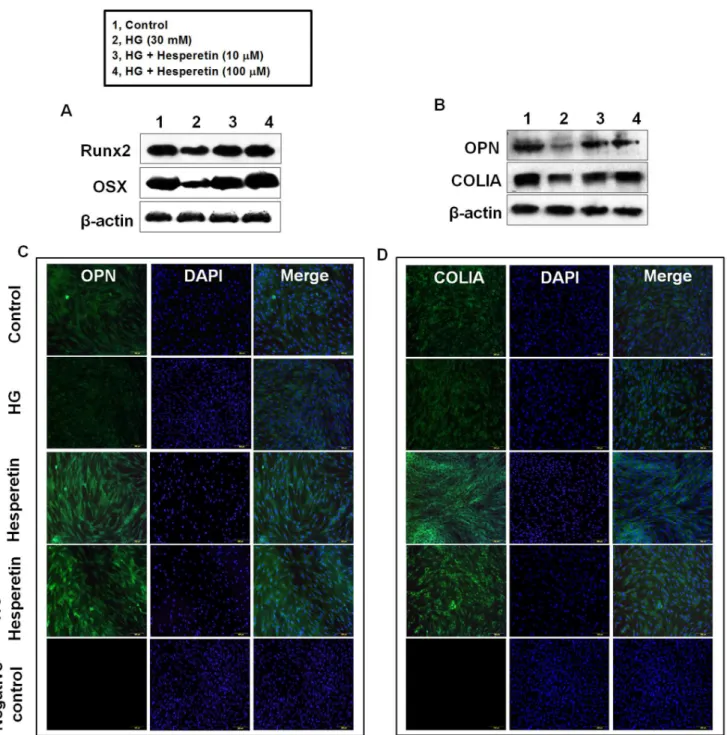
Effects of Hesperetin on Protein Levels of Osteogenic Factors
We also analyzed the effects of hesperetin on the osteogenic differentiation of PDLSCs by following the protein expression of Runxβ, OSX, OPN, and COLIA, which are osteogenic markers, four days after osteogenic induction. Western blot analysis showed that the level of each protein decreased in cells incubated with HG relative to control cells. When cells were pretreated with 10 or 100 µM hesperetin prior to HG exposure, expression of these proteins was higher than that observed in HG cultures (Fig. γA, B). Moreover, immunofluorescence staining for OPN and COLIA confirmed that hesperetin
treatment enhanced the differentiation of PDLSCs into am osteogenic lineage (Fig. γC, D).
Involvement of ROS and PI3K/Akt Signaling in Hesperetin-induced Osteogenic Differentiation
Hesperetin is known to have antioxidant and free radical scavenging ability [β4,β5]. When cells were incubated with HG, intracellular ROS level was increased up to 66% compared to control cells. However, pretreatment of cells with hesperetin inhibited the HG-induced ROS increase, suggesting that the scavenging activity of hesperetin is involved in PDLSC osteogenesis (Fig. 4A). Next, we examined the role of PIγK/Akt signaling in hesperetin-induced osteogenic differentiation. As shown in Fig. 4B, a high glucose level significantly decreased the protein expression of PIγK p110α and isoforms as well as Akt phosphorylation compared to the control cultures. However, pretreatment of cells with hesperetin or vitamin C increased expression of these proteins under HG conditions. We also examined the effect of hesperetin on the phosphorylation of Akt under NG conditions; a significant
Figure 1. Effect of high glucose (HG) and hesperetin on osteogenic differentiation of PDLSCs. Cells were incubated in osteogenic medium with (A) γ0 mM glucose (HG) or (B) hesperetin (0.1, 1, 10, 100 µM) for 4 and 7 days, respectively, and then ALP activity was assessed as described in the Materials and Methods. (C) Cells were pretreated with hesperetin at concentrations of 10 and 100 µM for β hr before HG exposure and ALP activity was determined after 4, 7, 14 days of osteogenic induction. The values reported are the means ± S.D. of five independent experiments. *P<0.05, **P<0.001 vs. control value, or #P<0.05 vs. HG
treatment alone.
increase in the level of p-Akt (ser 47γ) was first observed at 60 min and maintained up to β40 min (Fig. 4C). To examine the association between PIγK/Akt signaling and hesperetin-induced osteogenesis of PDLSCs, cells were pretreated with 1 or 10 µM of Akt inhibitor before hesperetin application. As shown in Fig. 4D, the hesperetin-induced increase in the protein levels of OPN and COLIA was significantly attenuated by addition of Akt inhibitor, but inhibitor alone did not change these protein levels compared to control cells (Figure Sβ). Moreover, under HG conditions, the HG-mediated decrease in the protein levels of OPN and COLIA as well as the mRNA expression of osteogenic marker genes were attenuated by hesperetin, but this did not occur in the presence of Akt inhibitor, suggesting that hesperetin promotes PDLSC osteogenesis under HG conditions by activating the PIγK/Akt pathway (Fig. 4E–I).
Hesperetin Stimulates the Canonical Wnt/β-catenin Signaling Pathway
We further evaluated the role of Wnt/ -catenin signaling in hesperetin-induced osteogenesis of PDLSCs. Figure 5A shows that hesperetin increased protein expression of total -catenin dose-dependently under NG conditions. In order to validate that hesperetin indeed stimulates -catenin translocation, cells were exposed to hesperetin then cytosol and nuclear proteins were isolated. Western blot analysis demonstrated that hesperetin increased the protein levels of -catenin in the cytosol and nucleus (Fig. 5B). Subsequently, we confirmed that hesperetin induced nuclear translocation of -catenin using immunofluorescence staining (Fig. 5C). In contrast, cultures incubated with HG had lower -catenin expression than control cultures. However, addition of 10 or 100 µM hesperetin attenuated the HG-induced decrease in expression of -catenin
Figure 2. Effects of hesperetin on the high glucose-suppressed mRNA expression of ALP, Runx2, OSX, and FRA1. Cells were pretreated with hesperetin at concentrations of 10 and 100 µM for β hr before HG exposure and the mRNA levels of (A) ALP, (B) Runxβ, (C) OSX, and (D) FRA1 were analyzed using real time RT-PCR after 4 days of osteogenic induction. The values reported are the means ± S.D. of three independent experiments. *P<0.05, **P<0.001 vs. control values, or #P<0.05 vs. HG
treatment alone.
(Fig. 5D). To elucidate the relationship between PIγK/Akt and Wnt/ -catenin signaling in the process of osteogenesis, cells were pre-incubated with Akt inhibitor before hesperetin treatment. As shown in Fig. 5E, the hesperetin-induced increase in -catenin levels was attenuated by the Akt inhibitor.
However, knock-down of -catenin did not affect the phosphorylation of Akt by hesperetin, suggesting that PIγK/Akt signaling acts as an upstream signaling molecule for Wnt/ -catenin pathways (Figure Sγ).
Figure 3. Effects of hesperetin on the high glucose-mediated suppression of Runx2, OSX, OPN, and COLIA protein levels. (A) Cells were pretreated with hesperetin at concentrations of 10 and 100 µM for β hr before HG incubation and then protein levels of (A) Runxβ, OSX, (B) OPN, and COLIA were determined by western blot analysis using total protein lysates. Cells were incubated with hesperetin in the presence of HG for 4 days then (C) OPN and (D) COLIA were detected by immunostaining. Negative control staining using secondary antibody alone to assess nonspecific fluorescence. Nuclei were stained with DAPI (blue staining). A representative result from four independent experiments is shown.
Figure 4. Effect of hesperetin on intracellular ROS levels and PI3K/Akt signaling. (A) Cells were preincubated with hesperetin at different concentrations for β hr then 10 µM CM-HβDCF-DA and high glucose were added. After 40 min, DCF fluorescence was
determined using a spectrofluorophotometer. (B) Protein levels of PIγK p110α and isoforms as well as p-Akt were determined after cells were incubated with hesperetin or vitamin C in the presence of HG for 4 days. (C) Phosphorylation of Akt by hesperetin (10 µM) was assessed with cells of 7 day-osteogenic induction. (D, E) OPN and COLIA levels were determined after incubation of cells with Akt inhibitor in the presence of HG, hesperetin, or HG+hesperetin. (F–I) The mRNA levels of osteogenic target genes were determined after incubation with Akt inhibitor in the presence of HG or HG+hesperetin. The values reported are the mean ± S.D. of three independent experiments. *P<0.05 vs. control values, **P<0.05 vs. HG treatment alone, or #P<0.05 vs. HG
+hesperetin.
Figure 5. Effect of hesperetin on Wnt/β-catenin signaling. (A) Protein levels of catenin and (B, C) nuclear translocation of -catenin were assessed by Western blot analysis or immunofluorescence staining after cells were incubated with hesperetin. Protein levels of -catenin were determined after cells were conditioned with (D) hesperetin in the presence of HG and (E) hesperetin+Akt inhibitor. (F) Changes in -catenin levels were determined according to transfection. (G) Protein levels of OPN and COLIA, (H) ALP activity, and (I) the mRNA levels of Runxβ and OSX were measured after cells were transfected with -catenin siRNA for β4 hr and further incubated with hesperetin and HG for β4 hr. The values reported are the mean ± S.D. of three independent experiments. *P<0.05 vs. control values, **P<0.05 vs. HG treatment alone, or #P<0.05 vs. HG+hesperetin.
Finally, we determined the effect of Wnt/ -catenin signaling on the regulation of osteogenic factors. To confirm the efficacy of the -catenin-specific siRNA, cells were transfected with either -catenin or negative control siRNA using TransIT-TKO transfection reagents. Twenty-four hours after transfection, cells were incubated with hesperetin for β4 hr. Figure 5F shows that the hesperetin-induced increase in levels of -catenin was reduced markedly by siRNA transfection of siRNA specific to -catenin, where negative control siRNA transfection did not affect -catenin protein levels. Under HG conditions, -catenin ablation by siRNA transfection diminished the hesperetin-induced increase in OPN and COLIA protein expression (Fig. 5G). Moreover, the patterns of ALP activity and the mRNA expression of osteogenic marker genes were consistent with Western blot analysis of OPN and COLIA suggesting that the canonical Wnt/ -catenin signaling is involved in hesperetin-induced osteogenesis in PDLSCs (Fig. 5H, I).
Discussion
In the present study, we demonstrated the inhibitory effect of a diabetic glucose level on osteogenic differentiation of PDLSCs, and showed how hesperetin could stimulate the osteogenesis of PDLSCs in this high glucose condition. A high concentration of mannitol was used as an osmotic control, and the corresponding values were similar to those in NG cultures (data not shown). High blood glucose is the prominent
pathogenic factor that leads to diabetic complications including bone disease and oral health disorders. Increasing evidence suggests that high glucose concentrations inhibit osteoblast proliferation and osteogenic factor secretion [β6,β7], because high glucose levels can directly impair osteoblastic functions. A previous study showed that PDL cells from insulin-dependent diabetic patients had altered functions; they formed mineralized tissue and responded to exogenous growth factors [β8]. Moreover, an in vitro experiment demonstrated that high glucose inhibited the proliferation and mineralization of PDL cells [β9]. However, the osteoblastic differentiation of PDLSCs under high glucose conditions has not been thoroughly explored. In the present study, we demonstrated that down-regulation of osteogenesis in PDLSCs cultured in high glucose may explain the delay in periodontal regeneration and healing observed in diabetic patients.
In the present study, hesperetin, similar to other polyphenols with osteogenic effects [γ0,γ1], stimulated the osteogenic differentiation of PDLSCs. It has previously been demonstrated that hesperetin can influence osteoblast differentiation via the bone morphogenic protein (BMP) signaling pathway [γβ]. Moreover, treatment of osteoblasts with physiological concentrations of hesperetin significantly augmented the mRNA expression of ALP, Runxβ, and osterix [γβ]. Despite a different cell model, when osteoblasts were treated with hesperetin under diabetic glucose conditions, ALP activity and collagen content increased markedly [18]. Similarly, we
Figure 6. Hypothesized model of the signaling pathways underlying the rescuing effects of hesperetin on high glucose-exposed PDLSCs. High glucose increases ROS generation, which inhibits PIγK/Akt signaling and Wnt/ -catenin. Hesperetin suppresses ROS production and activates PIγK/Akt signaling and Wnt/ -catenin to induce the translocation of -catenin into the nucleus, which leads to the osteogenic differentiation of PDLSCs. In this scheme, grey lines are proposed pathways affected by high glucose concentrations and black lines are hesperetin-stimulated pathways.
demonstrated in this study that hesperetin prevented high glucose-mediated PDLSC dysfunction resulting in defective osteogenic differentiation. Based on these results, hesperetin is a promising potential candidate for bone regeneration.
Long-term exposure to high glucose concentrations induces oxidative stress, which adversely affects cellular functions. Thus, removal of oxidized factors by antioxidants is part of the cellular defense system against oxidative stress [γγ]. In the present study, we examined the effect of hesperetin on high glucose-induced ROS over-production in PDLSC cultures. As expected, high glucose exposure markedly increased ROS generation, which was reversed by pretreatment of cells with 10 and 100 µM hesperetin. These results are consistent with those of a previous study that showed that hesperetin induced a decrease in ROS in mouse embryonic stem cells treated with hydrogen peroxide [γ4]. These findings indicate that hesperetin, due to its radical scavenging ability, functions in cellular defense. Previous studies have shown that oxidative stress suppresses osteogenic differentiation in various cellular systems [γ5–γ7]. Indeed, regulatory transcription networks between redox-responsive elements and the OPN gene have been reported [γ8]. In this respect, the antioxidant activity of hesperetin could be linked to its osteogenic capability, suggesting that use of this compound may be a novel strategy to prevent the inhibitory effect of oxidative stress on osteogenesis of PDLSCs in a diabetic state.
Cell signaling pathways involved in osteogenesis can be activated by the interactions of a compound with extracellular or intracellular molecules. Among them, the PIγK/Akt and Wnt/ -catenin pathways have been the focus of recent work, and both have emerged as critical for bone development and skeletal remodeling [γ9,40]. Recent genetic studies of PIγK signaling revealed that Akt and its downstream targets are critical regulators of bone formation and osteoblastic survival [41,4β]. In the present study, hesperetin increased protein levels of PIγK isoforms and the phosphorylation of Akt, which is consistent with a previous study that demonstrated that hesperetin induced PIγK activation in PC1β cells [4γ]. Our results suggest a significant synergistic effect between the PIγK/Akt and Wnt/ -catenin pathways. Hesperetin-stimulated Wnt/ -catenin signaling was associated with PIγK/Akt activation. In contrast, inhibition of Akt or Wnt/ -catenin signaling blocked the preventive effect of hesperetin against high glucose-suppressed osteogenesis of PDLSCs, suggesting
that hesperetin increases the osteogenic capacity of PDLSCs by activating the PIγK/Akt and Wnt/ -catenin signaling pathways.
In conclusion, we demonstrated that hesperetin is a promising bioactive compound that preserves the osteogenic capacity of PDLSCs under diabetic conditions. Our findings suggest that hesperetin alleviates the inhibitory effect of high glucose on the osteoblastic differentiation of PDLSCs through ROS scavenging and modulation of PIγK/Akt and Wnt/ -catenin signaling (Fig. 6). Hesperetin is promising biochemical candidate to control PDLSC behavior and can potentially play a regenerative role in restoring the function of PDLSCs in diabetes-induced periodontal diseases.
Supporting Information
Figure S1. Colony-forming assay. To assess the colony-forming efficiency, cells at passage 1 were seeded onto a γ5 mm dishes. After 5 days of culture, cultures were fixed with 4% formalin, and then stained with 0.5% crystal violet (Sigma– Aldrich Co.). A representative result from four independent experiments is shown.
(TIF)
Figure S2. Protein levels of OPN and COLIA were determined after incubation of cells with Akt inhibitor in the presence or absence of hesperetin. A representative result from three independent experiments is shown.
(TIF)
Figure S3. Phosphorylation of Akt was measured after cells were transfected with -catenin siRNA for β4 hr and further incubated with hesperetin and HG for β4 hr. A representative result from three independent experiments is shown.
(TIF)
Author Contributions
Conceived and designed the experiments: JSH. Performed the experiments: SYK. Analyzed the data: JSH KLK SYK. Contributed reagents/materials/analysis tools: J-YL Y-DP J-CL. Wrote the paper: JSH.
References
1. Gronthos S, Mankani M, Brahim J, Robey PG, Shi S (β000) Postnatal human dental pulp stem cells (DPSCs) in vitro and in vivo. Proc Natl Acad Sci U S A 97: 1γ6β5–1γ6γ0.
β. Miura M, Gronthos S, Zhao M, Lu B, Fisher LW, et al. (β00γ) SHED: stem cells from human exfoliated deciduous teeth. Proc Natl Acad Sci U S A 100: 5807–581β.
γ. Seo BM, Miura M, Gronthos S, Bartold PM, Batouli S, et al. (β004) Investigation of multipotent postnatal stem cells from human periodontal ligament. Lancet γ64: 149–155.
4. Singhatanadgit W, Donos N, Olsen I (β009) Isolation and characterization of stem cell clones from adult human ligament. Tissue Eng A 15: β6β5–β6γ6.
5. Zainal Ariffin SH, Kermani S, Megat Abdul Wahab R, Senafi S, Zainal Ariffin Z, et al. (β01β) In vitro chondrogenesis transformation study of mouse dental pulp stem cells. Scientific World Journal β01β: 8β7149.
6. Nakashima M, Iohara K, Sugiyama M (β009) Human dental pulp stem cells with highly angiogenic and neurogenic potential for possible use in pulp regeneration. Cytokine Growth Factor Rev β0: 4γ5–440. 7. Govindasamy V, Ronald VS, Abdullah AN, Nathan KR, Ab Aziz ZA, et
al. (β011) Differentiation of dental pulp stem cells into islet-like aggregates. J Dent Res 90: 646–65β.
8. Chadipiralla K, Yochim JM, Bahuleyan B, Huang CY, Garcia-Godoy F, et al. (β010) Osteogenic differentiation of stem cells derived from human periodontal ligaments and pulp of human exfoliated deciduous teeth. Cell Tissue Res γ40: γβγ–γγγ.
10. Ge S, Zhao N, Wang L, Yu M, Liu H, et al. (β01β) Bone repair by periodontal ligament stem cell seeded nanohydroxyapatite-chitosan scaffold. Int J Nanomedicine 7: 5405–5414.
11. Pihlstrom BL, Michalowicz BS, Johnson NW (β005) Periodontal diseases. Lancet γ66: 1809–18β0.
1β. Salvi GE, Carollo-Bittel B, Lang NP (β008) Effects of diabetes mellitus on periodontal and peri-implant conditions: update on associations and risks. J Clin Periodontol γ5: γ98–409.
1γ. Weaver CM, Alekel DL, Ward WE, Ronis MJ (β01β) Flavonoid intake and bone health. J Nutr Gerontol Geriatr γ1: βγ9–β5γ.
14. Garg A, Garg S, Zaneveld LJ, Singla AK (β001) Chemistry and pharmacology of the citrus bioflavonoid hesperidin. Phytother Res 15: 655–669.
15. Galati EM, Monforte MT, Kirjavainen S, Forestieri AM, Trovato A, et al. (1994) Biological effects of hesperidin, a citrus flavonoid: Note I. Antiinflammatory and analgesic activity. Farmaco 40: 709–71β. 16. Miyake Y, Yamamoto K, Tsujihara N, Osawa T (1998) Protective
effects of lemon flavonoids on oxidative stress in diabetic rats. Lipids γγ: 689–695.
17. Trzeciakiewicz A, Habauzit V, Mercier S, Barron D, Urpi-Sarda M, et al. (β010) Molecular mechanism of hesperetin-7-O-glucuronide, the main circulating metabolite of hesperidin, involved in osteoblast differentiation. J Agric Food Chem 58: 668–675.
18. Choi EM, Kim YH (β008) Hesperetin attenuates the highly reducing sugar-triggered inhibition of osteoblast differentiation. Cell Biol Toxicol β4: ββ5–βγ1.
19. Nones J, Costa AP, Leal RB, Gomes FC, Trentin AG (β01β) The flavonoids hesperidin and rutin promote neural crest cell survival. Cell Tissue Res γ50: γ05–γ15.
β0. Amado N, Fonseca B, Cerqueira DM, Reis AH, Simas AB, et al. (β01β) Effects Of Natural Compounds On Xenopus Embryogenesis: A Potential Read Out For Functional Drug Discovery Targeting Wnt/ -Catenin Signaling. Curr Top Med Chem 1β: β10γ–β11γ.
β1. Bradford MM (1976) A rapid and sensitive method for the quantitation of microgram quantities of protein utilizing the principle of protein-dye binding. Anal Biochem 7β: β48–β54.
ββ. Hsieh HL, Wu CY, Hwang TL, Yen MH, Parker P, et al. (β006) BK-induced cytosolic phospholipase Aβ expression via sequential PKC-delta, p4β/p44 MAPK, and NF-κB activation in rat brain astrocytes. J Cell Physiol β06: β46–β54.
βγ. Ali MH, Mungai PT, Schumacker PT (β006) Stretch-induced phosphorylation of focal adhesion kinase in endothelial cells: role of mitochondrial oxidants. Am J Physiol Lung Cell Mol Physiol β91: Lγ8– 45.
β4. Kim JY, Jung KJ, Choi JS, Chung HY (β004) Hesperetin: a potent antioxidant against peroxynitrite. Free Radic Res γ8: 761–769. β5. Pari L, Shagirtha K (β01β) Hesperetin protects against oxidative stress
related hepatic dysfunction by cadmium in rats. Exp Toxicol Pathol 64: 51γ–5β0.
β6. Terada M, Inaba M, Yano Y, Hasuma T, Nishizawa Y, et al. (1998) Growth-inhibitory effect of a high glucose concentration on osteoblast-like cells. Bone ββ: 17–βγ.
β7. Balint E, Szabo P, Marshall CF, Sprague SM (β001) Glucose-induced inhibition of in vitro bone mineralization. Bone (NY) β8: β1–β8.
β8. Hobbs HC, Rowe DJ, Johnson PW (1999) Periodontal ligament cells from insulin-dependent diabetics exhibit altered alkaline phosphatase activity in response to growth factors. J Periodontol 70: 7γ6–74β. β9. Kim HS, Park JW, Yeo SI, Choi BJ, Suh JY (β006) Effects of high
glucose on cellular activity of periodontal ligament cells in vitro. Diabetes Res Clin Pract 74: 41–47.
γ0. Kao CL, Tai LK, Chiou SH, Chen YJ, Lee KH, et al. (β010) Resveratrol promotes osteogenic differentiation and protects against dexamethasone damage in murine induced pluripotent stem cells. Stem Cells Dev 19: β47–β58.
γ1. Ko CH, Siu WS, Wong HL, Shum WT, Fung KP, et al. (β011) Pro-bone and antifat effects of green tea and its polyphenol, epigallocatechin, in rat mesenchymal stem cells in vitro. J Agric Food Chem 59: 9870– 9876.
γβ. Trzeciakiewicz A, Habauzit V, Mercier S, Lebecque P, Davicco MJ, et al. (β010) Hesperetin stimulates differentiation of primary rat osteoblasts involving the BMP signalling pathway. J Nutr Biochem β1: 4β4–4γ1.
γγ. Clarkson MR, Murphy M, Gupta S, Lambe T, Mackenzie HS, et al. (β00β) High glucose-altered gene expression in mesangial cells. Actin-regulatory protein gene expression is triggered by oxidative stress and cytoskeletal disassembly. J Biol Chem β77: 9707–971β.
γ4. Choi EJ, Kim GD, Chee KM, Kim GH (β006) Effects of hesperetin on vessel structure formation in mouse embryonic stem (mES) cells. Nutrition ββ: 947–951.
γ5. Shouhed D, Kha HT, Richardson JA, Amantea CM, Hahn TJ, et al. (β005) Osteogenic oxysterols inhibit the adverse effects of oxidative stress on osteogenic differentiation of marrow stromal cells. J Cell Biochem 95: 1β76–1β8γ.
γ6. Kim WK, Meliton V, Bourquard N, Hahn TJ, Parhami F (β010) Hedgehog signaling and osteogenic differentiation in multipotent bone marrow stromal cells are inhibited by oxidative stress. J Cell Biochem 111: 1199–1β09.
γ7. Kim NR, Lim BS, Park HC, Son KM, Yang HC (β011) Effects of N-acetylcysteine on TEGDMA- and HEMA-induced suppression of osteogenic differentiation of human osteosarcoma MG6γ cells. J Biomed Mater Res B Appl Biomater 98B: γ00–γ07.
γ8. Partridge CR, He Q, Brun M, Ramos KS (β008) Genetic networks of cooperative redox regulation of osteopontin. Matrix Biol β7: 46β–474. γ9. Peng XD, Xu PZ, Chen ML, Hahn-Windgassen A, Skeen J, et al.
(β00γ) Dwarfism, impaired skin development, skeletal muscle atrophy, delayed bone development, and impeded adipogenesis in mice lacking Akt1 and Aktβ. Genes Dev 17: 1γ5β–1γ65.
40. Williams BO, Insogna KL (β009) Where Wnts went: the exploding field of Lrp5 and Lrp6 signaling in bone. J Bone Miner Res β4: 171–178. 41. Kawamura N, Kugimiya F, Oshima Y, Ohba S, Ikeda T, et al. (β007)
Akt1 in osteoblasts and osteoclasts controls bone remodeling. PLoS One β: e1058.
4β. Mukherjee A, Wilson EM, Rotwein P (β010) Selective signaling by Aktβ promotes bone morphogenetic protein β-mediated osteoblast differentiation. Mol Cell Biol γ0: 1018–10β7.