Filipa Alexandra Barroso Gonçalves
Bachelor Degree in Human Biology
Dissecting the effect of Parkin
son’s
Disease related PINK1 mutations on kinase
activity
Dissertation to obtain the Master of Science Degree in Molecular Genetic and Biomedicine
Supervisor: Professora Doutora Vanessa Alexandra Morais
September 2016
-III-
Filipa Alexandra Barroso Gonçalves
Bachelor degree in Human Biology
Dissecting the effect of Parkin
son’s
Disease related PINK1 mutations on kinase
activity
Dissertation to obtain the Master of Science Degree in Molecular Genetic and Biomedicine
Supervisor: Professora Doutora Vanessa Alexandra Morais
Dissecting the effect of Parkinson’s Disease related PINK1 mutations on kinase activity
Copyright © Filipa Alexandra Barroso Gonçalves, Faculdade de Ciências e Tecnologia, Universidade Novas de Lisboa
The Faculty of Science and Technology and the NOVA University of Lisbon have the perpetual right, and without geographical limits, to archive and publish this dissertation through press copies in paper or digital form, or by other known form or any other that will be invented, and to divulgate it through scientific repositories and to admit its copy and distribution with educational or research objectives, non-commercial, as long as it is given credit to the author and editor.
-V-
Acknowledgments
I would like first to thank my supervisor Dr.ª Vanessa Morais for the opportunity to develop this work at her lab, in addition all the support, time, patient and energy spent teaching me. Her office door was always open when I had some question. In this all year she helped me becoming not only a
better scientist, but a better person. I couldn’t have a better advisor and mentor.
Very important too where mine two laboratory colleagues Elvira Leites and Andreia Pereira. To them I want to thank not only the help and upright advices, but the good environment, provided since the beginning of this journey. Loving theirs job and transmitting it to me, was a good encouragement at my hardest times.
A very big thanks to the most especial persons in my life, my parents, my little one and my family, that always, not matter what, have been there for me, giving me unfailing support and affection. I want to acknowledge too the courage and strength, always given, not only at this stage, but from the very early days of my life.
To all my friends, especially José Albuquerque besides the endless patient and help, he was always there for taking care of me; Catarina Lopes, for her good mood, keeping me always smiling; Margarida Ferreira, which support was very important in the beginning of this journey; and Vânia Martins, that proves herself to be more than family.
Finally I want to express my gratitude to everyone that directly or indirectly, contributed to this work.
-VII-
Abstract
Parkinson’s disease (PD), the second most common neurodegenerative movement disorder, affects approximately 2% of the population over 65. At present, there is only symptomatic but no causal cure for PD. Mitochondria are double membrane-bound organelles that are essential for energy production and cellular homeostasis in eukaryotic cells. Defects in this organelle are the underlying cause of several neurological disorders, namely PD. This mitochondrial connection has been furthered strengthened by the identification of mutations in the PINK1 gene that are linked to early-onset recessive PD. PINK1, a mitochondria targeted Ser/Thr kinase, regulates ATP production in healthy mitochondria by phosphorylating Complex I of the Electron Transport Chain. However, in damaged mitochondria PINK1 will phosphorylate Parkin and signal mitochondria for clearance via mitophagy. While understanding the regulation of PINK1 activity is pivotal to interpret how PINK1 executes its different functions in both healthy and damaged mitochondria it still remains unclear how PINK1 induced loss-of-function can affect the kinase activity and the overall (auto)phosphorylation status of PINK1.
To scrutinize the impact that the PINK1 clinical mutation have on PINK1 function, we systematically analysed five PD-causing clinical mutations G309D, L347P, E417G, H271Q and W437X. In order access their ability to phosphorylate the known PINK1 substrate Parkin and to (auto)phosphorylate PINK1 an in vitro phosphorylation assay was implemented. To determine their effect towards Parkin recruitment and sequential induction of mitophagy an immunofluorescence techniques was used where staining against Parkin and a mitochondria reside protein was performed.
Our results indicate that PINK1 is essential for Parkin recruitment, however the kinase activity is not required for this Parkin-mediated mitophagy pathway.
-IX-
Resumo
A doença de Parkinson (DP), é a segunda doença neurodegenerativa mais comum, afetando aproximadamente 2% da população acima dos 65 anos. Actualmente, o único tratamento que existe é sintomático. As mitocôndrias são organelos com duas membranas, essenciais para a produção de energia e homeostase celular nas células eucarióticas. Defeitos nestes organelos estão aproximadamente 1% da população acima dos 65 anos. Actualmente, não existe nenhuma mas apenas existe um tratamento sintomático. As mitocôndrias são organelos com duas membranas, essenciais para a produção de energia e homeostase celular nas células eucarióticas. Defeitos nestes organelos estão na base de diversas doenças neurológicas, nomeadamente na DP. Esta conexão com a mitocôndria foi reforçada com a identificação de mutações no gene PINK1 associadas à forma juvenil
recessiva de DP. PINK1 é uma cinase Ser/The que regula a produção de ATP numa mitocôndria saudável, através da fosforilação do complexo I da cadeia de transporte de electrões. No entanto, em mitocôndrias danificadas, a PINK1 fosforila a Parkin sinalizando a mitocôndria para degradação, através de um processo chamado mitofagia. Desta forma, a compreensão da regulação da actividade da PINK1 é essencial para a interpretação de como esta proteína executa as suas diferentes funções, tanto na mitocôndria saudável como na danificada. Ainda existem dúvidas sobre como é que a perda de função por parte da PINK1 afecta a actividade de cinase e, consequentemente a (auto)fosfoforilação.
De forma a clarificar o impacto de mutações na função da PINK1, foram analisadas cinco mutações clínicas, G309D, L347P, E417G, H271Q e W437X, que culminam em DP. Para aceder à sua capacidade de fosforilar Parkin, já conhecido substrato da PINK1, assim como a própria autofosforilação desta, foi implementado um ensaio in vitro. Adicionalmente, para determinar o
potencial efeito destas mutações no recrutamento da Parkin e posterior indução da mitofagia, recorreu-se a técnicas de imunofluorescência, utilizando marcação para ambas, Parkin e uma proteína mitocondrial endógena.
Os nossos resultados sugerem que PINK1 é necessária para o recrutamento da Parkin, mas a sua actividade de cinase não é imprescindível para a via de degradação mediada pela Parkin.
-XI-
Table of Contents
Acknowledgments ... V
Abstract ... VII
Resumo ... IX
Table of Contents ... XI
List of Figures ... XIII
List of Tables ... XV
1. General Introduction ... 1
Parkinson’s Disease ... 1
Mitochondria and PD ... 2
Genetics in PD ... 5
PINK1 ... 7
Parkin... 11
PINK1/Parkin pathway ... 13
2. Aims ... 15
3. Methods ... 17
Plasmids ... 17
Construction of Clinical PINK1 Mutants ... 17
Cell Culture and cell lines ... 19
Parkin expression and purification ... 20
Parkin recruitment ... 20
Human PINK1 purification and in vitro kinase assay ... 21
Statistical analysis ... 23
4. Results and Discussion ... 25
Parkin recruitment ... 25
In vitro PINK1 phosphorylation assay ... 35
Parkin expression and purification ... 35
hPINK1 expression and purification ... 36
5. General conclusion and future work ... 41
6. References ... 43
-XIII-
List of Figures
Figure 1.1 ... 2
Figure 1.2 ... 5
Figure 1.3 ... 9
Figure 1.4 ... 10
Figure 1.5 ... 11
Figure 1.6 ... 13
Figure 1.7. ... 14
Figure 3.1 ... 17
Figure 3.2 ... 20
Figure 4.1 ... 25
Figure 4.2 ... 26
Figure 4.3 ... 27
Figure 4.4 ... 28
Figure 4.5 ... 30
Figure 4.6 ... 30
Figure 4.7 ... 31
Figure 4.8 ... 32
Figure 4.9 ... 33
Figure 4.10. ... 35
Figure 4.11 ... 37
Figure 4.12 ... 37
Figure 4.13 ... 38
-XV-
List of Tables
Table 1.1 ... 6
Table 1.2 ... 10
Table 3.1 ... 18
Table 3.2 ... 18
Table 3.3 ... 21
Table 3.4 ... 22
-1-
1.
General Introduction
Parkinson
’s Dis
ease
In 1817 James Parkinson describes the disorder that bears his name, publishing as “An Essay on
the Shaking Palsy”. At the time, he dubbed the disorder as “Shaking Palsy”, once he observed what he
thought that might be a neurological illness, consisting of resting tremor and a peculiar form of progressive motor disability, attending the signs and symptoms seen in six individual, three of whom were merely seen on London’s streets (Parkinson, 2002). Later that century, these symptoms, including bradykinesia, muscular rigidity, resting tremor and postural and gait impairment, were refined by Jean-Martin Charcot (Charcot 1872).
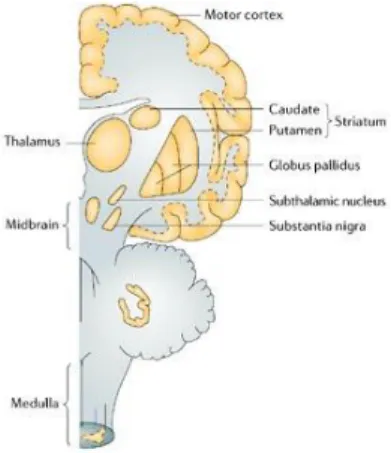
Currently Parkinson’s Disease (PD) is a known progressive multifactorial neurodegenerative disorder, characterized by the preferential loss of dopaminergic neurons in the region of the brain known as the substantia nigra, the disease’s pathological feature. Importantly, neurodegeneration is
not limited to the substantia nigra, the ventrolateral tier brain region which contains neurons that
project to the dorsal putamen of the striatum is also strongly affected with disease progression,
resulting in movement symptoms (Figure 1.1) (Farrer, 2006; Kalia & Lang, 2015). Clinical symptoms only appear when loss of dopaminergic neurons of the substancia nigra pars compact (SNpc DA) is
approximately 50-70% (Orth & Schapira, 2002; Chinta & Andersen, 2008).
Neurodegeneration causes an imbalance of excitatory (acetylcholine) and inhibitory (dopamine)
neurotransmitters and, it’s believed that lesions in this specific area leads to the characteristic motoric symptoms of PD (reviewed in Kalia & Lang, 2015). Pathologically, a second hallmark is fibrillary α -synuclein intracellular inclusions, where the insoluble α-synuclein aggregates form inclusion bodies within the cell body (Lewy bodies) and processes (Lewy neurites) of neurons (reviewed in Capriotti & Terzakis, 2016).
Presently, there is no cure for PD, the only existing treatment is symptomatic (Samii et al., 2004;
Capriotti & Terzakis, 2016). The most common pharmacologic treatment used is levodopa, since it provides the greatest symptomatic help, but it losses it effectiveness due to habituation. Another therapeutic options includes surgical options, from deep-brain stimulation to restorative treatment, but negative results have dampened enthusiasm in these approaches (Samii et al., 2004; Capriotti &
Terzakis, 2016). There has also been some substances describe as potential neuroprotective agents, such as Vitamin E, selegiline and coenzyme Q10, but no irrefutable evidences exists (Samii et al.,
2004).
Mitochondria and PD
The etiopathogenesis of sporadic cases is complex, but it is believed that genetic susceptibility and environmental factors contributes to this disease progression and both influence various mitochondrial aspects, such as bioenergetics, quality control, dynamics and transport (Moon & Paek, 2015). It is well known that aberrant mitochondrial forms and functions are connected with idiopathic (or sporadic) and familial PD (Henchcliffe & Beal, 2008). However, the mechanism still remains to be clarified. Mitochondrial dysfunctions are mainly characterized by the generation of reactive oxygen species (ROS), a decrease in the electron transport chain (ETC) Complex I activity, ATP depletion and cleaved caspase-3 activation (reviewed in Moon & Paek, 2015).
There is increasing evidence that link Complex I function to PD. Complex I is the first complex of the multimeric enzymatic system of the ETC, whose overall function is the generation of ATP.
Figure 1.1 - The main brain regions affected in Parkinson disease. Represented is a lateral brain’s
-3-
The role of mitochondria in PD became evident when it was discovered that the 1-methyl-4-phenyl-1,2,3,6-tetrahydropyridine (MPTP) metabolite, MPP+ (N-methyl-4-phenylpyridinium) inhibits Complex I of the ETC leading to DA loss in a way very similar to PD. After MPTP enters the cells through monoamine transporters, MPP+ binds and inhibits NADH CoQ10 reductase, decreasing ATP synthesis and increasing generation of free radicals (Orth & Schapira, 2002; Pesah et al., 2004).
Besides MPTP, rotenone also inhibits the Complex I by binding to the Complex I subunit ND-1 protein and leading to increased ROS levels. Rotenone induces a Parkinsonism syndrome in animal models and also in humans (Narendra, Tanaka, Suen, & Youle, 2008; Schapira et al., 1990). Another poisonous reagent is paraquat, whose toxicity is executed in a similar fashion as MPP+, causing generation of free radicals and oxidative stress (Schapira et al., 1990; Berry et al., 2010).
It is also worth noting that the brain consumes 20% of total resting body energy, therefore with such a high demand in mitochondria driven ATP production it is not surprising that hampered mitochondria will lead to a diseased brain (Orth & Schapira, 2002; MacAskill & Kittler, 2010). Further, SNpc DA have been characterized as a highly energy demanding population of neurons, thus it should be expected an increased mitochondria biogenesis, as well as increased basal oxidative phosphorylation (Henchcliffe & Beal, 2008). This feature must be due to the metabolic sustaining of their enormous axonal arborization, demonstrated by Pacelli et al., 2015. It was also shown that
mitochondrial reactive oxygen species (mROS) production is higher in these neurons due to the dopamine oxidative metabolism. Further, antioxidants such as reduced glutathione, are weakly synthesized in SNpc DA (Chinta & Andersen, 2008). This bioenergetics and morphological characteristics make SNpc DA more vulnerable to mitochondrial dysfunctions.
Then, what makes Mitochondria so special?
Known as the energy powerhouse of the cell, Mitochondria are double membrane organelles that have 4 distinct sub-mitochondrial compartments: the outer mitochondrial membrane (OMM), the intermembrane space (IMS), the inner mitochondrial membrane (IMM), and the matrix. The compartmentalization is crucial for vital mitochondrial functions. They actively sustain a highly
negative potential across their inner membrane (ΔΨ) that is maintained by four protein complexes I, II, III and IV of the mitochondrial ETC, that together with the F0/F1-ATP-synthase (Complex V), constitute the oxidative phosphorylation (OXPHOS) system. Maintenance of a highly negative potential across their inner membrane (ΔΨ) is essential for mitochondrial function and cell viability (Schapira, 2010).
coating. The mtDNA encodes 13 proteins, 2 ribosomal RNAs and 22 transfer RNAs. The mtDNA has some particularities: it is only inherited from the mother; exists approximately 8-10 mtDNA’s per mitochondrion, varying in different tissues; and the 13 proteins translated are all components of the ETC. Even having their own DNA, an overwhelming number of nuclear encoded proteins are targeted to the mitochondria, such as replication, transcription, translation and repair proteins (Orth & Schapira, 2002; Palikaras & Tavernarakis, 2014). The mtDNA does not have a complete repertoire of repair mechanisms to eliminate mutated DNA sequences, making it therefore rather vulnerable to mutations that accumulate with aging (Orth & Schapira, 2002). To prevent and reduce potential mitochondrial stress generators, these organelles have a mitochondrial unfolded protein response (UPRmt) system that deals with the accumulation of misfolded and toxic proteins (Moon & Paek, 2015). The UPRmt is composed by controlling chaperones and transcriptional proteases (Roberts
et al.,
2016). In addition, evidences suggest that vesicles derived from mitochondria engulf selected mitochondrial cargos and deliver them to peroxisomes for degradation. At the same time anti-oxidant pathways are activated, for example mitochondrial superoxide dismutase and glutathione (Roberts et
al., 2016). In addition to these processes, mitochondria are able to regulate their internal quality by
two processes: one that allows degradation of OMM proteins through the ubiquitin-proteasome system; and another one that uses the autophagy-lysosome pathway for elimination of mitochondria as whole organelles, known as mitophagy (Palikaras & Tavernarakis, 2014; Scarffe et al., 2014; Eiyama
& Okamoto, 2015)
Mitochondria have an important role in cellular processes by supporting cellular metabolic events, as iron-sulfur cluster biogenesis, amino acid synthesis and lipid metabolism. In order to maintain cellular homeostasis, mitochondria are able to regulate the calcium influx and inhibit apoptosis. Some of these cellular reactions may lead to oxidative stress with ROS formation, as superoxide anions, hydroxyl radical and hydrogen peroxide (Orth & Schapira, 2002). Mitochondria are fueled by pyruvate and fatty acids, which are used as carbon sources for the tricarboxylic acid cycle (or Krebs cycle) in the mitochondrial matrix.
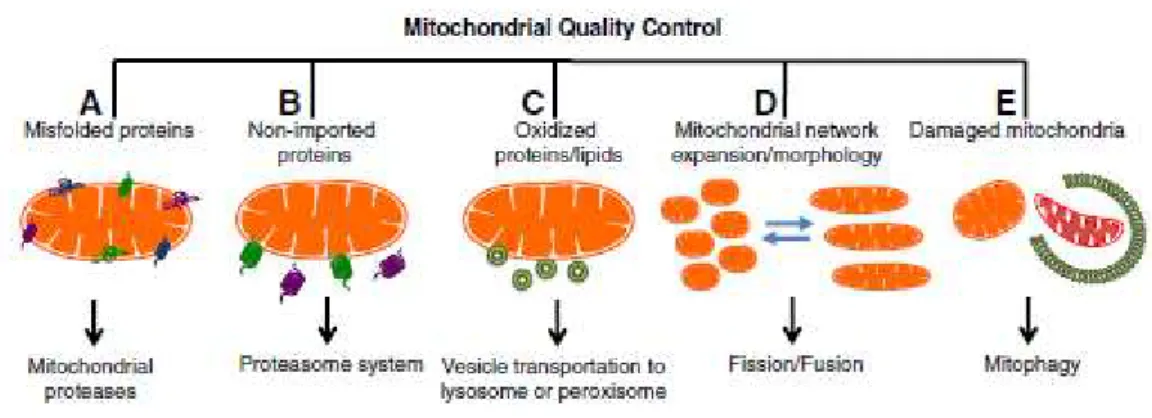
The aim of mitochondrial quality control is the maintenance of a healthy pool of mitochondria within the cell. This term is used to describe the coordination of mitochondrial dynamics, biogenesis and mitophagy (Scarffe et al., 2014). Mitophagy is a specialized mitochondria autophagy, or more
specifically macroautophagy, since it involves sequestration in a double membrane structure called autophagosome of mitochondria and selectively triggering them for clearance (Fig. 1.2) (Hattori et al.,
2014; Scarffe et al., 2014).
-5-
the cytosol and accumulates on OMM, where it oligomerizes into ring-like structures that will constrict the mitochondria through GTP hydrolysis, leading to the formation of new “daughter”
mitochondria. This process is essential in facilitating mitochondrial transport as well as the autophagic degradation of damaged mitochondria. On the other hand, fusion depends on the action of 3 GTPases: Mitofusins 1 and 2 (Mfn1/2) that are OMM proteins that tether organelles to the membrane for OMM fusion; and Optical atrophy 1 (Opa1), an IMM protein that mediates inner membrane fusion (Büeler, 2010; Roberts et al., 2016). Fusion process is crucial for maintain a functional mitochondrial
population within a cell, as mitochondria do not function in isolation but rather in a complex extensive network, its morphology undergoes continuous changes in response to metabolic stimuli and signaling pathways. Fusion also allows possible exchanges of contents between mitochondria (Detmer & Chan, 2007).
Figure 1.2 - Mitochondrial Quality Control. Mitochondria have different pathways to promote mitochondrial biogenesis and dynamics. (A) Proteolytic system; (B) Proteasome system; (C) Transportation to lysosome or peroxisome; (D) Fission/Fusion; (E) Mitophagy (Palikaras & Tavernarakis, 2014)
Genetics in PD
Nowadays, it is accepted that the involvement of mitochondria in PD is not only restricted to a decrease in ATP and increase in ROS production arising from the defective function of the respiratory chain. Defects in mitochondrial trafficking, dynamics, identification of mutations in genes involved in mitochondrial mitophagy) or defects in mitochondrial calcium buffering are emerging as mitochondrial dysfunctions related to PD (Aroso et al., 2016). Thus, the regulation of these
mechanisms is essential to maintain mitochondria healthy.
The past 15 years were marked by important discoveries which have led to a better understanding of the molecular pathogenesis of PD. Although 90% of cases are considered sporadic, the
identification of genes responsible for familial forms of PD where a clear “mendelian” autosomal
(where two mutated alleles are needed to originate the disease) of inheritance is observed have been crucial for a better understanding of the disease (Gasser, 2009; Scarffe et al., 2014; Kalia & Lang,
2015).
More than ten genes have been identified, and six of these genes were identified as mediating the autosomal dominant forms of PD, being the most common SNCA and LRRK2 (table1.1). The gene
SNCA encodes the α-synuclein protein, the principal constituent of Lewy bodies (Polymeropoulos et
al., 1996). So far, reports have identified three different missense mutations, as well as duplications
and triplications (Klein & Westenberger, 2012). Three missense mutations impair the amino-terminal
domain of α-synuclein causing misfolding and aggregation of the protein, a feature that is strongly correlated with PD (Klein & Westenberger, 2012; Recasens & Dehay, 2014).
The most frequent cause of autosomal dominant PD are mutations in LRRK2 (Klein &
Westenberger, 2012). This gene encodes the protein leucine-rich repeat kinase 2 LRRK2, a large multidomain enzyme, coupling kinase and GTPase activities with a number of protein/protein interaction domains (Paisán-Ruiza et al., 2013).
Autosomal recessive PD occurs less frequently but occurs in early-onset of the disease. In the form the associated genes are Parkin (Kitada et al., 1998), PINK1 (Valente et al., 2004) and DJ-1
(Bonifati et al., 2003)(table1.1), interesting all implicated within mitochondria pathways.
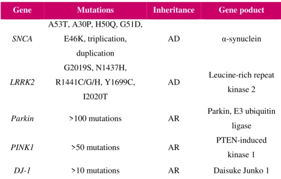
Table 1.1 – Most frequently genes implicated in monogenetic PD. Most frequently genes confirmed to be implicated in
autosomal dominant (AD) or autosomal recessive (AR) monogenetic PD. (adapted from Spatola & Wider, 2014).
Parkin, the second largest gene in human genome (Kitada et al., 1998; Klein & Westenberger,
2012), encodes an E3 ubiquitin ligase that catalyzes the ubiquitination of a range of proteins (Sarraf et
Gene Mutations Inheritance Gene poduct
SNCA
A53T, A30P, H50Q, G51D, E46K, triplication,
duplication
AD α-synuclein
LRRK2
G2019S, N1437H, R1441C/G/H, Y1699C,
I2020T
AD Leucine-rich repeat
kinase 2
Parkin >100 mutations AR Parkin, E3 ubiquitin
ligase
PINK1 >50 mutations AR PTEN-induced
kinase 1
-7-
al., 2013), particularly on damaged mitochondria (Narendra et al., 2008). In agreement, mutations in
this protein appear to lead to mitochondrial dysfunctions (Greene et al., 2003). Pathologically patients
with these alterations, although lacking α-synuclein aggregates, display clinical features of idiopathic (or sporadic) PD (Greene et al., 2003; Pesah et al., 2004).
PINK1, another autosomal recessive gene, encodes for the a serine/threonine (Ser/Thr) kinase
PINK1, whose mutations are less common than Parkin (Gasser, 2009). There is a genetic link between PINK1 and Parkin as mutant models for both proteins seem to have the same phenotypes, such as flight and climbing defects in the Drosophila model (Greene et al., 2003; Clark et al., 2006; Park et
al., 2006). Together, PINK1 and Parkin regulate mitochondrial quality control via clearance of
damaged mitochondria (Narendra et al., 2010).
Mutations in the DJ-1 gene are the least common and its function it is still not well understood
(Bonifati et al., 2003). The protein encoded is member of ThiJ/PfpI family and has H2O2 responsiveness, functioning as a sensor for oxidative stress and is an antioxidant (reviewed in Cheon, Chan, Chan, & Kim, 2012).
PINK1
Encoded by the PARK6 gene in chromosome 1p36, alterations in this protein are the second most common cause of early onset autosome recessive PD (Hatano et al., 2004; Bonifati et al., 2005;
Singleton et al., 2013; Requejo-Aguilar & Bolaños, 2016). Phosphatase and tensin homolog
(PTEN)-induced putative kinase 1 (PINK1) encodes a 581 amino acid protein with an N-terminal
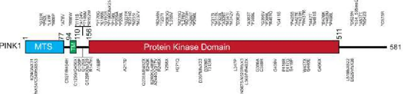
mitochondrial targeting sequence (MTS) spanning from residues 1-34, a conservative serine/threonine kinase domain from residues 150-513 and a C-terminal non catalytic region from residues 541-581 (Valente et al., 2004; Kondapalli et al., 2012). A hydrophobic patch formed by 11 amino acids can
also be found after the MTS. Bioinformatic analysis suggests that residues Gly-193 to Lys-507 form the ATP-binding cassette whereas residue Thr-313 is a autophosphorylation regulatory residue (Petit
et al., 2005).
PINK1 is ubiquitously expressed in all brain regions and in all cells types. Sporadic cases of PD or PD-related clinical mutations do not affect expression levels or localization pattern of PINK1 (Gandhi et al., 2006). PINK1 has been detected in both cytosol and mitochondria (Valente et al., 2004;
Beilina et al., 2005; Silvestri et al., 2005; Lin & Kang, 2008).
Drosophila PINK1 mutants exhibit male sterility, wing postural instability with rigidity that
kinase is essential for maintaining mitochondrial integrity and functions in vivo, as well as to dictate
Parkin localization through direct phosphorylation (Kim et al., 2008). PINK1 knockout mouse or
human dopaminergic neurons besides showing a high sensitivity to apoptosis also have abnormalities in mitochondrial morphology, a reduced membrane potential, and an increased ROS generation (Morais et al., 2009; Moon & Paek, 2015; Pacelli et al., 2015)
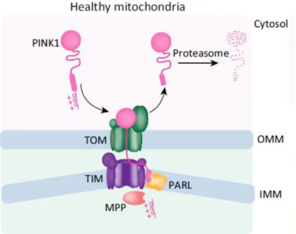
In healthy mitochondria, PINK1 is guided into mitochondria through the mitochondrial import machinery translocase of the outer membrane (TOM) and of the inner membrane (TIM) complexes, in a mitochondrial membrane potential dependent manner (Fig.1.3). PINK1 is translocated, partially, through TOM and TIM exposing the positively charged MTS to the matrix, which is removed by mitochondrial processing peptidase (MPP), and then cleaved by the inner mitochondrial membrane protease presenilin-associated rhomboid like protease (PARL) (Nguyen et al., 2016). Thus, in cells
normal conditions, three forms of PINK1 protein are detected: the full-length form (63 kDa) and two cleaved forms one form at approximately 55kDa that represents an intermediate PINK1 species produced by the inner mitochondrial membrane protease presenilin-associated rhomboid like protease (PARL); and 45kDa form cleaved by MPP‐mediated cleavage, between aminoacids Alanine 103 (Ala103) and Phenylalanine 104 (Phe104) (Deas et al., 2011; Meissner et al., 2011) and then
translocated to the cytosol where it is rapidly degraded through N-end rule pathway (Greene et al.,
2012; Song et al., 2013; Yamano & Youle, 2013; Voigt et al., 2016).
Complex I of the ETC is pivotal in generating the electrochemical gradient across IMM. Compromised Complex I activity has been related with PD sporadic cases (Schapira et al., 1990) and
further corroborated in PINK1 null mice (Morais et al., 2009). Vilain and co-workers suggested a
connection between PINK1 and Complex I where PINK1 is acting with or in parallel with Complex I (Vilain et al., 2012). The yeast Complex I Ndi1p rescued several phenotypes observed in Drosophila
PINK1 mutants further strengthening Complex I deficiency as the underlining cause of PINK1 related
-9-
Figure 1.3 - PINK1 processing in healthy mitochondria; firstly PINK1 is imported into OMM through TOM, and then IMM over TIM, where it is processed by MPP and PARL, exposing N-end rule substrate cytosolic and promoting PINK1
degradation (adapted from Nguyen, Padman, & Lazarou, 2016).
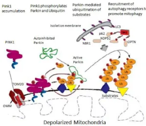
When mitochondria are depolarized, import of PINK1 within the mitochondria is inhibited and PINK1 is stabilized on the mitochondrial outer membrane triggering the damaged mitochondria for clearance via aPINK1/Parkin mediated mitophagy pathway (Narendra et al., 2010; Seirafi et al.,
2015). PINK1initiates mitophagy by phosphorylating Ubiquitin (Koyano et al., 2014), Mitofusin 2
(Chen & Dorn, 2013) and Parkin (Clark et al., 2006; Park et al., 2006; Matsuda et al., 2010). For this,
PINK1 has to be dimerizated and autophosphorylated on residues Ser228 and Ser402 (Okatsu et al., 2012; Aerts et al., 2015).
Approximately 50 pathogenic PINK1 mutations were identified; being that large amount of them located within the kinase domain, suggesting that this kinase activity plays a crucial role in the PD pathogenesis (Fig.1.4) (Rogaeva et al., 2004; Bonifati et al., 2005; Criscuolo et al., 2006; Kawajiri
et al., 2011). These homozygous point mutations R246X, H271Q, E417G and L347P, involving exons
3, 4, 5 and 6, and two nonsense mutations Q239X and R492X were identified in Asian families (Hatano et al., 2004). The residues implicated in H271Q, E417G and L347P mutations appear to be
highly preserved between PINK1 homologs (Hatano et al., 2004). Two missense mutations (E420K
and L489P) were described to abrogate PINK1 protective effect against cell death (Petit et al., 2005).
All patients shown early age onset, long disease duration and good response to L-dopa, therefore there does not exist any clinical features that will distinguish PINK1 mutations from Parkin or DJ-1
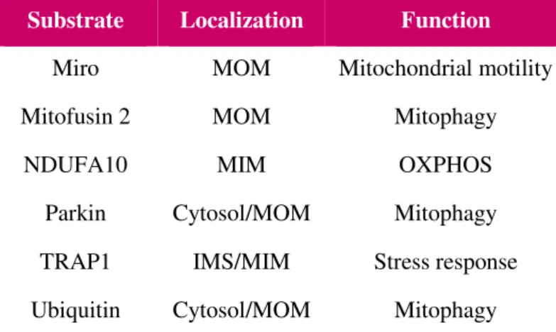
PINK1 is known to be included in numerous pathways, and to interact with several substrates related to mitochondrial homeostasis and mitochondrial quality control (Table 1.2) (Gómez-Sánchez et
al., 2016). PINK1 is also responsible for blocking the activation of apoptotic signalling pathways by
reducing caspase-3 activity and Cytochrome c translocation (Petit et al., 2005). Other studies have also
shown that PINK1 prevents the transport of defective mitochondria along microtubules by phosphorylating Miro (Weihofen et al., 2009).
PINK1 is pivotal in promoting cell survival by interacting with a mitochondrial molecular chaperone that protects against oxidative-stress-induced apoptosis, TNF receptor-associated protein 1 (TRAP1) (Pridgeon et al., 2007). The clinical PINK1 mutants G309D and L347P were unable to
phosphorylate TRAP1, in contrast with W437X clinical mutations where no difference was observed when compared to WT PINK1. These results suggest that cell survival may be affected by PINK1 function, especially in the case of the G309D and L347P clinical mutants.
It was also shown that kinase activity of PINK1 is involved in the regulation of mitochondrial apoptotic pathway, as its depletion increases cells susceptibility to oxidative stress induced apoptosis (Pridgeon et al., 2007). It is conceivable to assume that alterations in this domain may lead to
dysfunctions in kinase activity of this protein, and indeed L347P mutations appears to lead to a protein instability (Beilina et al., 2005). But studies about this possible dysfunction have been a little
controversial. In some studies the in vitro phosphorylation assay were performed with hPINK1 orthologues instead, such as Tribolium castaneum TcPINK1, that is remarkably more active than
hPINK1(Woodroof et al., 2011). Recently, it was shown that different PINK1 orthologues have
different substrate selectivity, so the extrapolation of obtained results back to human scenario need to be performed with caution (Aerts et al., 2016).
Table 1.2 – Resume of PINK1 Substrates mentioned. Besides (auto)phosphorylates itself, PINK1 as another substrates that
allows it to control pathways as mitophagy, mitochondrial motility and oxidative phosphorylation (OXPHOS). Only Miro,
-11-
TRAP1, Parkin and Ubiquitin are described as directly phosphorylated by human PINK1 (MOM, mitochondrial outer membrane; MIM, mitochondrial inner membrane; IMS, intermembrane space).
Substrate Localization Function
Miro MOM Mitochondrial motility
Mitofusin 2 MOM Mitophagy
NDUFA10 MIM OXPHOS
Parkin Cytosol/MOM Mitophagy
TRAP1 IMS/MIM Stress response
Ubiquitin Cytosol/MOM Mitophagy
Parkin
Mutations in the PARK2 gene, also known as
Parkin, are the most common cause of autosomal
recessive PD. The gene encodes a 465-amino acid E3 ubiquitin ligase, member of the RING1-in Between-RING-RING2 (RBR) family of E3’s, capable of mediating mono or poli-ubiquitination (Scarffe et al.,
2014; Koyano & Matsuda, 2015; Roberts et al., 2016).
An E3 ubiquitin-protein ligase, ubiquitinates proteins and labels them for degradation. Ubiquitination consists of a 76 amino acid polypeptide covalently conjugated to a lysine residue or N-terminal amino group of a substrate
protein allowing 3 enzymes to act sequentially: activating enzyme (E1), ubiquitin-conjugating enzyme (E2) and ubiquitin-protein ligase (E3) (Seirafi et al., 2015; Chin & Li, 2016).
The protein structure of Parkin is formed by the following independent domains (Fig.1.5): N-terminal ubiquitin-like (Ubl), four zinc-binding RING0, RING1, IBR (in-between RING), repressor element of Parkin (REP) and an N-terminal RING2 domain. The Ubl domain is involved in substrate recognition, binding SH3 and ubiquitin interacting motif (UIM) domains, proteasome association, and regulation of cellular Parkin levels and activity (Trempe, 2014; Koyano & Matsuda, 2015; Seirafi et
al., 2015). The IBR domain is attached through a flexible linker (Trempe, 2014). REP domain in line
with the catalytic center, is important in regulation of Parkin activity, once it is blocking E2 binding site on RING1 (Trempe, 2014; Seirafi et al., 2015).
Figure 1.5 - Parkin’s structure; 4 domains:
ubiquitin-like (Ubl), four zinc-binding RING0, RING1, IBR (in-between RING), repressor element of Parkin (REP) and an N-terminal RING2 domain. Catalytic Site (Cys431) and Phospho-site (Ser65 )
Parkin is a cytosolic protein, not only expressed in substantia nigra and others brain regions, but
also in many tissues, including heart, testis and skeletal muscle (Kitada et al., 1998). Results obtained
from the crystal structure, reveal that under basal conditions, Parkin exists as an autoinhibited form (Trempe, 2014). The catalytic active center, which accepts ubiquitin from E2 and transfers it onto substrates of Parkin is residue Cys 431 that lies in RING2 domain and beneath RING0 domain. This cysteine forms a thioester bond with ubiquitin, which is then transferred to the lysine residue of the substrate via an acyl transfer reaction (Koyano & Matsuda, 2015; Seirafi et al., 2015; Wauer et al.,
2015). The phosphorylation site is between IBR and RING2 domains, close to REP, in the autoinhibited conformation (Seirafi et al., 2015) (Fig.1.6).
Presumably, for Parkin activation it is needed some alterations that make both catalytic center and phosphorylation sites available. Reports have demonstrated that ubiquitin is necessary for Parkin activation, as it unlocks repression of the Cys 431 leading to destabilization of the Ubl domain and RBR core interactions (Koyano et al., 2014; Wauer et al., 2015).Structural analysis suggests a
conformational flexibility around Ser65, so after interaction with PINK1, the Ubl domain may undergo conformational changes, enabling PINK1 access (Kondapalli et al., 2012) (Fig .1.6).
Once activated, Parkin ubiquitinates several mitochondrial proteins that are involved in numerous mitochondrial dependent processes, such as mitochondrial motility, fission and fusion (FIS1; OPA1; MIRO, Mitofusins), small molecule transport (VDACs); apoptosis (MLC1 and BAX), mitochondrial autophagy (p62); and protein translocation (TOMM70) (Sarraf et al., 2013; Scarffe et al., 2014; Seirafi
et al., 2015).
Drosophila Parkin mutants show locomotor defects namely in flight and climbing due to
muscle degeneration; defects in spermatogenesis culminating in male sterility; female infertility most probably owing to functional or behavioral functions; fragmentation of mitochondrial cristae; and a reduced longevity and body size at eclosion, indicating defects of the growth and proliferating cell mechanisms (Greene et al., 2003; Pesah et al., 2004; Clark et al., 2006; Park et al., 2006).
As Parkin and PINK1 mutants have similar phenotypes one could suspect that they work in parallel genetic pathways or that one is able to regulate the other. Studies have revealed (Clark et al.,
2006; Park et al., 2006) that when Parkin is overexpressed the PINK1 null phenotype is restored.
However, PINK1 overexpression has no effect on Parkin-null phenotypes. Thus, these studies suggest
that both proteins function in a common genetic pathway, with PINK1 acting upstream of Parkin. And
-13-
Figure 1.6 –Model of Parkin Activation. In the cytosol, Parkin exists in a “closed” conformation, with RING0, Ubl and
REP, are obstructing, RING2 and E2 binding to RING1, respectively. Parkin is translocated to OMM thanks to the high affinity S65-phosphorylated ubiquitin. Because of this interaction, RING1 and IBR originates a displacement of the inhibitory
UBL and REP, and consequently Parkin structure to open. In the open conformation, E2 charged enzymes are able to bind to RING1 domain and expose RING2 catalytic cysteine to participate in ubiquitination; also, PINK1 is able to phosphorylate at S65 (Wauer, T.
et al. 2015)
Parkin is selectively recruited to Carbonyl cyanide m-chlorophenyl hydrazone (CCCP) treated depolarized mitochondria, and stimulates the autophagic removal of damaged mitochondria (Matsuda
et al., 2010; Koyano & Matsuda, 2015). Parkin mutated in the Ubl domain leads to a moderate loss in
mitophagy activity, while mutations in RBR conserved cysteines results in loss of RING2, exhibits a severe mitophagy defect; when the whole Ubl domain is truncated Parkin is also inhibited (Narendra
et al., 2010; Wauer et al., 2015).
Parkin is involved in the proteasome degradation of several substrates, prevents cytochrome c
release and α-synuclein aggregation, its loss of function causes accumulation of potentially toxic protein aggregates eventually involved in PD, since their role in protecting mitochondria is defected (Requejo-Aguilar & Bolaños, 2016).
PINK1/Parkin pathway
When mitochondria become depolarized, PINK1 accumulates stably on the OMM, due to interaction with TOM complex, dimerizes and is autophosphorylated on residues Ser228 and Ser402 (Okatsu et al., 2012; Aerts et al., 2015). Then, PINK1 phosphorylates Parkin at Ser65, a highly
conserved residue within Ubl domain, leading to Parkin E3 ligase activity (Narendra et al., 2008,
2010; Kondapalli et al., 2012; Kazlauskaite et al., 2014), and PINK1 also phosphorylates ubiquitin
(Koyano et al., 2014).
and recruit ULK1, DFCP1, WIPI1 and LC3 autophagy receptors (Lazarou et al., 2015; Seirafi et al.,
2015). OMM-bound PINK1 also mediates the recruitment of the autophagic protein p62 by Nrf2 and TFEB transcription factors (Ivankovic et al., 2016).
This process represents a positive feedback model (Fig1.7), where PINK1 phosphorylates a basal level of ubiquitin and subsequently Parkin is activated, increasing the amount of conjugated ubiquitin, which is then phosphorylated by PINK1 to recruit more Parkin, that will then in turn ubiquitinate other OMM substrates (Seirafi et al., 2015), namely Mfn1/2 (Gegg et al., 2010) that
regulates mitochondrial fusion.
In sum, this mitochondrial quality control allows taking away damaged and superfluous mitochondria and does not allow the accumulation of oxidized lipids, proteins and DNA, limiting the risk of apoptosis. The disruption of the PINK1/Parkin pathway results in an accumulation of abnormal mitochondria and overproduction of reactive oxygen species (ROS). This pathway regulates the autophagic degradation of damaged mitochondria, through ubiquitin-proteasome and autophagy pathways (Narendra et al., 2008, 2010; Matsuda et al., 2010).
Figure 1.7 – Model of PINK1/Parkin mitophagy induction;
-15-
2.
Aims
Both PINK1 and Parkin genes are known to be dysregulated in familiar forms of PD. PINK1
through phosphorylation of its downstream targets regulates multiple mitochondrial processes like dynamics and quality control. This pathway responsible for the specific removal of damaged mitochondria depends also on Parkin, a PINK1 substrate.
Given the importance of these encoded proteins in mitochondrial biology, it will be not surprising to find that their dysfunction is associated with damaged mitochondria.
In fact, some groups have described different levels of Parkin phosphorylation and recruitment in the presence of clinical mutant forms of PINK1. However, these results vary depending on the PINK1 specie that is used in these studies, making extrapolation of these findings into the disease context rather difficult to interpret.
-17-
3.
Methods
Plasmids
The plasmids used were pcDNA 3.1 hPINK1 WT (plasmid expressing human PINK1 wild type); pcDNA 3.1 hPINK1 KI (plasmid expressing hPINK1 kinase inactive); pcDNA 3.1 hPINK1 ΔN
WT (plasmid expressing a truncated form of hPINK1WT lacking the first 113 aminoacids); pcDNA 3.1 hPINK1 ΔN KI (plasmid expressing a truncated form of hPINK1 KI lacking the first 113 aminoacids). The cDNA of hPINK1 was also cloned into the pMSCV vector, a vector that has the low expressing promotor LTR,leading to near-to-endogenous PINK1 expression. The pGEX-4T-1 vector was used for the bacterial expression system. These constructs were previously described (Aerts et al.,
2015). The mutant Kinase Inactive form of human PINK1 consists of two point mutations of the residues K219 and D362 to Ala. These residues were predicted by computer modelling analysis as crucial residues of the catalytic pore of the kinase domain in PINK1 (Beilina et al., 2005).
All materials used in these experiments are described in appendix.
Construction of Clinical PINK1 Mutants
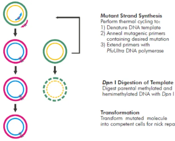
Mutant PINK1 constructs were inserted into pcDNA3.1 hPINK1-WT and hPINK1-ΔN using
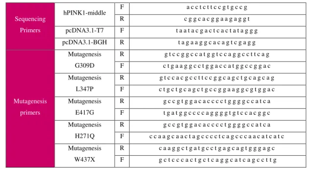
QuikChange II XL Site-Directed Mutagenesis (Agilent technologies). This technique allows the alteration, deletion and insertion of a base pair in our coding DNA sequence. In our case we performed single point mutations that gave rise to the following amino acid change: a glycine to aspartic acid in G309D, a leucine to proline for L347P, a glutamic acid to glycine in E417G, a histidine to glutamine for H271Q; and tryptophan to a STOP codon for W437X.
The protocol consists of a three step procedure (Fig.3.1). Before moving to the first, the mutagenic oligonucleotide primers must be designed according to the desired mutation. For their
design, the QuikChange Primer Design Program available online (at
www.agilent.com/genomics/qcpd) was used, where the melting temperatures, that should be ≥78°C,
are calculated with the following equation: . In addition primers should
be between 25 to 45 bases in length, with the desired mutation in the middle, a minimum GC content of 40% and end with a C or G bases. Primers obtained are described in table 3.1.
Table 3.1 - Primers used for quick change mutagenesis and sequencing; F=forward; R=reverse
Sequencing Primers
hPINK1-middle F a c c t c t t c c g t g c c g R c g g c a c g g a a g a g g t pcDNA3.1-T7 F t a a t a c g a c t c a c t a t a g g g pcDNA3.1-BGH R t a g a a g g c a c a g t c g a g g
Mutagenesis primers
Mutagenesis G309D
R g t c c g g c c a t g g t c c a g g c c t t c a g F c t g a a g g c c t g g a c c a t g g c c g g a c Mutagenesis
L347P
R g t c c a c g c c t t c c g g c a g c t g c a g c a g F c t g c t g c a g c t g c c g g a a g g c g t g g a c Mutagenesis
E417G
R g c c g t g g a c a c c c c t g g g g c c a t c a F t g a t g g c c c c a g g g g t g t c c a c g g c Mutagenesis
H271Q
R g c c g t g g a c a c c c c t g g g g c c a t c a F c c a a g c a a c t a g c c c c t c a g c c c a a c a t c a t c Mutagenesis
W437X
R c a a g g c t g a t g c c t g a g c a g t g g g a g c F g c t c c c a c t g c t c a g g c a t c a g c c t t g
First step is the Polymerase Chain Reaction (PCR) that is initiated by adding the following components into a PCR tube: reaction buffer (appropriate for the polymerase used), dNTP mix, QuikSolution reagent, milliQ water, pcDNA 3.1 hPINK1 ΔN WT, primers forward and reverse for each mutation and Pfu Turbo polymerase. To guarantee the kit’s effectiveness and absence of
contamination from any PCR reaction components, two control samples were used, one provided with the kit that consists in a control plasmid, and another one without the template pcDNA 3.1 hPINK1
ΔN WT, respectively. Reactions were performed with the cycling parameters mentioned in table 3.2.
Table 3.2 - Cycling parameters used for pcDNA 3.1 hPINK1 WT and ΔN mutagenesis reaction
Cycles Temperature Time
1 95°C 1 minute
18
95°C 50 seconds
60°C 50 seconds
68°C 7,5 minutes
1 68°C 7 minutes
-19-
Secondly, after the cycle is done, the parental non mutated DNA is digested with DpnI, leaving the PCR amplified DNA intact, therefore only mutated plasmid DNA will be transformed into XL-gold ultracompetent bacteria, which offers a highest transformation efficiency and ideal for large plasmid DNA. As pcDNA 3.1 plasmids carry an antibiotic resistance genes to ampicillin, we used LB agar plates with Ampicilin (100µg/mL) resistance. Transformed bacteria were incubated for overnight at 37°C. To proceed with the DNA extraction protocol, colonies had to be picked from the plates and
incubated with 3mL LB Broth Medium and 100µg/μL Ampicillin, in a shaker at 37°C and 225rpm, overnight. The protocol used for DNA extraction is according to the one described at QIAprep® Miniprep Handbook. Briefly, collected overnight bacterial cultures were spin and pellets were first, ressuspended in Ressuspension Buffer (50mM Tris-HCL pH8.0; 10mM EDTA; 100μg/mL RNaseA),
secondly lyzed with Lysis Buffer (200mM NaOH; 1%SDS), and finally neutralized with Neutralization Buffer (4,2M Gu-HCL; 0,9M potassium acetate; pH4,8). Then DNA is purified in QIAprep spin column, and eluted in Buffer EB (10mM Tris-HCL; pH 8,5) added to the column center. DNA concentrations and purity were measured using UV-Vis spectrophotometers NanoDrop™2000.
Based on incident and transmitted light intensity, spectrophotometer produces an optical density that correlates with Lambert–Beer law and determines the unknown concentrations.
Then the plasmids were analyzed via Sanger sequencing, where 5µg DNA and 2,5µg primer were used. All sequences obtained were analyzed using the GATC viewer program, which allows checking the chromatogram and DNA sequence. After the analysis, one colony with the desired mutation was selected and further expanded and purified using the Genopure Plasmid Midi Kit (Roche), in order to obtain a highly quality purified plasmid DNA. Briefly, the selected colony is inoculated in a bacterial culture of 50ml and incubated overnight at 37°C at 225rpm. The bacterial culture is centrifuged and pellets are ressuspended in Suspension Buffer (complemented with lyophilized enzyme RNase A), lysed and then neutralized. DNA was purified and washed, within a column, and then eluted with Elution Buffer. At that point, DNA is precipitated with isopropanol and washed in ethanol 70%. Concentrations were measured using NanoDrop™2000, as described above.
Cell Culture and cell lines
The HeLa-CrispR/Cas9-PINK1 cell line (here within referred to as HeLa PINK1 KO) were previously described (Aerts et al., 2015). Briefly, these cells were generated using clustered regularly
repeats guide RNA. Product generated, on all chromosomal copies, were an 84-bp deletion spanning the start codon of PINK1.
Other cell lines used were COS and HeLa WT. Cells were cultured at 37°C with 5% CO2 in
DMEM/F12 medium containing 10% fetal bovine serum (Life technologies). All cell lines were ideally manipulated with approximately 80% confluence.
Parkin expression and purification
The procedure is described in Aerts et al., 2015. Briefly, BL 21 bacteria were transformed
with pGEX-4T-1 expressing an N-terminal GST-tagged Ubl-domain of Parkin. Parkin expression was
induced with 100μM IPTG, a reagent that induces protein expression where the gene is under the control of the lac operator; and cells were incubated at 37°C with 280rpm of agitation for 2hours.
After a 15 minutes centrifugation, bacterial pellets were lyzed in 50mM Tris-HCl pH 7.5, 150mM NaCl, 1% Triton X-100, 2mM EDTA, 0.1% beta-mercaptoethanol, 0.2mM PMSF and 1mM benzamidine. GST-Ubl Parkin was purified using Glutathione Sepharose™ 4B (GE Healthcare), according to manufacturer’s instructions. Control samples were retained in every purification step,
quality and purity was evaluated via western blot. Briefly, this technique allows proteins separations based on molecular weight, by gel electrophoresis, producing a band for each protein. The proteins in the gel are then transferred to a nitrocellulose membrane, which is then incubated with antibodies to the protein of interest, and then develop (Mahmood & Yang, 2012). For Parkin membranes it was used for primary antibody rabbit anti-GST (1/1000; Sigma) and secondary antibody GARPO (1/10000; Bio-Rad).
Parkin recruitment
The procedure was adapted from Aerts et al., 2015. HeLa cells were plated in a 24-well plate,
as schematized in figure 3.2, on top of 13mm coverslips and transfected at approximately 80% confluence.
CCCP DMS
-21-
All the cell lines were transfected using FuGENE transfection reagent, agreeing to the
manufacturer’s instructions. This transfection protocol uses a ratio transfection reagent to DNA of 3:1. HeLa cells were transfected with mParkin-GFP and pcDNA3.1-hPINK1 WT or corresponding PINK1 mutants as described in table 3.3, and according to the manufacturer’s instructions.
Twenty-four hours post transfection, cells were treated with 10μM CCCP for 3hours or, as control, the equivalent volume of DMSO. CCCP is an uncoupling agent that is used to induce mitochondrial membrane depolarization by increasing membrane permeability to H+). Cells were washed 3 times in PBS+/+ (100 ml 10x PBS-/-; 2M CaCl
2; 1M MgCl2), fixed for 20 minutes in 4% formaldehyde in PBS+/+, washed 3 times in PBS+/+, permeabilized in 0,1% Triton X-100 in PBS+/+ for 10 minutes, and washed 3times in PBS+/+ afterwards. Cells were blocked for 1 hour in Blocking Buffer (0,2% gelatin, 2% fetal bovine serum, 2% bovine serum albumin, 0.3% Triton X-100 in PBS-/-) and 5% goat serum (Dako). Cells were stained using the mouse Turbo-GFP antibody (1/1000; Evrogen) and sheep Cytochrome c antibody (1/500; Sigma) for 2 hours. Cells were further washed 3 times with
PBS+/+, and further incubated with secondary antibodies Alexa 488 donkey anti-rabbit and Alexa 568 donkey anti-sheep (Life Technologies), in a 1/500 dilution. Images were acquired on a Zeiss LSM 710 confocal microscope, using a 40x objective, and analyzed with Image J and Photoshop software’s.
Table 3.3 - List of plasmids used to transfect HeLa cells
Plasmids
pCMV6 mParkin-GFP
pMSCV hPINK1 FL WT
pMSCV hPINK1 FL KI
pMSCV hPINK1 FL G309D
pMSCV hPINK1 FL L347P
pMSCV hPINK1 FL E417G
pMSCV hPINK1 FL H271Q
pMSCV hPINK1 FL W437X
Human PINK1 purification and in vitro kinase assay
The guidelines for this procedure were optimized and are described in Aerts et al., 2015.
COS-1 cells were transfected with the plasmids described in table 3.4, according to manufacturer’s
step, cells were lyzed in Lysis buffer (25mM Tris-HCl pH 7.5, 150mM NaCl, 5mM NaF, 1mM MgCl2, 1mM MnCl2, 0.5% Igepal-NP40 (Sigma), 50mg/L DNAse (Sigma), 50mg/L RNAse (Sigma), 1mM DTT), with 20% protease inhibitor cocktail for mammalian cell and tissue extraction (Sigma), 2X complete protease inhibitor (Roche), 4X PhosSTOP tablets (Roche) and homogenized using a 22-G needle in 5 strokes. Lysates were centrifuged during 25 minutes at maximal rpm, and then incubated for 45 minutes at 4°C with FLAG-magnetic beads (Sigma). The unbound fraction was removed, and
beads were washed 2 times with Lysis buffer and 3 times with kinase assay buffer (50mM Tris-HCl pH 7.5, 150mM NaCl, 10mM MgCl2, 3mM MnCl2 and 0.5mM DTT).
The kinase assay was executed immediately after the binding step where purified hPINK1-FLAG bound on the beads was incubated with 3,35μL of Parkin (2μg) , 10mM DTT and 100μM ATP containing 5μCi [-32P] ATP. Reactions were incubated for 1hour at 22°C.
Samples were analyzed by SDS-PAGE followed by Western blotting. For this, samples were incubated for 10 minutes at 70°C with Sample Buffer (77.8mM Tris-HCl pH6.8; 44,4% (v/v) glycerol;
4,4% LDS; 0,02% bromophenol blue and 4% β-mercaptoethanol). Samples were loaded on Mini-PROTEAN 7.5% Tris-Glycine Gels, the electrophoresis separation occurred for approx. 1 hour in Running Buffer (25mM Tris-HCl pH 8.3, 190mM glycine, 0.1% SDS). After SDS-PAGE, samples were transferred onto a PVDF 0.45µm membrane in Transfer buffer (25mM Tris-HCl pH 8.3, 190mM glycine, 20% methanol) for 1 hour at 100V. After transfer, PVDF membrane was stored in an autoradiography cassette with an amplifying film. Incorporation of radiolabelled phosphor was assed via a storage phosphor screen and development on Typhoon (GE Healthcare Life Sciences). Image studio lite software was used for signal quantification.
After radiolabelled phosphor quantification, the PVDF membranes were blocked for 1hour in 5% milk in TBS-T (50 mM Tris-HCl pH 7.5; 150 mM NaCl, 0.1% Tween-20), and incubated with agitation in primary antibody mouse anti-Flag M2 (1/5000; Sigma) and rabbit anti-GST (1/5000; Sigma), overnight.
Table 3.4 - Plasmids transfected on COS-1 cell line
Plasmids
pcDNA 3.1 hPINK1 ΔN WT
pcDNA 3.1 hPINK1 ΔN KI
pcDNA 3.1 hPINK1 ΔN G309D
pcDNA 3.1 hPINK1 ΔN L347P
pcDNA 3.1 hPINK1 ΔN E417G
pcDNA 3.1 hPINK1 ΔN H271Q
-23-
Statistical analysis
Statistical significance between the different test conditions was analysed using GraphPad Prism
-25-
4.
Results and Discussion
Parkin recruitment
The mutations studied have been previously identified as altered in PD, as earlier described in Chapter 1 (Fig.4.1). Quite a few studies have reported PINK1 G309D, L347P, H271Q and W437X mutants behaviour relatively to Parkin recruitment. However there is some contradiction in these reports due to the use of different experimental conditions (such as concentration or exposure time to CCCP) and quantification methods, so facts still remain unclear in the field.
.
In order to verify the impact of hPINK1 clinical mutants on Parkin recruitment, we transfected HeLa WT and HeLa-PINK1-KO cells with PINK1 constructs and with a GFP-tagged Parkin construct. Dual staining was performed in all cell lines studied. We confirmed that in basal conditions, Parkin is predominately located in the cytosol as expected (Fig.4.2B and Fig.4.2H), and does not colocalize with mitochondria (Fig.4.2C and Fig.4.2I). Although, when HeLa WT cells were treated with CCCP, a loss of mitochondrial network is observed when staining for a mitochondrial resident protein Cytochrome c and a perinuclear clustering of mitochondria is observed (Fig.4.2.D), and more
interestingly Parkin is recruited to the mitochondria (Fig.4.2F). This does not happen in HeLa PINK1 KO cells treated with CCCP where, in the absence PINK1, Parkin is not recruited to mitochondria (Fig.4.2L). This observation is in agreement with previous studies (Narendra et al., 2010) that show
that Parkin recruitment to depolarized mitochondria requires the presence of PINK1.
Figure 4.1 – Schematic representation of PINK1
D
MSO
Cyto. C
CCCP
Parkin Merge
E D
A B C
F
D
MSO
CCCP
K J
G H I
L
H
eLa WT
H
eLa
-PINK
1-KO
Figure 4.2 – Parkin recruitment to depolarized mitochondria in HeLa WT and HeLa-PINK1-KO. Cells were transfected with mParkin-GFP and treated with DMSO or 10μΜ CCCP in serum for 3h. Mitochondria were
-27-
Additionally, to further confirm that Parkin recruitment is dependent not only on the presence of the PINK1 protein but also requires an active form of PINK1, we transfected HeLa WT and HeLa-PINK1-KO cells with a kinase inactive mutant form of PINK1, the hPINK1 KI, and Parkin recruitment was evaluated. At present, there is not a known stoichiometry between PINK1 levels and Parkin recruitment (Seirafi et al., 2015), therefore, in order to quantify the percentage of cells where
Parkin recruitment is occurring we quantified cells that presented staining for Cytochrome c and
Parkin simultaneously. The quantified data for cells treated with DMSO and CCCP are represented in Fig. 4.3.
In DMSO conditions (Fig. 4.3A), Parkin recruitment shows no significant alterations between the different cell lines analysed. This is due to the fact that mitochondria are healthy with no defects at
the level of mitochondrial membrane potential (Δm).
When loss of Δm induced by CCCP treatment leads to a pool of damaged mitochondria represented by a loss in mitochondrial network and the formation of fragmented mitochondria (Fig. 4.3B), there is a complete different response from Parkin which is dependent on the presence of PINK1. In HeLa WT cells transfected with PINK1 WT and treated with CCCP, we did not observe a difference between transfected and non-transfected cells. On the other hand, Parkin recruitment is significantly decreased in the presence of hPINK1-KI (Fig.4.3.B). Parkin recruitment can be recovered in Hela-PINK1-KO cells by expressing PINK1 WT (Fig.4.4F), where 63,78% of recruitment is observed when compared to hPINK1 WT; but recruitment is not restored with PINK1 KI (Fig.4.3B; Fig4.4L), indicating that PINK1 has a crucial role in Parkin recruitment to mitochondria and cooperate functionally to clear damaged mitochondria via mitophagy.
A
B
Figure 4.3 – Quantification of Parkin recruitment to mitochondria. A. Parkin recruitment in HeLa WT and Hela PINK1 KO cell lines, non-transfected, and transfected with hPINK1 WT and KI, in DMSO conditions; #=Cell line not transfected. B. Parkin recruitment in HeLa WT and HeLa PINK1 KO cell lines, non-transfected, and transfected
.
CCCP
PINK
1 WT
PINK
1 K
I
CCCP
E D
A B C
F
K J
G H I
L
D
M
SO
Cyto. C Parkin Merge
D
M
SO
Figure 4.5 - Parkin recruitment to depolarized mitochondria in HeLa-PINK1-KO. HeLa-PINK1-KO cells
were cotransfected with mParkin-GFP and pcDNA 3.1 .ΔN PINK1 WT or pcDNA 3.1 .ΔN PINK1 KI, and treated
with DMSO or 10μM CCCP in serum for 3h. Mitochondria were immunostained for Cyto. C. The images in column on the right are merged images of the middle (Parkin staining) and left-hand (Mitochondria staining) columns. Scale bar=10μM.
Figure 4.4 - Parkin recruitment to depolarized mitochondria in HeLa-PINK1-KO. HeLa-PINK1-KO cells
were cotransfected with mParkin-GFP and pcDNA 3.1 .ΔN PINK1 WT or pcDNA 3.1 .ΔN PINK1 KI, and treated
with DMSO or 10μM CCCP in serum for 3h. Mitochondria were immunostained for Cyto. C. The images in column on the right are merged images of the middle (Parkin staining) and left-hand (Mitochondria staining)
-29-
In order to elucidate the impact of clinical PINK1 mutants on Parkin recruitment to mitochondria, we transfected HeLa-PINK1-KO with mParkin-GFP and hPINK1 clinical mutants, and investigated Parkin recruitment in the presence of CCCP treatment. Immunofluorescent images and consequent quantification was obtained for all studied mutants.
D
M
SO
Cyto. C
CCCP
Parkin Merge
E D
A B C
F
W437X
D
M
SO
CCCP
K J
G H I
L
L347P
Figure 4.5 - Parkin recruitment to depolarized mitochondria in PINK1 W437X and L347P mutants. HeLa-PINK1-KO cells were cotransfected with mParkin-GFP and pcDNA 3.1 .ΔN PINK1 W437X or pcDNA 3.1 .ΔN
PINK1 L347P ,and treated with DMSO or 10μM CCCP in serum for 3h. Mitochondria were immunostained for Cyto. C. The images in column on the right are merged images of the middle (Parkin staining) and left-hand (Mitochondria staining) columns. Scale bar=10μM.
-31-
K J
G H I
L
D
M
SO
Cyto. C
CCCP
Parkin Merge
E D
A B C
F
D
M
SO
CCCP
H
271Q
G
309D
Figure 4.6 - Parkin recruitment to depolarized mitochondria in PINK1 H271Q and G309D mutants. HeLa-PINK1-KO cells were cotransfected with mParkin-GFP and pcDNA 3.1 .ΔN PINK1 H271Q or pcDNA 3.1 .ΔN
PINK1 G309D, and treated with DMSO or 10μM CCCP in serum for 3h. Mitochondria were immunostained for
Cyto. C. The images in column on the right are merged images of the middle (Parkin staining) and left-hand (Mitochondria staining) columns. Scale bar=10μM.
G H I
In order to mimic depolarized mitochondria, cells were treated with CCCP. In this case all mutants showed Parkin recruitment and presented total or partial colocalization with the mitochondrial marker Cytochrome c (Fig.4.5D-F; Fig.4.5J-L; Fig.4.6D-F; Fig.4.6J-L; Fig.4.7D-F). However, the clinical mutants are not able to restore Parkin recruitment to levels comparable with hPINK1 WT, indicating that these mutations that occur in hPINK1 lead to the expression of a hampered active form of this kinase.
Transfected cells were scored for presence of Parkin-GFP (Figure 4.9) and further quantified for Parkin recruitment (Fig.4.8). All clinical mutants presented similar percentage of Parkin-GFP transfection, indicating that observed results within clinical mutants is not due to lower Parkin-GFP transfection efficiency. In cells treated with DMSO, PINK1 clinical mutants where able to significantly recruit Parkin when compared to non-transfected HeLa-hPINK1-KO cells, with the exception of PINK1 L347P. These results show that mitophagy can occur in healthy mitochondria, and that mitochondria turnover also occurs at basal levels. Nevertheless, this phenotype is massively increased with cells are treated with CCCP.
D
M
SO
Cyto. C
CCCP
Parkin Merge
E D
A B C
F
Figure 4.7 - Parkin recruitment to depolarized mitochondria in PINK1 E417G mutant.
HeLa-PINK1-KO cells were cotransfected with mParkin-GFP and pcDNA 3.1 .ΔN PINK1 E417G, and treated with DMSO or 10μM CCCP in serum for 3h. Mitochondria were
immunostained for Cyto. C. The images in column on the right are merged images of the middle
-33-
A
B
Figure 4.8 – Quantification of Parkin recruitment to mitochondria in PINK1 mutants. A. Parkin recruitment in Hela PINK1 KO cells, non-transfected, and transfected with hPINK1 WT, KI and mutants, in DMSO conditions; #: Cell line not transfected. B.Parkin recruitment in Hela PINK1 KO cell line, non-transfected, and transfected with hPINK1 WT, KI and mutants, in CCCP conditions; #: Cell line not transfected.
A
B
Figure 4.9 - Cells transfected with mParkin. A. Hela WT and HeLa PINK1 KO cells transfected with Parkin (%), in DMSO conditions. There is a significant difference between Hela WT transfected (#) and HeLa WT KO non-transfected (#). B. Hela WT and HeLa PINK1 KO cells transfected with Parkin (%), in CCCP conditions. There is a