Therapeutic Advances in Gastroenterology
Ther Adv Gastroenterol 2016, Vol. 9(6) 781 –794 DOI: 10.1177/ 1756283X16658223 © The Author(s), 2016. Reprints and permissions: http://www.sagepub.co.uk/ journalsPermissions.nav
http://tag.sagepub.com 781
Introduction
Infliximab (IFX) is a chimeric (human–murine) monoclonal IgG1 anti-tumor necrosis factor alpha (TNF-α) antibody used in the treatment of inflam-matory bowel disease (IBD) [Bendtzen, 2013].
Although IFX has profoundly improved the treat-ment of inflammatory diseases, not all patients respond to induction therapy, and up to 50% of patients experience the loss of clinical response over time (secondary loss of response) [Yanai and
Detection of anti-infliximab antibodies
is impacted by antibody titer, infliximab
level and IgG4 antibodies: a systematic
comparison of three different assays
Joana Afonso, Susana Lopes, Raquel Gonçalves, Paulo Caldeira, Paula Lago, Helena Tavares de Sousa, Jaime Ramos, Ana Rita Gonçalves, Paula Ministro, Isadora Rosa, Ana Isabel Vieira, Rosa Coelho, Patrícia Tavares, João Soares, Ana Lúcia Sousa, Diana Carvalho, Paula Sousa, João Pereira da Silva, Tânia Meira, Filipa Silva Ferreira, Cláudia Camila Dias, Yehuda Chowers, Shomron Ben-Horin and Fernando Magro on behalf Portuguese IBD Study Group (GEDII)
Abstract:
Background: There is scant information on the accuracy of different assays used to measure anti-infliximab antibodies (ADAs), especially in the presence of detectable infliximab (IFX). We thus aimed to evaluate and compare three different assays for the detection of IFX and ADAs and to clarify the impact of the presence of circulating IFX on the accuracy of the ADA assays. Methods: Blood samples from 79 ulcerative colitis (UC) patients treated with infliximab were assessed for IFX levels and ADAs using three different assays: an in-house assay and two commercial kits, Immundiagnostik and Theradiag. Sera samples with ADAs and undetectable levels of IFX were spiked with exogenous IFX and analyzed for ADAs.
Results: The three assays showed 81–96% agreement for the measured IFX level. However, the in-house assay and Immundiagnostik assays detected ADAs in 34 out of 79 samples, whereas Theradiag only detected ADAs in 24 samples. Samples negative for ADAs with Theradiag, but ADA-positive in both the in-house and Immundiagnostik assays, were positive for IFX or IgG4 ADAs. In spiking experiments, a low concentration of exogenous IFX (5 µg/ ml) hampered ADA detection with Theradiag in sera samples with ADA levels of between 3 and 10 µg/ml. In the Immundiagnostik assay detection interference was only observed at concentrations of exogenous IFX higher than 30 µg/ml. However, in samples with high levels of ADAs (>25 µg/ml) interference was only observed at IFX concentrations higher than 100 µg/ ml in all three assays. Binary (IFX/ADA) stratification of the results showed that IFX+/ADA- and IFX-/ADAs+ were less influenced by the assay results than the double-positive (IFX+/ ADAs+) and double-negative (IFX-/ADAs-) combination.
Conclusions: All three methodologies are equally suitable for measuring IFX levels. However, erroneous therapeutic decisions may occur when patients show double-negative (IFX-/ADAs-) or double-positive (IFX+/ADAs+(IFX-/ADAs-) status, since agreement between assays is significantly lower in these circumstances.
Keywords: anti-infliximab antibodies, anti-Infliximab antibody methodologies, infliximab trough levels, therapeutic drug monitoring
Correspondence to: Fernando Magro, MD, PhD Gastroenterology Department, Medical School, Centro Hospitalar São João, Alameda Prof. Hernâni Monteiro, 4200-319 Porto, Portugal fm@med.up.pt Joana Afonso, MSc Filipa Silva Ferreira, MD
Department of Pharmacology and Therapeutics, Faculty of Medicine, University of Porto, Porto, Portugal MedInUP, Centre for Drug Discovery and Innovative Medicines, University of Porto, 4200 Porto, Portugal
Susana Lopes, MD Rosa Coelho, MD Patrícia Tavares, MD
Gastroenterology Department, Centro Hospitalar São João, Porto, Portugal
Raquel Gonçalves, MD João Soares, MD
Gastroenterology Department, Hospital de Braga, Braga, Portugal
Paulo Caldeira, MD Ana Lúcia Sousa, MD
Gastroenterology Department, Centro Hospitalar do Algarve, Faro, Portugal Paula Lago, MD Gastroenterology Department, Centro Hospitalar do Porto, Porto, Portugal
Helena Tavares de Sousa, MD Gastroenterology Department, Centro Hospitalar do Algarve, Portimão, Portugal Departament of Medicine e Medical Biosciences, University of Algarve, Faro, Portugal Jaime Ramos, MD Diana Carvalho, MD Gastroenterology Department, Centro Hospitalar de Lisboa, Lisboa, Portugal Original Research
Hanauer, 2011; Steenholdt et al. 2013]. When managing loss of response, clinicians may empiri-cally intensify treatment with the existing drug (increase dosage and/or increase frequency), switch to another TNF-α antagonist or switch to a totally different class of drug. This empirical approach has disadvantages: risk of irreversible tis-sue damage while the physician searches for an effective new drug, and significant economic con-sequences of unsuccessful trial and errors [Bendtzen and Svenson, 2011; Steenholdt et al. 2014a]. A more astute strategy is probably to use therapeutic drug monitoring (TDM), which ena-bles clinicians to identify patients in whom a medi-cation or change in medimedi-cation is likely to be effective [Roblin et al. 2014; Steenholdt et al. 2014b; Yanai et al. 2015]. Indeed, a rational evidence-based and tailored therapy according to individual needs may reduce delays in effective treatment [Bendtzen, 2013; Steenholdt et al. 2014b].
Awareness of the value of TDM has led to the development of different techniques for assessing levels of infliximab and anti-infliximab antibodies (ADA) in patients, but these different methodol-ogies have distinctive limitations and may yield different results. This potential bias may have a significant impact on TDM results and interpre-tation. There is still little information allowing us to compare different assays, in particular in rela-tion to ADAs detecrela-tion, which is likely to be sub-ject to interference by detectable levels of IFX [Casteele et al. 2012; Kopylov et al. 2012; Steenholdt et al. 2013].
In order to incorporate therapeutic drug moni-toring into clinical practice it is pertinent to
recognize the potential for assay heterogeneity and accuracy. Therefore, the objective of this study was to evaluate and compare three different methodologies used to detect IFX and ADA and to clarify the importance of detectable IFX levels when measuring ADA levels namely on the accu-racy of ADA assays.
Methods
Patients and sera
Trough blood samples were collected from 79 IFX treated ulcerative colitis (UC) patients. Blood samples were centrifuged, and the serum collected and stored at –80°C. This was a multi-center, open-label, single-arm trial. Study partici-pants were recruited from ten IBD centers in Portugal. The trial was conducted in accordance with the Declaration of Helsinki and Ethical Principles of Good Clinical Practice and was approved by the local Ethics Committees. All participants gave their written informed consent. Evaluation of IFX levels
IFX levels were evaluated using a sandwich enzyme-linked immunosorbent assay (ELISA) from three different sources (Figure 1A): one in-house ELISA and two commercial ELISA kits. The upper limit of the measurement for the three assays was calculated as the highest concentra-tion of the standard curve × sample diluconcentra-tion fac-tor used.
IFX levels were evaluated using the in-house ELISA as previously described by Baert and
Figure 1. (A) Infliximab assays. (B) Anti-infliximab antibodies assays.
Ana Rita Gonçalves, MD
Gastroenterology Department, Centro Hospitalar Lisboa Norte, Lisboa, Portugal
Paula Ministro, MD Paula Sousa, MD
Gastroenterology Department, Hospital de S. Teotónio, Viseu, Portugal
Isadora Rosa, MD, PhD João Pereira da Silva, MD
Gastroenterology Department, Instituto Português de Oncologia de Lisboa, Lisboa, Portugal
Ana Isabel Vieira, MD Tânia Meira, MD
Gastroenterology Department, Hospital Garcia de Orta, Almada, Portugal
Cláudia Camila Dias, MSc
Department of Pharmacology and Therapeutics, Faculty of Medicine, University of Porto, Porto, Portugal
Yehuda Chowers, MD, PhD
Gastroenterology Department, Rambam Health Care Campus and Bruce Rappaport School of Medicine, Technion Israel Institute of Technology, Israel
Shomron Ben-Horin, MD, PhD
IBD Service, Department of Gastroenterology, Sheba Medical Center and Sackler School of Medicine, Tel-Aviv University, Israel
colleagues with some modifications [Baert et al. 2003; Ben-Horin et al. 2011]. Briefly, 100 ml of 1:100 diluted serum was added to a plate pre-coated with 750 ng/ml TNFα (Peprotech, Rocky Hill, NJ, USA) and incubated for 60 min. Following washing, horseradish peroxidase (HRP) labelled goat anti-human Fc fragment antibody (MP Biomedicals, Solon, OH, USA) at a concentration of 0.62 µg/ml was added for 60 min. After washing, a 3-min reaction was per-formed with tetramethylbenzidine (TMB) sub-strate and stopped with H2SO4, 2 M. The results were read at 450/540 nm using a Power Wave 340 (Biotec Instruments). The infliximab con-centration was quantified using a standard curve constructed using exogenous infliximab (Schering Plough, NJ, USA). The lower limit of quantifica-tion was 0.1 µg/ml.
IFX levels were also evaluated in parallel using the commercial TNFα-Blocker ELISA (Immundiagnostik AG, Germany) and Lisa-Tracker Premium Infliximab ELISA (Theradiag, France) kits according to the instructions pro-vided by the manufacturer. The lower limit of quantification was 0.5 and 0.1 µg/ml, for Immundiagnostik and Theradiag, respectively. Evaluation of ADA levels
Antibodies to infliximab were evaluated using three distinct methodologies (Figure 1B), one in-house assay [anti-human lambda chain assay (AHLC)], and two commercially available kits [a semi-quantitative assay, TNFα- Blocker ADA (Immundiagnostik AG, Germany); and a quanti-tative assay, Lisa-Tracker Premium Infliximab ELISA (Theradiag, France)]. The upper limit of the measurement for the three assays was calcu-lated as the highest concentration of the standard curve × sample dilution factor used.
In-house AHLC. ADA levels were determined in all samples using an in-house ELISA as previ-ously described by Ben-Horin and colleagues [Ben-Horin et al. 2011]. The AHLC is a sandwich ELISA that uses anti-human lambda chain conju-gate antibody in the detection step, taking advan-tage of the exclusively kappa chain composition of IFX [Kopylov et al. 2012]. Briefly, IFX was added to a plate precoated with TNFα (Peprot-ech, Rocky Hill, NJ, USA). After Diluted serum was added and incubated for 60 min at room temperature. Goat anti-human lambda chain HRP-labeled antibody (Serotec, Oxford, UK)
was added and incubated for 60 min, at room temperature. After a 6-min reaction with TMB the reaction was stopped with H2SO4. Absor-bances were read at 450/540 nm and the results are expressed as µg/ml-equivalent (µg/ml-e) after normalization against results obtained using a standard curve of goat anti-human F(ab’)2 frag-ment antibody (MP Biomedicals). For the pur-pose of brevity, the results are thereafter expressed as µg/ml, rather than µg/ml-e. The lower limit of quantification was 1.2 µg/ml.
Immundiagnostik semi-fluid phase enzyme immu-noassay. ADA levels were determined in a semi-fluid phase enzyme immunoassay (SFPE) using a commercial kit (TNFα-Blocker ADA) purchased from Immundiagnostik (Germany) according to the instructions provided by the manufacturer. The SFPE uses an initial acid buffer treatment to dissociate the IFX–ADA immune complexes and two IFX conjugates with different conjugates. Acidified samples are incubated with IFX–biotin conjugate which immobilizes ADAs on the plate, and IFX–peroxidase conjugate is used for detec-tion [Imaeda et al. 2012].
Theradiag bridging ELISA. ADA levels were deter-mined in a bridging ELISA (BE) performed using a commercial kit (Lisa-Tracker Premium Inflix-imab ELISA) purchased from Theradiag (France) according to the instructions provided by the manufacturer. The BE uses a double-antigen bridge: ADAs create a bridge between IFX immo-bilized on the plate and IFX enzyme-linked con-jugate. A total of 4 out of 79 samples determined with Theradiag were higher than the upper limit of the kit: in these cases the upper limit was con-sidered as the result. The lower limit of quantifi-cation was 10 ng/ml.
Exogenous IFX in ADA-positive sera samples Exogenous IFX (Schering Plough, NJ, USA) was used to spike ADA-positive sera samples that contained undetectable levels of IFX.
Evaluation of exogenous IFX incubation time. The incubation time of exogenous IFX in sera was evaluated. Sera samples with ADAs and unde-tectable levels of IFX were incubated with sev-eral concentrations of IFX (5, 10, 15, 30, 100 and 300 µg/ml) for different lengths of time (0, 0.5, 3, 6, 12 and 24 h). Levels of ADAs were then determined using the AHLC assay as described above.
Evaluation of stratified ADA levels (four groups) with exogenous IFX spiking using AHLC methodol-ogy. Four groups with undetectable IFX (<0.1 µg/ml) were stratified according to ADA-positive concentration as previously measured by AHLC assay: group A (ADAs within 1.7–3 µg/ml); group B (ADAs within 3–10 µg/ml); group C (ADAs within 10–25 µg/ml); group D (ADAs >25 µg/ ml). Sera were preincubated with several concen-trations of exogenous IFX (5, 10, 30, 100 and 300 µg/ml) for 30 min, at room temperature. Levels of ADAs were then determined using the in-house AHLC ELISA as described above.
Evaluation of ADA levels with exogenous IFX spiking in two groups of sera samples using three different methodologies. Two groups of ADA-positive sera with undetectable IFX (<0.1 µg/ml) were selected from sera previously measured by AHLC assay: high levels of ADAs (>25 µg/ml) and intermediate levels of ADAs (>3 µg/ml and <10 µg/ml). Sera were preincubated with several concentrations of exogenous IFX (5, 10, 15, 30, 100 and 300 µg/ml) for 30 min, at room tempera-ture. Levels of ADAs were then determined using the three different methodologies: in-house AHLC, Immundiagnostik SFPE and Theradiag BE as described above.
Measurement of ADA IgG4 in the sera
Sera samples containing ADAs were evaluated for ADA IgG4. ADA IgG4 levels were determined using an ELISA developed and described by Bendtzen and colleagues and adapted for IgG4 detection [Bendtzen and Svenson, 2011; Steenholdt, 2013]. Briefly, diluted serum was added and incubated for 60 min at room tempera-ture to a plate precoated with anti-IgG4 antibody (Serotec, Oxford, UK). Plates were then washed and HRP-labeled infliximab was added for 60 min, at room temperature. A 5-min reaction with TMB substrate was performed and stopped with 2 M H2SO4. Absorbances were read at 450/540 nm and the results were expressed as µg/ml-e after normalization against results obtained using a standard curve of goat anti-human F(ab’)2 frag-ment antibody (MP Biomedicals).
Statistical analysis
The intraclass correlation coefficient (ICC) was used to determine the quantitative agreement between IFX levels evaluated by the three assays. ICC within 0.4 and 0.75 was considered
moderate, lower than 0.4 poor and higher than 0.75 excellent. Agreement between assays was also assessed using Bland–Altman analysis, in which the mean differences and limits of agree-ment are descriptive. The KAPPA coefficients of 0–0.20 were considered to indicate slight, 0.21–0.40 fair, 0.41–0.60 moderate, 0.61–0.80 substantial and 0.81–1 almost perfect agreement [Landis and Koch, 1977]. The mean difference (bias) and its 95% confidence interval (CI) were computed. Limits of agreement (LOAs) were defined as mean difference ± 2 standard devia-tions of difference (LOA = bias ± SDD). Ninety-five per cent of differences are expected to lie between LOAs.
The quantitative agreement between assays for ADA could not be assessed because data were reported using different and arbitrary units (AU/ml).
Kappa coefficient and percentage agreement were therefore used to determine the qualitative agree-ment between IFX levels, ADAs and status (IFX+/ADAs-; IFX+/ADAs+; IFX-/ADAs-; IFX-/ADAs-). Correlations between assays were determined only for IFX levels using linear cor-relation analysis expressed as Pearson’s correla-tion coefficient (Pearson’s r).
Results IFX levels
A total of 79 sera samples were evaluated for IFX levels using three types of ELISA. Samples were collected from multicentric cohort of UC patients, composed of 44% men and with a median [inter-quartile range (IQR)] age of 35 (23–48) years. All patients were in the maintenance phase (after 14 weeks). A total of 35% of the patients were in IFX monotherapy and the remain cohort in concomi-tant therapy [38% azathioprine (AZA); 12% 5-aminosalicylic acid (5-ASA); 7% AZA + 5ASA; 1% methotrexate; 1% corticosteroids; 2% AZA + 5-ASA + corticosteroids; 1% AZA + corticoster-oids + antibiotics].
Quantitative agreement was calculated using the ICC and mean differences (Table 1). Compari-sons showed that ICC varied from a moderate agreement of 0.694 (0.618–0.805) between Immundiagnostik and Theradiag up to an excel-lent agreement of 0.957 (0.933–0.972) between the in-house ELISA and Immundiagnostik kit.
Mean differences (bias) confirmed the ICC evalu-ation. Moreover, a Bland and Altman plot analysis (graphics not shown) indicate that the significant bias between in-house and Theradiag is a result of dispersion of values along all of infliximab concen-tration, even for minor concentrations. Regarding in-house and Immundiagnostik, bias is more sig-nificant for concentrations higher than 15 µg/ml. The Pearson correlation between different assays was also calculated: 0.84 (p<0.001) for in-house versus Theradiag; 0.79 (p < 0.001) for Theradiag versus Immundiagnostik and 0.92 (p<0.001) for in-house versus Immundiagnostik.
In order to quantify the qualitative agreement of the different methods, two different cutoffs were applied to the measured concentrations: one used the limit of detection determined by the manufac-turers for each assay as the cutoff, conveying an analytical perspective; and the other used clini-cally relevant cutoffs, as defined in the literature [Kopylov et al. 2012; Ungar et al. 2015].
Regarding the analytical approach, IFX was con-sidered positive when above 0.1 µg/ml using the in-house and Theradiag methods. For the Immundiagnostik kit, positivity was considered for IFX concentrations above 0.5 µg/ml. The in-house ELISA technique detected trough levels in 61 of 79 samples, a detection rate similar to that verified using the Immundiagnostik method (78%). Using Theradiag the IFX trough levels were detected in fewer sera (71%). The accuracy and KAPPA coef-ficients for these methods using the analytical cut-off are depicted in Table 2(A). In-house and Theradiag ELISA showed a moderate agreement (0.509), whereas the in-house/Immundiagnostik and Immundiagnostik/Theradiag had a substan-tial agreement (0.69 and 0.726, respectively). Moreover, a good accuracy was found among the three assays (81–88%).
Considering the clinical approach, IFX was defined as positive for concentrations above 1 µg/ml. The cutoff was settled taking in consid-eration cutoffs reported previously in the litera-ture [Kopylov et al. 2012; Ungar et al. 2015]. The in-house ELISA detected positive IFX trough levels in 48 of the 79 samples (61%), while the Immundiagnostik kit did so in 45 of the 79 samples (57%) and the Theradiag kit did so in 49 of the 79 samples (62%). The data regarding IFX trough levels obtained using the different assays is shown in Table 2(B).
The accuracy and KAPPA coefficient between the three different assays were calculated (Table 2B). All assays showed excellent agreement regarding IFX levels, with the KAPPA coefficient ranging from 0.835 (0.709–0.960) between Immundiagnostik and Theradiag results, to 0.922 (0.836–1.000) between the in-house assay and Immundiagnostik results. A high level of accuracy was found between the three assays, with the in-house and Immundiagnostik assays showing the highest accuracy (96%).
ADA levels
The ICC and mean difference for ADAs could not be evaluated because the different assays used different units of measurement (µg/ml for in-house assay and Theradiag and AU/ml for Immundiagnostik).
From an analytical point of view, ADAs positivity was defined as levels above 1.2 µg/ml for in-house method, 0.01 µg/ml for the Theradiag kit and 10 AU/ml for Immundiagnostik kit. All techniques have identified the same number of ADAs-positive samples (46%). Accuracy and KAPPA coefficient were calculated and are depicted in Table 3(A): a good accuracy was found for the
Table 1. Intraclass correlation (ICC) and mean differences between different assays.
ICC 95% CI Differences
Mean 95% CI
IFX
In-house ELISA/ Theradiag ELISA 0.762 0.627–0.848 3.99 2.63–5.36
In-house ELISA/ Immundiagnostik ELISA 0.957 0.933–0.972 –0.43 –1.31 to 0.44
Immundiagnostik ELISA / Theradiag ELISA 0.694 0.618–0.805 4.43 2.76–6.10
three assays (82–85%), whereas KAPPA coeffi-cient shown a moderate agreement.
A clinical approach to the ADAs positivity is shown in Table 3(B). Samples were considered ADA-positive when levels exceeded 1.7 µg/ml for the in-house (AHLC) as applied by Kopylov and colleagues [Kopylov et al. 2012]. For Theradiag the same cutoff was applied. For the Immundiagnostik (SFPE) assay ADA-positive were considered when >10 AU (cutoff defined by the manufacturer for positivity). The in-house and Immundiagnostik assays detected ADAs in 34 out of 79 samples (43%), while Theradiag detected ADAs in 24 out of 79 samples (30%). Although the in-house and Immundiagnostik assays detected the same number of samples with ADAs not all samples matched. In fact, ADAs were con-firmed by both the in-house and Immundiagnostik assay in 28 out of the 34 samples (82%). Regarding the comparative performance of Theradiag with the in-house and Immundiagnostik assays, of the 24 samples with ADAs identified by Theradiag, 21 (87.5%) were also positive in both the in-house
and Immundiagnostik assays. Nevertheless, the in-house and Immundiagnostik assays considered positive 13 and 12 samples, respectively, that were negative with Theradiag.
Moderate agreement was found between the in-house and Theradiag assays, with a KAPPA coef-ficient of 0.602 (0.435–0.769) while the in-house and Immundiagnostik assays showed good agree-ment with a KAPPA coefficient of 0.692 (0.531– 0.852). Accuracy ranged from 81% to 85% among the different pairs of assays (Table 3B). IFX and ADA status
Figure 2 depicts the levels of IFX and ADAs eval-uated by all assays. The vertical dotted line sepa-rates samples positive for IFX from samples negative for IFX. The horizontal dotted line sepa-rates samples positive for ADAs from samples negative for ADAs. Both analytical and clinical perspectives are represented in Figure 2. From the analytical point of view, 71–77% of total sam-ples are included in IFX+ group, and 45% in anti-IFX+. On the other hand, from a clinical perspective all the three assays identified 25% of samples positive for ADAs and IFX-negative.
Table 2. Agreement between different assays
regarding levels of IFX: (A) analytical approach and (B) clinical approach.
Agreement
Accuracy KAPPA
[95% CI] (A) IFX
(>0.1 µg/ml in-house and Theradiag; 0.5 µg/ml Immundiagnostik) In-house ELISA/ Theradiag ELISA 81% 0.509[0.295–0.722] In-house ELISA/ Immundiagnostik ELISA 87% 0.697 [0.526–0.868] Immundiagnostik ELISA / Theradiag ELISA 88% 0.726 [0.559–0.893] (B) IFX (>1 µg/ml) In-house ELISA/ Theradiag ELISA 92% 0.840[0.716–0.963] In-house ELISA/ Immundiagnostik ELISA 96% 0.922 [0.836–1.000] Immundiagnostik ELISA / Theradiag ELISA 92% 0.835 [0.709–0.960] CI, confidence interval; ELISA, enzyme-linked immuno-sorbent assay; IFX, infliximab.
Table 3. Agreement between different assays
regarding levels of ADAs: (A) analytical approach and (B) clinical approach.
Agreement
Accuracy KAPPA
[95% CI] (A) ADAs
(>1.2 µg/ml for in-house and >0.01 µg/ml Theradiag; >10 AU for Immundiagnostik)
In-house/ Theradiag 85% 0.695 [0.539–0.851] In-house/ Immundiagnostik 82% 0.643[0.472–0.814] Theradiag / Immundiagnostik 82% 0.635[0.463–0.807] (B) ADAs
(>1.7 µg/ml for in-house and Theradiag; >10 AU/ ml for Immundiagnostik) In-house/ Theradiag 81% 0.602 [0.435–0.769] In-house/ Immundiagnostik 85% 0.692[0.531–0.852] Theradiag / Immundiagnostik 83% 0.653[0.492–0.814] AU, arbitrary units; CI, confidence interval; ELISA, enzyme-linked immunosorbent assay; IFX, infliximab.
However, the group of samples that had a double-positive status for both IFX and ADAs were iden-tified almost exclusively by the in-house and Immundiagnostik assays. Theradiag detected only three samples that were positive for ADAs in the presence of the drug. However, these three samples were considered IFX-negative by the other two assays.
Thereafter, we focused on the double status of IFX and ADAs. As the IFX/ADAs group defini-tion depends directly on the cutoffs applied, the two different approaches (analytical and clinical) were taken into consideration. Tables 4 and 5 show the agreement between assays for different combinations of IFX/ADA status from an analyti-cal and clinianalyti-cal perspective, respectively.
IFX+/ADAs-. The analytical approach has shown that 50% of samples had an IFX+/ADAs-status (Table 4). Agreement between assays was consid-ered good (85%).
The clinical perspective exhibited a strong agree-ment ranging between 75.6 and 100% (Table 5). The poorest agreement between assays was observed when using Theradiag as the reference assay and Immundiagnostik as the confirmatory assay (75.6%).
IFX+/ADAs+. The double-positive samples obtained using the cutoffs given by the analytical perspective is shown in Table 4. The agreement was low (31.3–50%). The lowest agreement rate
occurred for Theradiag and Immundiagnostik (considering Theradiag as the standard assay). In contrast, for the double-positive status (IFX+/ ADAs+) defined using the clinical approach, there was a significant discrepancy between Theradiag and the in-house and Immundiagnostik assays. The in-house assay detected a total of 11 double-positive sera and Immundiagnostik a total of 10 IFX+/ADAs+. Theradiag was able to detect three double-positive samples but neither were in agreement with the other assays. In fact, in those three samples, both the in-house and Immundiagnostik assay detected ADAs whereas IFX was considered undetectable.
IFX-/ADAs-. Using the analytical perspective (Table 4) only two or three double-negative sam-ples were detected. Furthermore, Theradiag and the other two assays never agreed. The in-house and Immundiagnostik kit agreed in one out of three samples.
The detection of samples with double-negative status (IFX-/ADAs-) by clinical approach showed similar levels of agreement between pairs of assays (Table 5). When considering the in-house and Immundiagnostik assay there was 62.5% agree-ment, but when considering Theradiag as the ref-erence assay, there was a lower level of agreement between methodologies (50–55.6%).
IFX-/ADAs+. From an analytical perspective, Immundiagnostik and Theradiag had a good agreement (75–90%) concerning IFX-/ADAs+. A very good agreement was also obtained for this status between the in-house and Immundiagnos-tik assays when considering in-house the stan-dard test.
From the clinical point of view, IFX-/ADAs+ sta-tus showed good agreement (more than 84%) between the in-house and Immundiagnostik assays. The in-house and Theradiag assays showed a lower level of agreement, although it was still considered good at 90.5% and 79.2%, respectively.
Exogenous IFX in ADA-positive samples
To evaluate the effect of IFX on in-house, Immundiagnostik and Theradiag assessment of ADAs in sera, an experiment was designed that involved spiking with exogenous IFX. The influ-ence of exogenous IFX incubation time was assessed previously and no differences were found
Figure 2. Levels of infliximab (IFX) and anti-infliximab
antibodies (ADAs) determined by all methodologies. Dotted lines represent cutoffs for analytical approach (A, B, D, E and F) and clinical approach (C and D). Cutoffs for IFX levels: (A) 0.1 µg/ml (in-house and
Theradiag); (B) 0.5 µg/ml (Immundiagnostik); (C) 1 µg/ml (all assays).
Cutoffs for ADAs levels: (D) 1.7 µg/ml (in-house and Theradiag) and 10 AU/ml (Immundiagnostik); (E) 1.2 µg/ml (in-house); (F) 0.01 µg/ml (Theradiag).
(data not shown). An incubation time of 30 min was selected for further experiments.
Evaluation of ADA levels with exogenous IFX spik-ing in four groups of sera samples. To evaluate whether different intrinsic concentrations of ADAs are important when assessing the influence of exogenous IFX, we used stratified concentra-tions of ADA levels in sera (four different groups as evaluated previously: 1.7–3, 3–10, 10–25 and >25 µg/ml) and spiked them with exogenous IFX. The evaluation was performed using the in-house assay only. The results are presented in Fig-ure 3. In-house assay lost the capability to detect levels of antibodies in sera samples with ADA lev-els between 1.7 and 3 µg/ml at concentrations of IFX over 5 µg/ml. The influence of exogenous IFX was also evident in the 3–10 µg/ml group, but
loss of antibody detection capacity was only observed beyond 100 µg/ml IFX, which generally exceeds the therapeutic concentrations of inflix-imab in vivo even during induction phase [Ade-dokun et al. 2014]. A decreased ADA detection capacity was observed in the two upper groups (10–25 and >25 µg/ml) in a concentration-dependent manner, but in both groups the in-house assay was able to detect antibodies even with 300 µg/ml of exogenous IFX. Nevertheless, a significant reduction of detection capacity of 75% and 61% was observed in the 10–25 µg/ml and >25 µg/ml groups, respectively.
Evaluation of ADA levels with exogenous IFX spiking in two groups: high and intermediate levels of ADAs. Taking into account the above-mentioned results, we decided to explore the performance of
Table 4. Agreement between the three assays for all combinations of IFX/ADAs status (analytical approach).
Agreement
Accuracy KAPPA
[95% CI] In-house assay versus Theradiag assay(a) Theradiag assay versus In-house
assay(b) IFX + ADA–
85% (n = 40) IFX + ADA +38.1% (n = 21) IFX + ADA –85% (n = 40) IFX + ANTI +50% (n = 16) 68% 0.502[0.355–0.649] IFX - ADA –
0% (n = 2) IFX – ADA +75.0% (n = 16) IFX - ADA –0% (n = 2) IFX – ADA+57.1% (n = 21) In-house assay versus Immundiagnostik(c) Immundiagnostik assay versus
In-house assay(d) IFX + ADA –
85.0% (n = 40) IFX + ADA +35.0% (n = 20) IFX + ADA –85.0% (n = 40) IFX + ADA +58.3% (n = 12) 72% 0.566[0.425–0.707] IFX - ADA –
33.3% (n = 3) IFX – ADA +93.8% (n = 16) IFX - ADA –33.3% (n = 3) IFX – ADA +62.5% (n = 24) Immundiagnostik assay versus Theradiag
assay(e) Theradiag assay versus Immundiagnostik(f)
IFX + ADA –
84.6% (n = 39) IFX + ADA +41.7% (n = 12) IFX + ADA –82.5% (n = 40) IFX + ADA +31.3% (n = 16) 77% 0.562[0.415–0.707] IFX - ADA –
0.0% (n = 2) IFX – ADA +75.0% (n = 24) IFX - ADA –0.0% (n = 1) IFX – ADA +90.0% (n = 20)
(a)In-house assay was considered the reference assay and it was evaluated whether Theradiag confirmed the double status result. (b)Theradiag assay was considered the reference assay and it was evaluated whether in-house confirmed the double status result. (c)In-house assay was considered the reference assay and it was evaluated whether Immundiagnostik confirmed the double status result. (d)Immundiagnostik assay was considered the reference assay and it was evaluated whether in-house confirmed the double status result. (e)Immundiagnostik assay was considered the reference assay and it was evaluated whether Theradiag confirmed the double status result. (f)Theradiag assay was considered the reference assay and it was evaluated whether Immundiagnostik confirmed the double status result.
the three assays under the interference of exoge-nous IFX. The two most representative groups were used: samples with ADAs within 3–10 µg/ml (intermediate) and those with >25 µg/ml (high level). Figures 4 and 5 present the results of the groups spiked with exogenous IFX at different con-centrations (0, 5, 10, 15, 30, 100 and 300 µg/ml). Table 6 shows the half maximal inhibitory con-centrations (IC50).
Figure 4 (high levels of ADAs) illustrates that exogenous IFX only interfered with ADA detec-tion at concentradetec-tions higher than 100 µg/ml. Spiking 100 µg/ml of exogenous IFX in sera sam-ples with high levels of ADA resulted in a decline in ADA detection of 87%, 46% and 16% in the Theradiag, in-house and Immundiagnostik assays, respectively. Nevertheless, in-house showed a
higher IC50 than Theradiag, at 143.6 µg/ml versus 74.9 µg/ml, respectively, indicating that it was less affected than Theradiag by the presence of IFX. In the Immundiagnostik assay the influence of exogenous IFX showed a lower decline in ADA detection. In fact, 300 µg/ml decreased ADA detection by 17% (the IC50 was therefore impos-sible to determine), while in in-house and Theradiag assays, ADA detection decreased by 75% and 93%, respectively. However, all method-ologies were able to detect ADAs, in sera origi-nally positive for high levels of ADAs, even with 300 µg/ml of exogenous IFX.
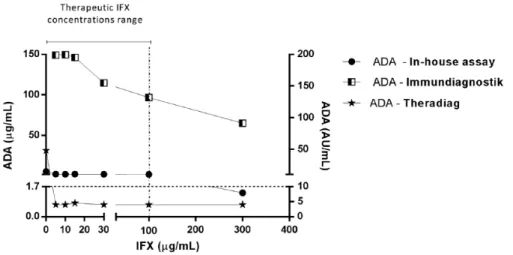
Figure 5 shows exogenous IFX interference in sera samples with intermediate levels of ADAs. The presence of exogenous IFX decreased ADA detection in Theradiag and in-house with the
Table 5. Agreement between the three assays for all combinations of IFX/ADAs status (clinical approach).
Agreement
Accuracy KAPPA
[95% CI] In-house assay versus Theradiag
assay(a) Theradiag assay versus In-house assay
(b)
IFX + ADA–
100% (n = 35) IFX + ADA +0% (n = 11) IFX + ADA –77.8% (n = 45) IFX + ANTI +0% (n = 3) 76% 0.620[0.489–0.751] IFX - ADA –
62.5% (n = 8) IFX – ADA +79.2% (n = 24) IFX - ADA –55.6% (n = 9) IFX – ADA+90.5% (n = 21) In-house assay versus
Immundiagnostik(c) Immundiagnostik assay versus In-house assay(d) IFX + ADA –
83.8% (n = 37) IFX + ADA +54.5% (n = 11) IFX + ADA –88.6% (n = 35) IFX + ADA +60% (n = 10) 81% 0.716[0.591–0.841] IFX - ADA –
62.5% (n = 8) IFX – ADA +95.7% (n = 23) IFX - ADA –62.5% (n = 8) IFX – ADA +84.6% (n = 26) Immundiagnostik assay versus
Theradiag assay(e) Theradiag assay versus Immundiagnostik(f) IFX + ADA –
100% (n = 34) IFX + ADA +0% (n = 10) IFX + ADA –75.6% (n = 45) IFX + ADA +0% (n = 3) 76% 0.625[0.491–0.758] IFX - ADA –
66.7% (n = 6) IFX – ADA +76.9% (n = 26) IFX - ADA –50% (n = 8) IFX – ADA +100% (n = 20)
(a) In-house assay was considered the reference assay and it was evaluated whether Theradiag confirmed the double status result. (b) Theradiag assay was considered the reference assay and it was evaluated whether in-house confirmed the double status result. (c) In-house assay was considered the reference assay and it was evaluated whether Immundiagnostik confirmed the double status result. (d) Immundiagnostik assay was considered the reference assay and it was evaluated whether in-house confirmed the double status result. (e) Immundiagnostik assay was considered the reference assay and it was evaluated whether Theradiag confirmed the double status result. (f) Theradiag assay was considered the reference assay and it was evaluated whether Immundiagnostik confirmed the double status result.
lower concentration (5 µg/ml) of exogenous IFX. For Theradiag this decrease led to the inability to detect ADAs and consequently it was impossi-ble to determine the IC50. In-house showed an IC50 of 0.88 µg/ml while Immundiagnostik showed a higher value of 140.1 µg/ml. At 300 µg/ml the interference was stronger in Theradiag (97%) while in in-house detection decreased by 74%. In-house failed to detect ADAs at IFX concentrations of 300 µg/ml, which are above the therapeutic levels achieved in vivo. Above 30 µg/ml IFX the Immundiagnostik assay was influenced by an additive concentration effect but was able to detect ADAs even at the higher concentration (300 µg/ml) of exogenous IFX (total decrease of 60%).
Figure 3. Stratified anti-infliximab antibodies (ADA)
levels in the presence of exogenous infliximab (IFX), evaluated by in-house assay. Therapeutic IFX concentrations range was considered between 0 and 100 µg/ml [Adedokun et al. 2014].
Figure 4. High anti-infliximab antibodies (ADAs) levels in the presence of exogenous infliximab (IFX) evaluated
by in-house, Immundiagnostik and Theradiag assay. Therapeutic IFX concentrations range was considered between 0 and 100 µg/ml [Adedokun et al. 2014].
Figure 5. Intermediate anti-infliximab antibodies (ADAs) levels in the presence of exogenous infliximab (IFX)
evaluated by in-house, Immundiagnostik and Theradiag assay. Therapeutic IFX concentrations range was considered between 0 and 100 µg/ml [Adedokun et al. 2014].
IgG4 ADAs
In order to evaluate the influence of IgG4 ADAs, samples positive for ADAs were tested for the presence of IgG4. IgG4 ADAs were found in 54.8% of the ADA-positive samples. Figure 6 shows the levels of IFX and IgG4 in samples that were negative with Theradiag but ADA-positive for both the in-house and Immundiagnostik assays. Of the eight samples negative for antibod-ies with Theradiag, five presented positive levels of IFX and three presented IgG4 ADAs.
Discussion
Several methodologies are available for use in drug monitoring. However, different methodologies return different results. There is still little informa-tion comparing different assays, in particular for ADA detection, which is susceptible to interfer-ence in the presinterfer-ence of detectable levels of IFX. The aim of this study was to evaluate and com-pare three different assays for detecting IFX and
ADAs and to clarify the importance of the pres-ence of detectable levels of IFX on the accuracy of the ADA assays.
Cutoffs for a qualitative evaluation of the assays are difficult to establish, particularly for ADAs assays. In fact, ADAs results are expressed in dif-ferent units by the difdif-ferent assays. In addition, Immundiagnostik defines a cutoff for positivity that is different from the limit of detection and not suggested by all commercial assays. The manufac-turers’ instruction for Theradiag do not contain any suggestion for a cutoff for ADAs positivity besides the limit of detection of 0.01 µg/ml. Van Schouwenburg and colleagues have shown that BE assays (Theradiag methodology) performance is depend not only of the amount of antibody in the serum but also of the affinity of ADAs to the assay [Van Schouwenburg et al. 2016]. This indicates a possible source of intervariability of the assay, namely for values near the limit of detection. In fact, our results show that with Theradiag 70% of the values considered to be positive, with analytical approach, are between 0.01 and 0.04 µg/ml. This might be a reflection of the intervariability of the assay, rather than a faithful quantification of the presence of antibodies. Taking this into considera-tion, we have decided to perform two different approaches in the qualitative analyses of the results: an analytical one, in which cutoffs were defined by the manufacturers’ instructions; and a clinical one, in which the cutoffs were defined according to the literature.
From an analytical perspective, IFX assays showed a substantial agreement and a good accuracy, although the in-house and Theradiag methods had only a moderate agreement. On the other hand, upon applying a cutoff of 1 µg/ml, the behavior of the three assays is very similar.
Table 6. Infliximab (IFX) inhibition effect on ADAs at intermediate and high levels in different assays.
IC50 (µg/ml) Intermediate levels
3 µg/ml > ADA <10 µg/ml High levelsADA >25 µg/ml
In-house (AHLC) 0.88 143.60
Immundiagnostik (SPFE) 140.10 NA
Theradiag (BE) NA 74.89
IC50, half maximal inhibitory concentration; NA, IC50 value not determined because either 50% inhibition fell outside the
highest concentration or because inhibition was 100% above the lowest concentration of IFX; ADA, infliximab anti-bodies; AHLC, anti-human lambda chain; SPFE, semi-fluid phase enzyme; BE, bridging enzyme-linked immunosorbent assay.
Figure 6. Levels of IFX and IgG4 in samples that were
ADA-negative in Theradiag and ADA-positive in both the in-house and Immundiagnostik assays.
This suggests that using the limit of detection as a cutoff may decrease the quality agreement between assays.
The behavior of the three assays in terms of IFX determination was very similar. In fact, an accu-racy of over 92% for IFX was observed when ana-lyzing the agreement between the three assays. An agreement of 100% (data not shown) was observed between the in-house and Immundiagnostik assays for IFX-negative sam-ples, while the Theradiag assay identified four IFX-positive samples that the other two assays returned as negative. Casteele and colleagues also evaluated three IFX assays based on the sandwich ELISA principle and found good correlation between them [Casteele et al. 2012]. The authors used the same commercial kit as used here (Theradiag). In fact, Theradiag returned 11 posi-tive samples that were negaposi-tive according to the other two assays, showing that this assay can detect nonspecific binding. Overall, high titers in one assay were also high in the other two assays. Nevertheless, the Immundiagnostik and in-house assays were able to detect higher levels of IFX in sera samples, which may indicate the use of a more specific anti-human IgG antibody in the detection step in these assays than in Theradiag. The agreement between the three assays seemed to be more qualitative than quantitative. The three methodologies use the same ELISA sand-wich principle, but there are some idiosyncratic differences that can explain the small discrepan-cies in quantitative evaluation: different detection limits and test sensitivity; reagent stability; differ-ent calibration standards and curve adjustmdiffer-ent; and different sample dilution, manipulation and processing (e.g. different incubation times). Nevertheless, from a clinical point of view, the information given by the three assays (presence of positive/negative levels; low/therapeutic/high lev-els of the drug) showed acceptable interassay agreement. However, clinicians should be aware that changing drug level assays during patient fol-low up could induce errors in the interpretation of results and subsequent therapeutic strategies. ADAs were evaluated using three methodologies: in-house AHLC, Immundiagnostik SFPE and Theradiag BE. These three assays have different methodological principles. The in-house AHLC is a sandwich ELISA that uses anti-human lambda chain conjugate antibody in the detection step, benefitting from IFX kappa chain exclusive composition [Kopylov et al. 2012]. The Theradiag
BE uses a double-antigen bridge: ADAs create a bridge between IFX immobilized on the plate and IFX enzyme-linked conjugate. The Immundiagnostik SFPE uses an initial acid buffer treatment to dissociate the IFX–ADA immune complexes and two IFX conjugates with different types of conjugates. Acidified samples are incu-bated with an IFX–biotin conjugate, which immobilizes ADAs to the plate, whilst an IFX– peroxidase conjugate is used for detection. A total of 79 samples of sera were compared using the three assays. The analytical approach showed the same moderate agreement for the three assays than the clinical approach. However, and from the analytical perspective, Theradiag was able to detect 37 positive ADAs, whereas from a clinical perspective only 24 patients were considered to be positive for ADAs. Conversely, the in-house assay detected almost the same number of ADA-positive patients with the two approaches (34 for analytical versus 37 for clinical). The results obtained using a clinical perspective with the Theradiag kit suggest a high rate of false-negative outcomes. This high prevalence of false-negative outcomes with Theradiag was reported previ-ously by other authors [Imaeda et al. 2012; Steenholdt et al. 2013]. Kopylov and colleagues reported that BE was unable to detect ADAs in the presence of IFX [Kopylov et al. 2012]. Our results showed that 22 out of 24 ADA-positive samples were IFX-negative. Only three patients showed a double-positive status with Theradiag, however neither was confirmed as IFX-positive in the in-house or Immundiagnostik assay. These samples were probably false positive for IFX. In fact, the disagreement of the in-house and Immundiagnostik assay with Theradiag for dou-ble-positive status (IFX+/ADAs+) was 100%. Since Theradiag uses labeled IFX as the detec-tion antibody, the presence of IFX may compete with the detection of the ADAs. On the other hand, the BE is unable to detect monovalent IgG4 ADAs, contributing to the false-negative results. Our results were consistent with these two statements and showed that samples positive for ADAs with the in-house and Immundiagnostik assays, but negative with Theradiag, one of two situations occurred: (i) sera samples were IFX-positive; or (ii) ADAs were IgG4 antibodies. The inability to detect antibodies in the presence of the drug is important as two recent studies have shown that patients with IFX levels >3 µg/ml but with positive ADA have significantly higher levels of C-reactive protein (CRP) and less mucosal
healing [Casteele et al. 2015; Yanai et al. 2015], indicating a reduced control of inflammation mediated by these antibodies even when drug lev-els are adequate.
In order to understand the influence of drug pres-ence on ADA detection, we designed an experi-ment using IFX-negative sera that were incubated with different concentrations of exogenous IFX. The in-house assay was slightly affected by 5 µg/ml exogenous infliximab but was able to detect anti-bodies up to 100 µg/ml of IFX. Kopylov and col-leagues described the same drug concentration dependency for the AHLC assay [Kopylov et al. 2012]. However, our results permit us to define which concentration of IFX decrease the assay capacity to determine ADAs. The Immundiagnostik assay showed the best behavior in the presence of exogenous IFX. Interference was observed above 100 µg/ml of exogenous IFX but the assay was able to detect ADAs even at the higher concentration of 300 µg/ml of IFX. However, the results obtained in sera with high levels of ADAs were surprising and to the best of the authors’ knowledge have not been reported previously. Even the Theradiag BE, which is widely described as being unable to detect antibodies in the presence of the drug, was able to identify ADA-positive samples in the presence of high levels of exogenous IFX. The results indi-cated that assays are not only limited by the levels of drug in the sera but also by the concentration of ADAs. These phenomena could be explained as follows: (i) in Theradiag, ADAs bind to all free drug in serum but there is still enough ADAs to bind to the plated IFX and bridge it to the conju-gated one; (ii) in Immundiagnostik high levels of ADAs are most likely to diminish free IFX interference.
In summary, for IFX drug level determination, the three methodologies are equivalent, however the agreement between them seems to be more qualitative than quantitative. Regarding ADAs, despite being equally effective, the three assays show significant differences. Disagreement increased when samples had a double-positive or double-negative status, probably related to the specific limitations of each assay. The choice of cutoff to discriminate positive versus negative also enhances disagreement. This led us to define two different approaches with different cutoffs and present both evaluations. Clinical approach seems to highlight inabilities of the assays. In fact, Theradiag showed inability to detect positive ADA levels in samples with IFX (<5 µg/ml) or a
high percentage of IgG4 ADAs. IgG4 ADAs are monovalent and unable to create a bridge with the labeled IFX used in detection step. In contrast, both the in-house assay and the Immundiagnostik assay are able to detect ADAs in the presence of IFX up to levels of >100 µg/ml which conform with the therapeutic concentrations observed in patients receiving IFX. Unexpectedly, in samples with high levels of ADAs (>25 µg/ml), the pres-ence of IFX was not as important and even Theradiag was able to detect antibodies. Nevertheless, Immundiagnostik was least affected by IFX, followed by the in-house assay. In conclu-sion, our results indicated that assays are not only limited by the levels of drug in the sera but also by the concentration of ADAs.
Clinicians must be aware when optimizing treat-ment that binary (IFX/ADAs) stratification of results as positive and negative can differ accord-ing to the assay used. There is a strong agreement between assays for IFX+/ADAs- and IFX-/ ADAs+ status. The choice of assay will probably have little influence on therapeutic decisions to change the class of drug (IFX+/ADA-) or change anti-TNFα antibody (IFX-/ADAs+). However, erroneous therapeutic decisions may occur when patients show double-negative or double-positive status, since agreement between assays is signifi-cantly lower in these circumstances. Treatment intensification (IFX-/ADAs-) and change of drug class or concomitant use of immunomodulators (IFX+/ADAs+) should take into account the fact that the results are assay dependent.
Acknowledgements
We thank Sandra Dias for her contribution as the GEDII coordinator.
Joana Afonso performed all laboratory experi-ments and participated in the data analysis, inter-pretation and drafting of the manuscript. Susana Lopes, Raquel Gonçalves, Paulo Caldeira, Paula Lago, Helena Tavares de Sousa, Jaime Ramos, Ana Rita Gonçalves, Paula Ministro, Isadora Rosa, Ana Isabel Vieira, Rosa Coelho, Patrícia Tavares, João-Bruno Soares, Diana Carvalho, Paula Sousa and Tânia Meira participated in patient recruit-ment and data collection. Filipa Silva Ferreira par-ticipated in data analysis and interpretation. Claudia Camila Dias performed the statistical analysis and participated in data analysis and inter-pretation. Yehuda Chowers and Shomron Ben-Horin revised the manuscript critically for important intellectual content. Fernando Magro conceived and designed the study, participated in
data analysis, interpretation and drafting of the manuscript, and revised it critically for important intellectual content. All authors read and approved the final version of the manuscript.
Funding
This work was supported by the Portuguese Group of Studies in Inflammatory Bowel Disease (GEDII), Portugal, and partially by the European Regional Development Fund (ERDF); COMPETE (Operational Programme ‘Thematic Factors of Competitiveness) and Fundação para a Ciência e a Tecnologia, Portugal (FCT) [grant number EXPL/IVC-PEC/1302/2013].
Conflict of interest statement
The authors declare that there is no conflict of interest.
References
Adedokun, O., Sandborn, W., Feagan, B., Rutgeerts, P., Xu, Z., Marano, C. et al. (2014) Association between serum concentration of infliximab and efficacy in adult patients with ulcerative colitis.
Gastroenterology 147: 1296.
Baert, F., Noman, M., Vermeire, S., Van Assche, G., D’haens, G., Carbonez, A. et al. (2003) Influence of immunogenicity on the long-term efficacy of infliximab in Crohn’s disease. N Engl J Med 348: 601–608. Ben-Horin, S., Yavzori, M., Katz, L., Kopylov, U., Picard, O., Fudim, E. et al. (2011) The immunogenic part of infliximab is the F(Ab ‘)(2), but measuring antibodies to the intact infliximab molecule is more clinically useful. Gut 60: 41–48.
Bendtzen, K. (2013) Personalized medicine: theranostics (therapeutics diagnostics) essential for rational use of tumor necrosis factor-alpha antagonists. Discov Med 83: 201–211.
Bendtzen, K. and Svenson, M. (2011) Detection and
Quantification of Antibodies to Bipharmaceuticals: Practical and Applied. Chichester: John Wiley & Sons Ltd.
Casteele, N., Buurman, D., Sturkenboom, M., Kleibeuker, J., Vermeire, S., Rispens, T. et al. (2012) Detection of infliximab levels and anti-infliximab antibodies: a comparison of three different assays.
Aliment Pharmacol Therapeut 36: 765–771.
Casteele, N., Khanna, R., Levesque, B., Stitt, L., Zou, G. Singh, S. et al. (2015) The relationship between infliximab concentrations, antibodies to infliximab and disease activity in Crohn’s disease. Gut 64: 1539–1545.
Imaeda, H., Andoh, A. and Fujiyama, Y. (2012) Development of a new immunoassay for the accurate
determination of anti-infliximab antibodies in
inflammatory bowel disease. J Gastroenterol 47: 136–143. Kopylov, U., Mazor, Y., Yavzori, M., Fudim, E., Katz, L. Coscas, D. et al. (2012) Clinical utility of antihuman lambda chain-based enzyme-linked immunosorbent assay (ELISA) versus double antigen ELISA for the detection of anti-infliximab antibodies.
Inflamm Bowel Dis 18: 1628–1633.
Landis, J. and Koch, G. (1977) Measurement of observer agreement for categorical data. Biometrics 33: 159–174.
Roblin, X., Rinaudo, M., Del Tedesco, E., Phelip, J., Genin, C., Peyrin-Biroulet, L. et al. (2014) Development of an algorithm incorporating pharmacokinetics of adalimumab in inflammatory bowel diseases. Am J Gastroenterol 109: 1250–1256. Steenholdt, C. (2013) Use of infliximab and anti-infliximab antibody measurements to evaluate and optimize efficacy and safety of infliximab maintenance therapy in Crohn’s disease. Danish Med J 60(4): B4616.
Steenholdt, C., Ainsworth, M., Tovey, M., Klausen, T., Thomsen, O., Brynskov, J. et al. (2013)
Comparison of techniques for monitoring infliximab and antibodies against infliximab in Crohn’s disease.
Ther Drug Monitor 35: 530–538.
Steenholdt, C., Bendtzen, K., Brynskov, J., Thomsen, O. and Ainsworth, M. (2014a) Clinical implications of measuring drug and anti-drug antibodies by different assays when optimizing infliximab treatment failure in Crohn’s disease: post hoc analysis of a randomized controlled trial. Am J Gastroenterol 109: 1055–1064. Steenholdt, C., Brynskov, J., Thomsen, O., Munck, L., Fallingborg, J., Christensen, L. et al. (2014b) Individualised therapy is more cost-effective than dose intensification in patients with Crohn’s Disease who lose response to anti-TNF treatment: a randomised, controlled trial. Gut 63: 919–927.
Ungar, B., Anafy, A., Yanai, H., Ron, Y., Yavzori, M., Picard, O. et al. (2015) Significance of low level infliximab in the absence of anti-infliximab antibodies.
World J Gastroenterol 21: 1907–1914.
Van Schouwenburg, P., Kruithof, S., Wolbink, G., Wouters, D. and Rispens, T. (2016) Using monoclonal antibodies as an international standard for the measurement of anti-adalimumab antibodies. J
Pharm Biomed Anal 120: 198–201.
Yanai, H. and Hanauer, S. (2011) Assessing response and loss of response to biological therapies in IBD.
Am J Gastroenterol 106: 685–698.
Yanai, H., Lichtenstein, L., Assa, A., Mazor, Y., Weiss, B., Levine, A. et al. (2015) Levels of drug and antidrug antibodies are associated with outcome of interventions after loss of response to infliximab or adalimumab. Clin Gastroenterol Hepatol 13: 522–530.
Visit SAGE journals online http://tag.sagepub.com