UNIVERSIDADE DA BEIRA INTERIOR
Ciências da Saúde
Gene Therapy
Development of a new nanocarrier system for
mitochondrial gene therapy
João Miguel Almeida Santos
Tese para obtenção do Grau de Mestre em
Ciências Biomédicas
(2º ciclo de estudos)
Orientador: Prof.ª Doutora Diana Costa
Co-orientador: Prof.ª Doutora Fani Sousa
The content of this work is the sole responsibility of the author:
____________________________________________________________
"The fundamental cause of trouble in
the world is that the stupid are
cocksure while the intelligent are full
of doubt."
Acknowledgments
Over a year has passed, one year of dedication to an MsC thesis that is finally coming to an end. What I feel proud about is not the work done but the experience and knowledge that I have gained during the past year. The truth is, not everything went according to planned and there were times when constant failures lead me to think that the best I could do was just give up. Yet I was able to reach the end thanks to the support I have been receiving from friends, family and colleagues and for those people I dedicate this page of my thesis.
I would like to thank my parents, Jorge and Laurinda, and my little brother André for believing in me and giving me the opportunity to get my MsC degree.
To both my supervisors, Prof.ª Diana Costa, for always being there when I had any doubt or problem and for never letting me go astray, and Prof.ª Fani Sousa, who even though could not always be in the University due to familiar issues, made sure to always be present and help even if from far away.
Then I would like to thank the members from Biotechnology research group that helped in any sort of way even if just for taking a simple doubt. They were important during the first few months of my work and there are some noteworthy people I would like to mention such as Profª Angela Sousa, Patricia Pereira and Diana Bicho for contributing to the development of the thesis.
Outside from research I would like to thank Engª Ana Duarte for accompanying me during the SEM images acquisition and Catarina Ferreira for accompanying me during the Confocal Microscopy visits.
The development of this thesis has some important results thanks to the CICECO research group from Universidade de Aveiro for whom I am really appreciated, as they gladly accepted and helped me even though they had no obligation. Although all group members deserve my appreciation, I would like to dedicate a great part of it to Profª Ana Luisa for being the one who accompanied during the few days I stayed there.
To all my friends for being present and giving me support with special thanks to Tiago Roxo and Julieta Oliveira who helped me a lot during the in vitro transfection tests.
To finish, I would like to dedicate this thesis to the person which I believe to be the most important one in my life and where I got most of my strength to never give up, my girlfriend Carina Tomás.
To all those people who may have contributed to my success and the success of this thesis I want you to know that I feel really appreciated and that none of this would ever had been possible without your help.
Resumo
As mitocôndrias são organelos únicos pois possuem o seu próprio genoma, o ADN mitocondrial (ADNmt). Apesar de bastante pequeno quando comparado com o ADN nuclear (ADNn), mutações ao nível do ADNmt são bastante frequentes devido à falta de mecanismos de protecção e de reparação. Como consequência, citopatias e doenças associadas à mitocôndria são bastante frequentes afectando essencialmente órgãos e tecidos onde existe muito dispêndio de energia como é o caso dos músculos e do cérbero. Logo, o desenvolvimento de um novo e eficiente protocolo para terapia génica mitocondrial (MGT) é visto como uma proposta aliciante.
Durante esta tese de Mestrado, tentamos criar um novo nanosistema que consiga entregar eficazmente ADN plasmídico (pDNA) à mitocôndria para que no futuro possa ser usado em terapia génica mitocondrial (MGT).
Assim, este projecto de investigação pode ser dividido em três etapas principais:
1. O isolamento e purificação de três plasmídeos (pUC19, pVAX1-LacZ e pcDNA3-myc-FLNa S2152A);
2. A síntese e caracterização de nanopartículas com afinidade para a mitocôndria; 3. O estudo da capacidade das nanopartículas efectuarem transfecção celular e
dirigirem-se à mitocôndria;
As nanopartículas desenvolvidas, através do método de co-precipitação oferecem-nos qualidades únicas como a sua biocompatibilidade, alta eficiência de encapsulamento de ADN e baixo custo de produção.
A transfecção celular foi alcançada com sucesso sendo que, tais resultados, podem contribuir em grandes avanços na correcção de defeitos mitocondriais, oferecendo-nos uma nova estratégia terapêutica no combate a diversas patologias desde o cancro, às doenças de Parkinson e Alzheimer.
Palavras Chaves
Resumo Alargado
As mitocôndrias são organelos únicos que possuem o seu próprio genoma, o ADN mitocondrial, uma molécula com dupla cadeia circular com aproximadamente 16.5 mil pares de base (kbp) e que codifica 13 polipéptidos, 2 ácidos ribonucleicos ribossomais (rARN) e 22 ácidos ribonucleicos de transferência (tARN).
Apesar de conhecidas como as principais geradoras de energia dos seres vivos, sobre a forma de ATP, as mitocôndrias são também importantes por desempenharem outras funções relevantes para o bem-estar do organismo como a produção e regulação de espécies reactivas de oxigénio, regulação da entrada de cálcio na célula e apoptose.
Ainda que o ADNmt pareça insignificante quando comparado com o DNA nuclear (ADNn) a verdade é que mutações ao nível do ADNmt são bastante frequentes e estima-se que este seja, pelo menos, 10 vezes mais mutável que o nuclear. Tal discrepância é essencialmente devido à falta de mecanismos de protecção e reparação, onde o único conhecido até ao momento é o reparo por excisão de bases, “Base Excision Repair” (BER).
Assim, torna-se preponderante a investigação de novas formas terapêuticas tendo como alvo as doenças mitocondriais e, em especial, a manutenção da integridade do genoma mitocondrial. Pondera-se cada vez mais a incorporação da terapia génica como uma abordagem adequada a deficiências mitocondriais, surgindo assim a terapia génica mitocondrial (MGT). Para o sucesso desta terapia, é necessário o desenho de um vector adequado que seja capaz de incorporar um tamanho ilimitado de ADN inserido, seja facilmente produzido e em larga escala, pudesse ser direccionado para organelos celulares específicos, não permitisse a replicação autónoma do ADN e pudesse garantir uma expressão génica a longo prazo. Adicionalmente, o vector seria biocompatível e não-imunogénico.
Apesar do sucesso desta terapia ao nível nuclear, os avanços na área mitocondrial têm sido escassos. Alguns progressos importantes na formulação de um vector baseado em ADN mitocondrial foram reportados, mas contudo, apresentando limitações ao nível da transfecção celular.
Tendo em conta esta lacuna e a pouca investigação centrada na terapia génica mitocondrial, o objectivo desta tese passa por desenvolver um novo sistema à escala nano para a entrega de ADN plasmídico (ADNp) à mitocôndria numa abordagem terapêutica inovadora para tratamento de doenças mitocondriais.
Este projecto de investigação inclui 3 etapas fundamentais: 1. Isolar e purificar três plasmídeos com diferentes tamanhos;
2. Desenvolver e caracterizar novas nanopartículas com características adequadas para uso em terapia génica;
Foram amplificados, numa cultura de Escherichia coli, e isolados com sucesso três plasmídeos (pUC19, pVAX1-LacZ e pcDNA3-myc-FLNa S2152A) com recurso ao kit Qiagen.
Procedemos em seguida à criação de nanopartículas contendo rodamina123 (rho123). Estas nanopartículas têm como ideia a encapsulação de ADN plasmídico (ADNp) e rho123 através de um simples método de co-precipitação de carbonato de cálcio (CaCO3). Criaram-se dois protocolos, destinados à entrega de 1µg e 10µg de pDNA. Procedemos, de seguida, à sua caracterização de modo a averiguar qual o mais eficiente para futuros estudos.
A caracterização envolveu a identificação da morfologia através da microscopia electrónica de varrimento (SEM), análise da capacidade de encapsulamento de ADNp por parte das nanopartículas, determinação do tamanho médio das nanopartículas, do potencial ζ e do poder protector das nanopartículas perante a presença de nucleases.
Por último, procedemos ao crescimento de uma cultura celular de fibroblastos humanos, avaliámos a viabilidade celular através do ensaio MTT e procedemos à avaliação in
vitro da eficiência de transfecção por microscopia confocal.
As nanopartículas apresentam uma forma praticamente circular e um potencial ζ entre os valores de -8mv e -10mv para ambos os sistemas. No caso do sistema de 1µg, a percentagem de encapsulamento varia entre os 50% e os 60%, o tamanho médio entre os 300nm e os 400nm e as nanopartículas encontram-se bastante degradadas na presença de 5µg/ml de DNAse. Nos sistemas de 10µg, a percentagem de encapsulamento varia entre os 55% e os 70%, o tamanho médio entre os 350nm e os 550nm e as nanopartículas só começam a degradar-se para valores superiores a 10µg/ml de DNAse. Quando comparados, os resultados obtidos durante o teste da protecção sugerem que o sistema de 10µg seja o mais indicado para fins terapêuticos.
A transfecção e transporte para a mitocôndria foi observada com sucesso através de estudos de microscopia confocal. Apresentamos deste modo um novo método para a entrega de ADNp à mitocôndria que apresenta vantagens únicas em aspectos como: encapsulamento, biocompatibilidade e custo de produção.
O trabalho desenvolvido ao longo desta tese pode contribuir fortemente para o sucesso da terapia génica mitocondrial (MGT) e talvez num futuro próximo, as nanopartículas desenvolvidas possam oferecer uma nova estratégia terapêutica no combate a diversas patologias, das quais, se destacam as doenças de Parkinson e Alzheimer consideradas grandes flagelos da vida moderna.
Abstract
Mitochondria are unique organelles that have their own genome, the mitochondrial DNA (mtDNA). Although quite small compared to nuclear DNA (nDNA), mutations in mtDNA are quite frequent due to the lack of protection and repair mechanisms. Per consequence, cytopathies and diseases are quite common and mostly associated with high energy demanding tissues such as muscles and the brain. Therefore, the development of a new and efficient mitochondrial gene therapy protocol is seen as a promising approach.
During this MSc thesis we try to bring together a new nanocarrier system with the ability to deliver plasmid DNA into the mitochondria, for future application in mitochondrial gene therapy (MGT).
Hence, the development of this research project can be divided itself into three main stages:
1. Isolation and purification of three plasmid DNAs (pUC19, pVAX1-LacZ and pcDNA3-myc-FLNa S2152A);
2. Synthesis and characterization of nanoparticles with mitochondria affinity; 3. In vitro study of mitochondrial transfection ability.
The newly developed nanoparticles, created through a co precipitation method, offer us unique features such as: biocompatibility, plasmid DNA (pDNA) encapsulation efficiency and low manufacturing cost.
We were able to successfully achieve transfection into the mitochondria which may result in a huge step in the correction of mitochondrial defects, offering new therapeutic strategies for a variety of pathologies ranging from cancer to Parkinson and Alzheimer´s diseases.
Keywords
Figure List
Figure 1 – Human Mitochondrial DNA. Figure 2 – mtDNA replication
Figure 3 – Electron transport chain of mitochondria
Figure 4 – Mitochondrial sources of ROS and mitochondrial ROS targets Figure 5 – Mitochondrial Ca transport pathways
Figure 6 – Apoptosis intrinsic pathway Figure 7 – Mitochondrial genetic bottleneck Figure 8 – Base Excision Repair
Figure 9 - Severe mitochondrial disease condition Figure 10 - The mitochondrial theory of aging Figure 11 – Transfection Schematic
Figure 12 – Mitochondria Specific Nanotechnology Figure 13 – plasmid UC19 mapping
Figure 14 – plasmid VAX1-LacZ mapping
Figure 15 – plasmid cDNA3-myc-FLNa S2152A mapping Figure 16 - CaCO3-pDNA-Rho123 nanoparticles synthesis. Figure 17 – CaCO3 co-precipitation
Figure 18 – pUC19 Nanoparticles Morphology Figure 19 – pVAX1-LacZ Nanoparticles Morphology
Figure 20 – pcDNA3-myc-FLNa S2152A Nanoparticles Morphology
Figure 21 – Encapsulation Efficiency of CaCO3-pDNA-Rho123 nanoparticles Figure 22 – CaCO3-pDNA-Rho123 nanoparticles Size
Figure 23 - Understanding nanoparticles ζ potential Figure 24 – ζ Potential of CaCO3-pDNA-Rho123 nanoparticles
Figure 25 – Agarose gel electrophoresis of pUC19 nanocarriers followed by incubation with DNAse I for 1h at 37ºC.
Figure 26 – Agarose gel electrophoresis of pVAX1-LacZ nanocarriers followed by incubation with DNAse I for 1h at 37ºC.
Figure 27 – Agarose gel electrophoresis of pcDNA3-myc-FLNa S2152A nanocarriers followed by incubation with DNAse I for 1h at 37ºC.
Figure 28 - Cell Viability after incubation of fibroblast cells with CaCO3-pDNA-Rho123 nanoparticles for all three pDNA, for 24 and 48 hours.
Table List
Table 1 - Pathology associated to mitochondrial genome Table 2 – Confocal Microscopy Probes
Lista de Acrónimos
AP APE ATP BER Ca2+ CaCl2 CaCO3 CARD Caspase Cl- Co3 2-DA DNA DMEM DQAsomes EE H2O2 IMM IMS kbp kDA KSS LHON LP-BER LS MELAS MERRF MIDD MMR MOMP mtBER mtDNA Apurinic/apyrimidinic Apurinic/apyrimidinic endonucleases Adenosine triphosphateBase excision repair Calcium
Calcium Chloride Calcium Carbonate
Caspase Recruitment Domain
Cysteinyl aspartate-specific proteases Chloride
Carbonate Dalton
Deoxyribonucleic acid
Dulbecco’s Modified Eagle Medium DeQuAlinium-based liposome-like vesicles Encapsulation Efficiency
Hydrogen Peroxide
Inner mitochondrial membrane Inner mitochondrial space Kilobase Pair
KiloDalton
Kearns-Sayre syndrome
Leber hereditary optic neuropathy Long-pathway BER
Leigh syndrome
Mitochondrial encephalopathy, lactic acidosis, and stroke-like episodes Myoclonic Epilepsy with Ragged Red Fibers
Maternally inherited diabetes and deafness Mis-match Repair
Mitochondria Outer Membrane Permeabilization Mitochondrial base excision repair
NARP nDNA NER O2 •-OC OMM OXPHOS pDNA polγ PS PFA PTP Rho123 RNA ROS
Neuropathy, ataxia, retinitis pigmentosa Nuclear DNA
Nucleotide excision repair Superoxide anion
Open Circular
Outer mitochondrial membrane Oxidative Phosphorylation Plasmid DNA
Polymerase Gamma Pearson syndrome Paraformaldehyde
Permeability Transition Pore Rhodamine123
Ribonucleic acid
Reactive oxygen species rRNA SC SEM SOD SP-BER TEM Ribosomal RNA Supercoiled
Single Electron Microscopy Superoxide Dismutase Short-pathway BER
Transmission Electron Microscopy tRNA Transfer RNA
Índex
Introduction ... 1
1.Understanding the mitochondria ... 2
1.1.Functions of mitochondria ... 4
1.1.1.Generation of energy ... 4
1.1.2.Generation and regulation of ROS ... 5
1.1.3.Calcium Regulation ... 6
1.1.4.Regulate Apoptose ... 7
2.Maintaining mtDNA Integrity ... 8
2.1.Inheritance and clonal expansion ... 9
2.2.Repair mechanisms of the mitochondria ... 10
2.3.Mutations and Diseases ... 12
2.3.1.Cytopathies ... 13
2.3.2.Mutations and Mitochondrial Diseases ... 15
2.3.3.Aging ... 15
3.Mitochondrial Gene Therapy ... 17
3.1.Non-Viral Gene Therapy ... 19
3.2.Traversing Mitochondrial Membrane ... 20
3.3.Nanotechnology ... 21
3.4.Salt as an efficient nanocarrier ... 22
Aims of the Project ... 24
Materials and Methods ... 25
1.Materials ... 25
1.1.Reagents ... 25
1.2.Plasmid ... 25
2.Methods ... 27
2.1.Bacterial Growth and Plasmid Purification ... 27
2.2.Agarose Gel Electrophoresis ... 27
2.6.Nanoparticles Size ... 28
2.7.ζ Potential ... 29
2.8.Protection Test ... 29
2.9.Cell Culture Growth and Maintenance ... 29
2.10.Cell Viability ... 30
2.11.In Vitro Transfection ... 31
2.12.Statical Analysis ... 31
Results ... 32
1.Plasmid Purification and Amplification ... 32
2.Synthesis of CaCO
3-pDNA-Rho
123Nanoparticles ... 32
3.Scanning Electron Microscopy ... 34
4.Encapsulation Efficiency ... 36
5.Nanoparticles Size ... 37
6.ζ Potential ... 37
7.Protection Test ... 39
8.MTT Assay ... 41
9.Confocal Microscopy ... 42
Discussion ... 45
Conclusions and Future Perspectives ... 48
Introduction
The human genome is the complete set of human genetic information stored within the 23 chromosome pairs of the cell’s nucleus but also the small closed double-stranded circular molecule situated inside the mitochondria, the mitochondrial DNA (mtDNA). Responsible for encoding 13 proteins as well as 2 ribosomal RNA (rRNA) and 22 transfer RNA (tRNA) (1), damage to mtDNA ends up affecting high energy-demanding tissues like brain, heart and muscles.
In recent reports, it was shown that 1 in 4 healthy individuals inherit a mixture of wild-type and variant mtDNA, process known as heteroplasmy (2). Even less frequently, less than 1 in 200 inherit a potentially pathogenic variant of mtDNA (3) whereas 1 in 10.000 manifest clinically a mtDNA disease (4). It is presumed that the mutation rate of mtDNA is at least 10 times bigger than nuclear DNA (nDNA), leading to a wide range of metabolic and neuromuscular syndromes. In addition, mutations and/or polymorphism variance in mtDNA genes are related with Parkinson and Alzheimer’s diseases, diabetes and a greater susceptibility to develop cancer.
Gene therapy consists of the introduction of genetic material into cells for a therapeutic purpose with the objective of correcting or treating a deficient gene responsible for a disease (5). Over the last decades, gene therapy has had remarkable advances becoming a feasible treatment option (6). With such achievements, new strategies and shifts should be made to further improve the conventional therapy systems.
The first reference to gene therapy was presented in 1963, when Joshua Lederberg suggested the control of nucleotide sequences in human chromosomes, coupled with selection and integration of the desired genes (7). This idea ended up being accomplished only in 1980 with the introduction of two functional genes into mammalian cells (8). Virus-based vectors were once the most commonly used gene delivery systems in gene therapy because of their highly efficient infection rate and ability to integrate therapeutic genes into the host chromosome to ensure stable and long term gene expression. However, the initial enthusiasm for the use of viruses in gene therapy has diminished due to its lack of safety associated with fatal adverse effects reported in previous studies (9). Meanwhile clinical trials, concerning the usage of non-viral vectors, has been constantly growing day-by-day due to its improved safety features, high cell viability and rapid manufacturing timeline (10).
The interest of incorporating gene therapy into mitochondria started with the observation of patients suffering from myopathy (11) and optic neuropathy (12), both studies observed for the first time mutations in mtDNA. The incorporation of a therapeutic vector, to
A successful mitochondrial gene therapy (MGT) would lead to the cellular uptake and intracellular targeting of a therapeutic nucleic acid or protein specifically to the mitochondrial compartment (13). The design and development of an adequate mitochondrial transfection vector is quite relevant and deserves more attention from scientific community once this first step can compromise the entire gene therapy operation.
Nanomedicine plays an important role in biotechnology with the creation of new gene delivery systems. Recent nanocarriers ensure protection against enzymatic degradation, are able to by-pass the innate immune system, have good biodistribution, reduced side-effects, safety, no toxicity, reduced inflammation and ensure the therapeutic payload to be released in the desired intracellular compartment (14).
1. Understanding the mitochondria
Mitochondria are membrane-enclosed organelles found in most eukaryotic cells that range from 0.5 to 1 micrometer (μm) in diameter. Responsible for producing most part of cell’s energy in the form of adenosine triphosphate (ATP), mitochondria play an essential role in the life cycle of the cell (15).
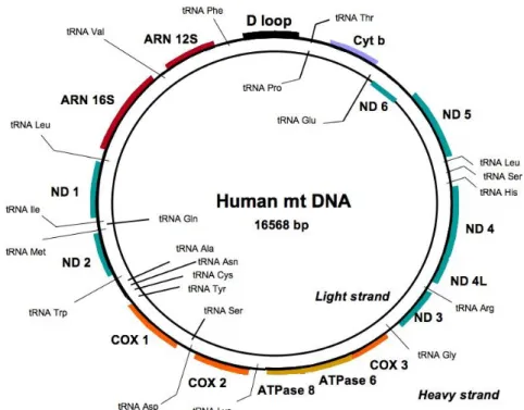
Human genetic information can be found in the form of nDNA but we should not neglect the existence of a second form of genome found inside the mitochondria, the mtDNA. Of nearly 17kb or 10.000 KDa, circular double-stranded mtDNA is present in hundreds to thousands of copies in each cell (16) (Figure 1). MtDNA consists predominantly of coding DNA, with the exception of the control region that has mainly regulatory functions.
A unique feature of mitochondria is the possession of proteins encoded from two separate genomes. While all mtDNA encoded proteins are mitochondria exclusive, most of mitochondrial proteins are nuclear encoded, synthesized in the cytosol and finally directed to the mitochondria by specific targeting sequences (17).
Figure 1. Human Mitochondrial DNA. Circular double-stranded molecule of 16,568 (bp) in length
consisting predominantly of coding DNA that codes for 13 polypeptides, 2 ribosomal RNAs (rRNAs), and 22 transfer RNAs (tRNAs) (Adapted from (18)).
The control region is a three stranded D-loop of nearly 600bp that promotes the origin of mtDNA replication. The leading strand has historically been termed Heavy (H) while, logically, the other strand was labeled as Light (L). The D loop is thus defined as a three-stranded structure with the nascent leading H strand defining the origin of leading-strand replication (OH) at its 5 ' end (19). DNA synthesis occurs unidirectionally, after the growing of H strand has elongated to two-thirds or more of its total length, the origin of lagging L-strand replication is exposed on the displaced parental H strand and initiation of daughter L-strand synthesis begins leading into two distinct progeny circles being segregated. This process is finalized by the synthesis of a new D loop (20).
1.1. Functions of mitochondria
Mitochondria are cytosolic double-membrane organelles that have been considered the powerhouse of the cell. Mitochondria, however, participate in a high number of other cellular processes such as ion homeostasis, redox signaling, apoptotic and necrotic cell death, as well as the control of cell cycle and cell growth (22, 23)
Even though the mitochondria contribute to a huge number of cellular processes, four crucial functions to the cell homeostasis are enlightened.
1.1.1. Generation of energy
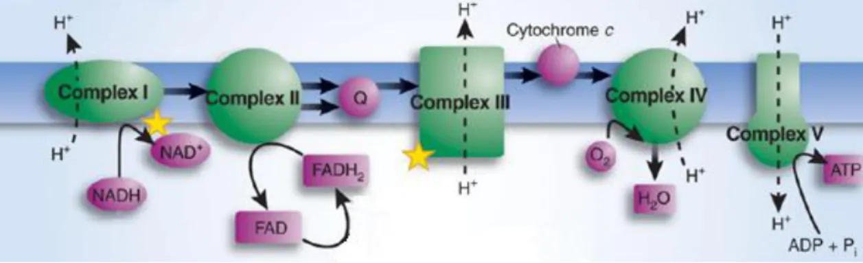
All biological and molecular events require energy to function properly. Energy is available in the form of ATP (adenosine triphosphate) which is mainly produced through aerobic cellular respiration of carbohydrate and glucose, which represent most of the source of biological energy of the human body. On the other side, reduced energy levels threaten cellular homeostasis and integrity (24). The principal source of ATP production is the oxidative phosphorylation (OXPHOS) which takes place in the mitochondria (25).
OXPHOS begins with the entry of electrons into the respiratory chain through complex I (NADH ubiquinone oxidoreductase) (26) or complex II (Succinate ubiquinone oxidoreductase) (27). Electrons from complex I or complex II are subsequently transferred to complex III (Ubiquinol cytochrome c oxidoreductase) then to cytochrome c, the second mobile electron carrier, and finally to complex IV (Cytochrome c oxidase). Complex IV is the terminal enzyme in the electron transfer chain reducing O2 to H2O by using the delivered electrons (28). This whole process ends up creating a membrane potential that promotes the conformational change of complex V (ATP synthase) resulting in the generation of ATP (29) (Figure 3).
All 13 polypeptides encoded by the mtDNA are subunits of the OXPHOS system: complex I (7 polypeptides), complex III (1 polypeptide), complex IV (3 polypeptides) and complex V (2 polypeptides) (30).
Figure 3. Electron transport chain of mitochondria. The function of the electron transport chain is to produce a transmembrane proton electrochemical gradient as a result of the redox reactions ending
with the production of ATP. (Adapted (31))
1.1.2. Generation and regulation of ROS
Reactive oxygen species (ROS), are oxygen derivatives, that are oxidized and easily converted into radicals (32). A wide range of mitochondrial ROS-induced damages have been described, which can lead, either individually or collectively, to a cellular energetic catastrophe
All the mitochondrial enzyme complexes can generate ROS or at least contribute to their appearance (33). ROS are produced by mitochondria during oxidative metabolism through the one-electron reduction of molecular oxygen (O2), forming superoxide anion (O2•-). Superoxide is the proximal ROS produced by mitochondria and is converted to hydrogen peroxide (H2O2) through the action of superoxide dismutases (SODs) both within the mitochondria and in the cytosol (34).
H2O2 generated in mitochondria may act as a signaling molecule in the cytosol (35) however another possibility is to infuse within the cell and be eliminated by cytosolic or mitochondrial antioxidant systems such as catalase, glutathione peroxidase, and thioredoxin peroxidase (36).
Figure 4. Mitochondrial sources of ROS and mitochondrial ROS targets. ROS generators (red) and ROS
targets (yellow) are present all over mitochondria. Aconitase and complex I through IV are both sources and targets of ROS. (Adapted from (37).
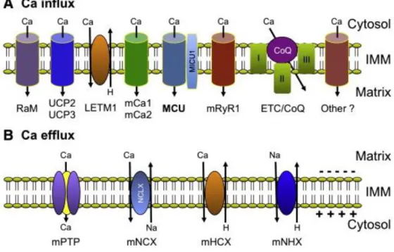
1.1.3. Calcium regulation
Calcium (Ca2+) is a highly versatile second messenger that controls critical cellular responses in all eukaryotic organisms (38). Mitochondria has the ability to act a Ca2+ buffer therefore having an important part in regulating cytosolic Ca2+ signals (39).
Ca2+ uptake by mitochondria alters the activity of mitochondria in multiple ways. An increase in the free Ca2+ concentration within the mitochondrial matrix results in the increase of [Ca2+]
mit which leads to a larger respiratory rate, H+ extrusion and ATP production. However, prolonged increases in [Ca2+]
mit can induce the opening of the mitochondrial permeability transition pore (PTP) leading to mitochondrial swelling, cytochrome C release, and cell death by apoptosis (40).
Figure 5. Mitochondrial Ca transport pathways. A) Mitochondrial Ca uptake and mechanisms and
pathways located at the IMM. B) Mitochondrial Ca extrusion mechanisms and pathways located at the IMM. (Adapted from (41))
1.1.4. Regulate Apoptosis
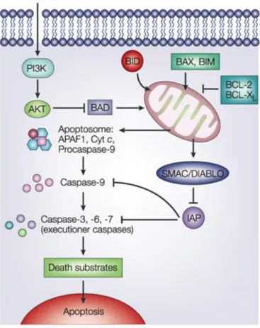
Apoptosis is a programmed form of cell death, characterized by the activation of cysteinyl aspartate-specific proteases (caspases) and the systematic breakdown of dying cells into easily phagocytized apoptotic bodies. (42, 43)
There are two alternative pathways that initiate apoptosis: one is mediated by death receptors on the cell surface (extrinsic pathway) while the other is mediated by the mitochondria (intrinsic pathway). The intrinsic pathway is activated in response to intracellular stressors, induced by several stimuli including DNA damage (44).
These stress signals trigger the mitochondria outer membrane permebilization (MOMP) resulting of the activation of certain proapoptotic BCL-2 family members (45). MOMP then facilitates the release of cyt c which interacts with Apaf-1 stimulating it into a caspase-activating complex known as the “Apaf-1 apoptosome”. The apoptosome subsequently recruits the initiator procaspase-9 through caspase recruitment domains (CARDs) present in the N-termini of both Apaf-1 and procaspase-9. Once bound, active caspase-9 then processes the effector caspase-3 and induces death (46).
Figure 6. Apoptosis intrinsic pathway. Activation of apoptosis starts with MOMP, generally as a result
of the activation of pro-apoptotic members. MOMP then facilitates the release of several pro-apoptotic factors from the mitochondria into the cytoplasm where they promote cell death (47).
2. Maintaining mtDNA integrity
Mammals normally inherit their mtDNA just prior to fertilization (48) and immediately after mtDNA replication (49). Such method of inheritance supposedly tend to make copies of identical mtDNA, (homoplasmy) (50). However, recent studies reveal that is not unusual for two different copies of mtDNA, wild-type and mutated, to be present inside of the mitochondria, process known as heteroplasmy (2, 51).
MtDNA is highly mutable due to the limited mtDNA protection and repair mechanisms but also because of the proximity to the electron transport chain, ROS formation site and even nuclear genome (52) which may contribute to the high diversity in the levels of mutated mtDNA. However, that does not explain the high rate of mutation and the high number of mutated mtDNA copies present in some populations. Furthermore, it is quite hard to explain why a woman with two children, one is healthy, while the other infant may have a devastating and fatal disorder.
2.1. Inheritance and clonal expansion
It is widely accepted that in the cells of most animals, mtDNA is inherited solely from the mitochondria of the oocyte, making the inheritance, with few exceptions, maternal. The maternal mode of mtDNA transmission gives rise to a genetic asymmetry between females and males, whereas the nuclear genome is inherited in equal measure through males and females, enabling selection to act on male and female traits in both sexes (53).
Although the maternal inheritance of mtDNA is conserved in many species, it is not understood why paternal mitochondria and mtDNA should be eliminated from zygotes. Three possible explanations are suggested to why there is no paternal inheritance: 1) paternal mitochondria and/or mtDNA could be heavily damaged by reactive oxygen species produced during spermatogenesis and the long swim of the sperm. 2) Uniparental inheritance may prevent further heteroplasmy. 3) Uniparental inheritance may be the mechanism to prevent further potentially deleterious mtDNA (54).
Yet, uniparental inheritance makes it even stranger why a healthy mother gives rise to a child with high percentage of mutated mtDNA. One hypothesis, the Mitochondrial Genetic Bottleneck, possibly the most well accepted explanation, does however explain why this strange phenomenon may happen (55-57). The Bottleneck suggests that a healthy mother can give birth to an unhealthy child as long as she has some mutated mtDNA. The idea is that in the primary oocyte, a small number of mother’s mitochondria are randomly selected. Once the oocyte becomes mature, an expansion of the few randomly chosen mtDNA will have occurred. Since the sperm does not contribute with mitochondria, if those randomly selected mtDNA correspond to a high percentage of mutated mtDNA, it means that the offspring will definitively harbor a high percentage of mutated mtDNA and will probably manifest a severe disease even though the mother did not have any symptoms (Figure 7).
The degree of heteroplasmy can vary between tissue to tissue and individual to individual. When the percentage of mutant copies reaches above a critical point, the normal cell phenotype fluctuates and a new diseased phenotype may appear. This process is known as the threshold effect (58). While the necessary value to reach the threshold varies for different tissues and the type of mutation, it does explain why some offspring’s have some diseases while the mother does not.
Fig.7. Mitochondrial genetic bottleneck. During production of primary oocytes, a selected
number of mtDNA molecules are transferred into each oocyte. Oocyte maturation is associated with the rapid replication of this mtDNA population. This may lead to a random shift of mtDNA mutational load between generations and is responsible for the variable levels of mutated mtDNA.(Adapted from (59))
2.2. Repair mechanisms of the mitochondria
When comparing mitochondrial genome to nuclear genome, we may think that since smaller, mutations at mtDNA should be less likely to occur. Actually, mtDNA is presumed to be ten times more mutable than nDNA. In comparison to nDNA, mtDNA has no real protection mechanism like the chromatine to pack DNA. Moreover, the close proximity to ROS formation sites makes mtDNA errors quite frequent.
In order to maintain genomic integrity, different DNA repair pathways have evolved. Without efficient cellular DNA repair mechanisms, DNA stability and cellular survival are seriously compromised. DNA repair mechanisms have been extensively investigated in the nucleus, where different repair pathways occur: nucleotide excision repair (NER); base excision repair (BER) and mismatch repair pathway (MMR).
Although these mechanisms have mostly been investigated in the nucleus, our knowledge regarding mitochondrial DNA repair pathways has significantly increased during the last decade (60).
Over the last two decades it was confirmed that mitochondria do possess effective DNA repair mechanisms (61), and the understanding of how these mechanisms function has significantly increased in the last few years. The first DNA repair pathway that was described to actively take place in mammalian mitochondria was the BER pathway. Today, MMR, thought to occur exclusively in the nucleus has been described to take place in mammalian mitochondria (62, 63). However, there has not been space to conclude the existence of MMR in mtDNA and how does it works.
BER is the primary and best known pathway described for repair of small DNA modifications in the mitochondrial genome caused by alkylation, deamination, or oxidation (64, 65). It starts with recognition of the damage followed by enzymatic processing steps that aim to remove the lesion and restore genomic integrity (66). Although nDNA also possess a BER mechanism, mitochondria base excision repair (mtBER) has rather unique features (67). BER facilitates the repair of damaged DNA via two general pathways: the short-pathway BER (spBER) leads to the repair of a single nucleotide while the long-pathway BER (lpBER) produces a repair of at least two nucleotides (68).
The first step of BER is catalyzed by DNA glycosylases, which are responsible for initial recognition of the lesion. Some DNA glycosylases may be bifunctional and possess AP lyase activity (69). Mitochondrial and nuclear DNA glycosylases are both encoded by the same nDNA gene however generated by alternative splicing and transcription (70). Repair of these lesions promote the migration of various enzymatic processes to induce DNA single-strand breaks and spontaneously generation of purinic/apyrimidinic (AP) sites. Among those processes, AP endonuclease 1 (APE1) is the most important and indispensable enzyme to the cleavage of AP sites and continuation of mtBER (71, 72).
Once the AP site has been processed by APE1, the following step in the BER pathway is catalyzed by a DNA polymerase gamma (polγ), which inserts the correct nucleotide(s) in the generated gap. Polγ is the only known DNA polymerase in mammalian mitochondria and it is responsible for all aspects of mtDNA synthesis, including all replication and recombination of the mitochondrial genome (73). During the SP-BER, one single nucleotide is incorporated into the gap by polγ, while the LP-BER involves the incorporation of several nucleotides and additional enzymes (74).
The final step of the mitochondrial BER pathway is the nick sealing catalyzed by a DNA ligase. While two DNA ligases are described in the nucleus (I and III), in mammalian mitochondria only DNA ligase III has been detected, acting both in replication and repair (75) (Figure 8).
Figure. 8. Base Excision Repair in mitochondria. Major steps: 1) recognition and removal of the
modified base; 2) processing of the generated AP site; 3) incorporation of the correct nucleotide(s); 4) strand ligation (60)
2.3. Mutations and Diseases
The first association of mtDNA with human disease was in 1988, with the observation of pacients suffering from myopathy (11) and optic neuropathy (12). Originally considered rare, recent epidemiological studies indicate that pathogenic mtDNA mutations are a significant cause of human disease, affecting millions of individuals all around the world. Currently more than 250 different provisional or confirmed pathogenic changes have been reported
Mitochondrial diseases represent a genetically and clinically heterogeneous group of inherited metabolic disorders characterized by impaired energy production. Their heterogeneity is due in part to the biochemical complexity of mitochondrial respiration and the fact that 2 genomes, 1 mitochondrial and 1 nuclear, encode the protein subunits of the respiratory chain complexes, as well as, their import and assembly proteins (77). One interesting observation is that mitochondrial genome only code proteins involved with organelle gene expression, electron transport and oxidative phosphorylation. As a consequence, mutations in mtDNA will mostly present a deficit in ATP production.
The clinical presentations of mitochondrial diseases are highly variable and the symptoms are often initially vague and non-specific. A mitochondrial disease should be considered in patients of any age with apparently unexplained combinations of symptoms and signs, rapid progressive course and multi-organ involvement, generally affecting brain and the muscles due to their high energy demand (78).
2.3.1. Cytopathies
Mitochondrial cytopathy is a term used to describe a number of diseases which have their appearance due to disturbance in mitochondrial metabolic pathways (79). Mitochondrial diseases are quite complex and correspond to a group of heterogeneous multisystem disorders that mostly affect the function and sometimes the structure of an organ, usually the brain muscle and the heart (80). According to epidemiological studies, at least one in 8000 people under the age of 65 suffers or is at risk of having a mitochondrial disorder in the future (81).
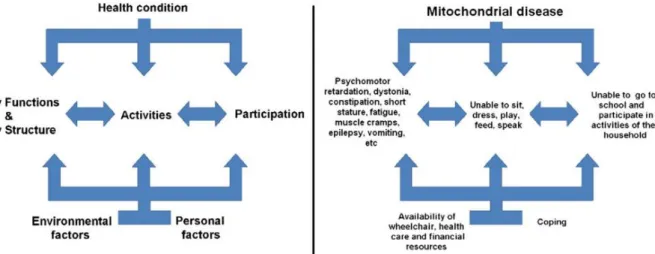
Inherited and sporadic mitochondrial cytopathies may have quite varied effects since the mutation rate of mtDNA varies from organ to organ within the body. Commonly, minor exercise intolerance is often observed in pacients with no serious illness or disability. Severe cases often involve into more complex disorders (Figure 9)
.
Figure 9. Severe mitochondrial disease condition. Symptoms, limitations and disabilities of a
typical patient suffering from mitochondrial disease (right side). (Adapted (82))
Tissues known to be affected by mitochondrial diseases are (79, 82-84):
1. The muscles: Usually manifest lack of energy and exercise intolerance due to the low muscle tone (Hypotonia). Cramps and muscle pains are also observed.
2. The brain: A wide variety of mental problems such as: dementia, mental disorders, development delay and neuro-psychiatric disturbances. Stroke and stroke-like episodes are quite common.
3. The heart: myopathy, heart blocks and cardiac dysrhythmia are the most likely causes associated to the heart
4. The kidneys: Proximal renal tubular dysfunction (Fanconi syndrome) resulting in the loss of electrolytes.
5. The eyes: Optic neuropathy and retinitis pigmentosa are the common conditions usually leading to some vision loss and in some cases permanent loss.
6. The ears: Sensory-neural hearing loss leading to deafness
7. Endocrine System: A wide variety of conditions such as weight loss, diabetes, hypoparathyroidism and exocrine pancreatic dysfunction.
Although, the amount of research behind mitochondrial cytopathies has been growing during the past years, there is currently no established treatment for mitochondrial disorders since the association of mitochondria and disease is quite recent and because it usually refers to multi-systemic symptoms. At this moment, the best approach for treatment are: pharmacological and nutritional agents, diets supplemented with vitamins and co-factors and exercise based therapy (85).
2.3.2. Mutations and mitochondrial diseases
Mutations all over mitochondrial genome have been observed and some specific diseases are often associated to mutations in specific mitochondrial genes and mitochondrial tRNA, while rRNA mutations appear to be less frequent compared to the other two.
Somatic mutations, mutations that were not present in the germ cells but occurred through time, may occur in the mitochondria. Point mutations and deletions are the most common. Point mutations occur essentially due to three factors: base substitution caused by errors in polymerase y (86); proximity to ROS formation sites and lack of histones. No nucleotide excision repair (NER) mechanisms are present, as BER is the only repair mechanism known to work at the site of mitochondria. On the other side, no elucidative studies are able to explain the mechanism behind the origin of mtDNA delections (21, 87).
Mutations in mtDNA genes are a quite frequent cause of mitochondrial cytopathy resulting in a huge variety of clinical phenotypes associated with severe metabolic dysfunctions, including progressive cardiomyopathy, encephalopathy, leukodystrophy, Leigh´s syndrome or ragged red fibbers syndrome and premature age-related symptoms (11, 88-96). In addition, mutations and/or polymorphism variance in mitochondrial genes are related with Parkinson, Huntington´s and Alzheimer´s diseases, diabetes and the greater susceptibility to develop cancer (97-111).
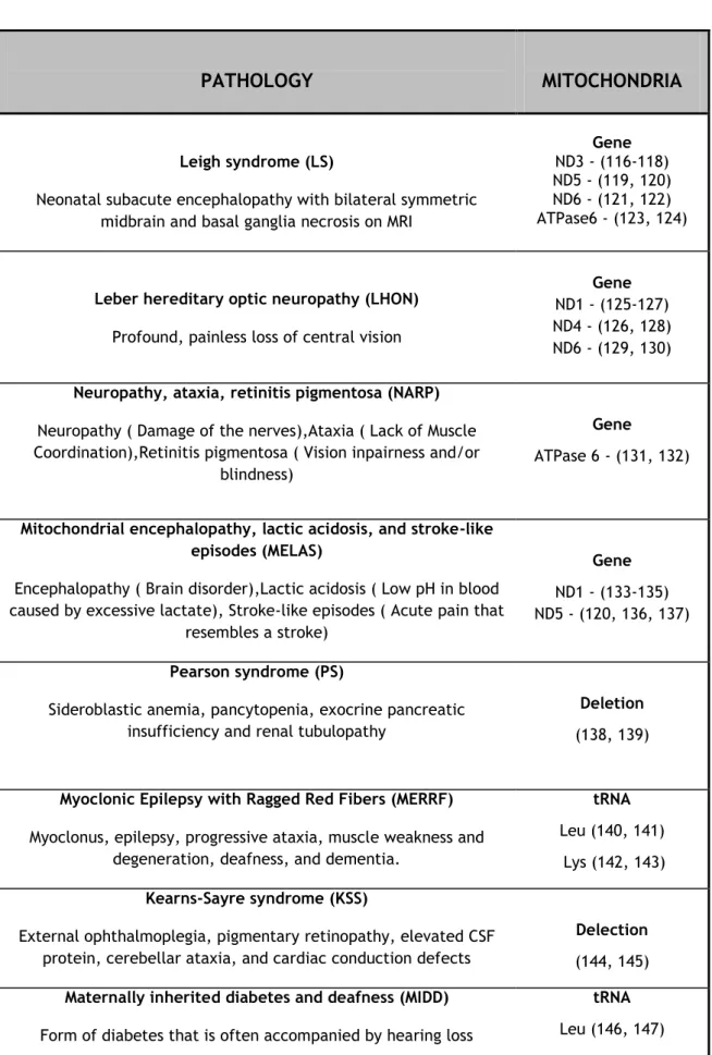
Although, approximately 200 different mitochondrial diseases have been reported (112), the most important diseases that are caused due to defects on the mtDNA are summarized in Table 1.
2.3.3. Aging
Aging is a degenerative process that is characterized by a decline in physiological function and an increased chance of developing a disease and death. These changes, that occur in all organisms, are believed to be associated with the metabolic activity and therefore with the mitochondria.
The initial idea originated in 1956, when Harman proposed the theory of “Free Radical Theory of Aging” (113). The idea assumes that free radicals, produced from normal metabolism, could be the cause of aging and aging-related degenerative diseases. Later on, the same Harman, suggested that mitochondria could be the main producer and the major target of free radicals and therefore, an organelle responsible for aging (114).
As mentioned before, mitochondria are the main cellular energy sources that generate ATP through the process of OXPHOS located at the IMM (25). While the OXPHOS system has
Table 1. Pathology associated to mitochondrial genome. (Adapted from (115))
PATHOLOGY
MITOCHONDRIA
Leigh syndrome (LS)
Neonatal subacute encephalopathy with bilateral symmetric midbrain and basal ganglia necrosis on MRI
Gene
ND3 - (116-118) ND5 - (119, 120) ND6 - (121, 122) ATPase6 - (123, 124)
Leber hereditary optic neuropathy (LHON)
Profound, painless loss of central vision
Gene
ND1 - (125-127) ND4 - (126, 128) ND6 - (129, 130)
Neuropathy, ataxia, retinitis pigmentosa (NARP)
Neuropathy ( Damage of the nerves),Ataxia ( Lack of Muscle Coordination),Retinitis pigmentosa ( Vision inpairness and/or
blindness)
Gene
ATPase 6 - (131, 132)
Mitochondrial encephalopathy, lactic acidosis, and stroke-like episodes (MELAS)
Encephalopathy ( Brain disorder),Lactic acidosis ( Low pH in blood caused by excessive lactate), Stroke-like episodes ( Acute pain that
resembles a stroke)
Gene
ND1 - (133-135) ND5 - (120, 136, 137)
Pearson syndrome (PS)
Sideroblastic anemia, pancytopenia, exocrine pancreatic insufficiency and renal tubulopathy
Deletion
(138, 139)
Myoclonic Epilepsy with Ragged Red Fibers (MERRF)
Myoclonus, epilepsy, progressive ataxia, muscle weakness and degeneration, deafness, and dementia.
tRNA
Leu (140, 141) Lys (142, 143)
Kearns-Sayre syndrome (KSS)
External ophthalmoplegia, pigmentary retinopathy, elevated CSF protein, cerebellar ataxia, and cardiac conduction defects
Delection
(144, 145)
Maternally inherited diabetes and deafness (MIDD)
Form of diabetes that is often accompanied by hearing loss
tRNA
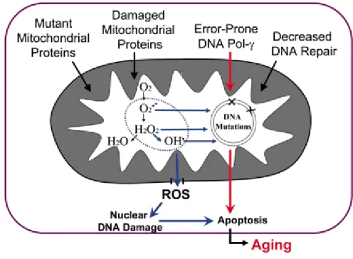
The Mitochondrial theory of aging proposes that during the course of the years, mtDNA suffers somatic mutations causing a decline in mitochondrial bioenergetics function contributing to aging. During normal conditions, mitochondria produce low levels of ROS and that low amount of ROS can be easily removed by antioxidants and free radical scavenging enzymes. However, mitochondria lack protection mechanisms and oxidative damage cause damage to the mtDNA leading to the appearance of mutant mtDNA. The accumulation of mutant type mtDNA usually results in the dysfunction of the respiratory chain, leading to an increased production of ROS and even more oxidative damage to the mtDNA. This becomes a vicious cycle, resulting in the decline of cellular and tissue functions resulting in the low amount of energy leading to apoptosis (148, 149) (Figure 10).
Figure 10. The mitochondrial theory of aging. Multiple factors may cause mtDNA mutations
which then leads to an increased production of ROS species. ROS species then contribute to even more mutations leading to a never ending process of aging (149).
3. Mitochondrial Gene Therapy
Over the last decades we have seen the harmful effects mitochondrial diseases may have in a patient’s life. Health care professionals have a great dilemma at hands since classical interventions have revealed unsuccessful in the fight against mitochondrial disorders.
Nowadays, it is well recognized that the future approach of mitochondrial medicine may involve the targeting of mitochondria. Focusing on the development of new therapies to correct mitochondrial disorders has become an active and expanding research field (150).
Introducing a mitochondrial gene into mitochondrial matrix might turn out to be the only approach to achieve permanent cure against mtDNA diseases. Up until today, two possible strategies have been suggested to fight against such disorders.
One good strategy is to avoid the threshold effect. As stated before, mitochondrial diseases generally manifest when mutant mtDNA reaches above a critical point in cells. If wild-type mtDNA, isolated from the pacient’s healthy tissues, were to be administered into the damaged tissue, swapping with mutated mtDNA may occur and the threshold level may not be reached and the disease not manifested (151).
The other approach is the introduction of a therapeutic mitochondrial gene into the matrix which is seen as a major hurdle. Although electroporation (152) and biolistic transformation (153) have been suitable methods utilized to introduce DNA into the mitochondria, no evidence supports their success for human MGT. Endocytosis is nowadays seen as the most promising solution due to the ability of mitochondria to receive exogenous DNA.
An adequate mitochondrial gene therapy (MGT) system should compromise a minimum number of requisites:
1) The carrier system should initially be taken up by the host cell through an internalization mechanism such as endocytosis. Once inside the cell, our carrier should target the mitochondria instead of other intracellular organelle such as the nucleus, acting as a mitochondriotropic agent.
2) The genetic material inside must be able to traverse both OMM and IMM and reach the matrix where it may meet its target. The target may vary from a vast list which comprises all forms of nucleic acids (mtDNA, rRNA, tRNA) depending on specific aim. 3) The carrier system should bring beneficial effect to the mitochondria. The introduction of genetic material may be beneficial by correcting either a mtDNA mutation or modulation of gene expression. However, compromising the integrity or survival of the cell even if coupled with good results may not be a viable MGT. 4) The mitochondrial transfection vector should ensure long and sustained gene
3.1. Non-Viral Gene Therapy
Gene therapy has evolved during the last decade becoming a trend as a therapeutic approach against a big number of incurable diseases. The possibility of trying to treat these patients, by providing replacement copies of the defective gene, leads to a huge impact in the traditional clinical management (5).
The idea was initially originated in 1963, when Joshua Lederberg suggested that it would be possible to control a nucleotide sequence in human chromosomes, coupled with selection and integration of the desired genes (7). This idea ended up being accomplished only in 1980 with the introduction of two functional genes into mammalian cells (8).
The two main types of gene therapy are somatic cell gene therapy and germ-line gene therapy. Germ-line gene therapy is an interesting concept which consists in the introduction of a gene into reproductive cells (sperm or eggs) or later on the zygote, which results in the transmission of a beneficial gene into the offspring. Even more interesting is that this therapy can be extended into the mitochondria. As shown by Tachibana and colleagues, mtDNA can effectively be replaced in oocytes reproducing embryonic stem cells similar to controls (154). As fascinating it may sound, ethical aspects puts a halt, resulting in less and less studies around this concern and therefore no imminent application in humans, seems to be, even possible (155). Therefore, most of the research, nowadays, is centered on somatic cells.
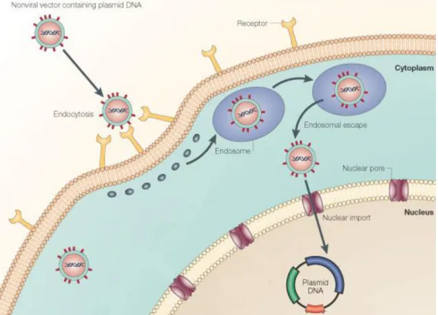
The delivery of genetic material into the cell can be accomplished by using viral and non-viral vectors. Viral vectors exploit the intrinsic ability of the viruses to target the nucleus. These vectors were once the most commonly used gene delivery systems in gene therapy, because of their highly efficient infection rate and ability to integrate therapeutic genes into the host chromosome ensuring sustained gene expression (156). However, several disadvantages presented by virus vectors such as, the given antigenicity, possibility of virus recombination (157), potential oncogenic effects (158, 159), difficulty in large scale production and instability of storage along with fatal cases associated with a severe inflammatory response have instigated the search for new vectors. When targeting mitochondria is concerned, the choice becomes easier. Since no virus is known to transfect the mitochondria, the intrinsic ability of the viruses becomes obsolete. As a result non-viral vectors, such as the plasmid DNA (pDNA), become the only viable option as transformation is considered (Figure 11).
The pDNA, discovered by Joshua Ledberg in 1952 (160) is a small circlular double-stranded DNA molecule that can replicate independently within the cell. Usually ranging from 1kbp to 1.000kbp, plasmids are widely used as non-viral vectors since they are able to integrate exogenous DNA and then replicate this exogenous within the cell target.
genome or just to stay in the nucleus with no integration occurring within the host genome (161).
Figure 11. Transfection Schematics. Non-viral vectors are capable of receptor-mediated
endocytosis. In the cytoplasm, endosomal escape and nuclear import, ending with the delivery of pDNA carrying a therapeutic gene into the nucleus. Adapted from (162)
Non-viral gene therapy has evolved to the point that there are huge varieties of methods to deliver the pDNA, ranging from conventional methods such as injection of naked DNA and electroporation to the usage of nanoparticles (163) and gels (164).
3.2. Traversing mitochondrial membrane
As is well known, to the proper function of the mitochondria both mtDNA and nDNA play important roles. In order to correct mutated mitochondrial genes, gene therapy emerges as a viable possibility; but are we able to deliberately introduce nucleic acids at the site of mitochondria in a transfection process?
Targeting DNA into mitochondria should present itself as a hard challenge involving several obstacles. Mitochondrial membranes are of lipophilic nature preventing the entrance of big molecules such as peptides and DNA unless there is an active transport mechanism (165). Moreover, if it ends up entering the cell via the endocytic pathway it may become entrapped in the endosome and eventually end in the lysosome where degradation takes place (165).
Milana Koulintchenko and colleagues first demonstrated that plant have a transmembrane potential-dependent mechanism of plamid-like DNA uptake into mitochondria (166) and later, through five different assays that mammalian mitochondria possess a natural competence for DNA uptake (167). Further observation of both works proposed that DNA intake in both plants and mammals is achieved through voltage-dependent anion channel and that mammalian can intake both ss (single-stranded) and ds (double-stranded) DNA while plants can only internalize ds DNA (166, 167). As previously described, mitochondria are able to successfully integrate exogenous DNA and although the idea behind how DNA can transverse OMM has started to become understood and accepted until today the mechanisms behind the IMM are still unknown.
Transfection of plamid DNA into the mitochondria has been successfully achieved (168, 169) but transformation, incorporation and expression of transfected DNA has only been reported in Saccharomyces cerevisiae, Chlamydomonas reinhardtii (170) and Candida
glabrata (171) while in mammals it is still a pipe dream.
3.3. Nanotechnology
A major research thrust in the biochemical/pharmaceutical technology is still the development of efficient and safe controlled release systems for the sustained delivery of drugs and bioactive agents. To be used therapeutically, these systems should be able to deliver the drug and/or gene at a specified rate and time period. Furthermore, they can be targeted to a particular organelle or cell type.
Nanotechnology provides appropriate knowledge and tools for the design and creation of new suitable biocompatible formulations for gene delivery purposes. This technology can be applied to mitochondrial gene therapy providing nanosystems to carry genetic information to mitochondria, since mitochondria size range from 0,5 to 1 (μm) in diameter (15). Furthermore, the idea that nanosystems have unique physical and biological properties that might be used to overcome the problems of gene delivery, has gained interest in recent years because they can ensure protection against enzymatic degradation, are able to by-pass the innate immune system, have good biodistribution, reduced side-effects, safety, no toxicity,
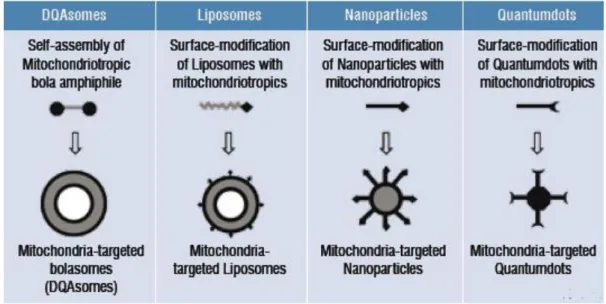
and liposomes. Unfortunately, mitochondria targeted therapy represents a significant challenge and only a few nanocarriers are, at the moment, seen as possible candidates (Figure 12). For instance, considerable improvements have been made with dequalinium cationic vesicles (DQAsomes) with mitochondria affinity as vehicles for drugs and DNA to this organelle. These mitochondriotropic vesicles bind and condense plasmid DNA and release it when in contact with cardiolipin-rich liposomes mimicking mitochondrial membranes (172, 173).
Figure 12. Mitochondria-specific Nanotechnology. : DQAsomes (DeQuAlinium-based liposome-like
vesicles), nanolipid vesicles, solid nanoparticles and quantum dots are the typical nanocarrier systems used when approaching the mitochondria (Adapted from (174))
3.4. Salt as an efficient nanocarrier
Although the development of efficient methods to produce gene delivery vehicles for gene therapy purposes started decades ago, it has not still reached a point where we can say that we have found the perfect nanocarrier system. Several promising gene delivery systems including polymeric (175), silica based (176) and liposomal (177) have been developed. However, the biocompatibility displayed by these systems is not satisfactory.
One of our main concerns, when designed the mitochondrial nanocarrier, was its biocompatibility and biodegradability. The nanocarrier system, proposed in this thesis, is based on calcium carbonate (CaCo3) and was prepared through the technique of co-precipitation of calcium (Ca2+), an inorganic cation, with carbonate (CO
32-), an inorganic anion.
Calcium is essential for living organisms since it functions as an important signal for many cellular processes. It is the major material used in mineralization of bone and teeth and it is
required for the normal function of all muscles and nerves. On the other side, carbonate works as a buffer in the blood.
Calcium carbonate is widely used medicinally as an inexpensive dietary calcium supplement. Calcium carbonate is composed of three important elements that are present in almost all organic and inorganic materials: carbon, oxygen and calcium. CaCO3 is a white, non-toxic and odorless salt (178) that allure many scientists to its usage as a nanocarrier system due to their good biocompatibility, biodegradability, wide range of resources, size and low cost (179).
Although the usage of CaCO3 nanocarriers has been presented as a plausible choice for gene therapy (180-183), its application as a MGT has not, until now, been demonstrated.
Aims of the Project
In addition to the cell nucleus, mitochondrion is the unique organelle with its own genome-the mitochondrial DNA. Mitochondrial genome is considered a hotspot for mutations due to the lack of protection and repair mechanisms. Mutations in mtDNA genes are a quite frequent cause of mitochondrial cytopathy and conventional treatments are, in most of the cases, inefficient. Mitochondrial gene therapy, thus, emerges as a new perspective to correct such anomalies.
Although MGT is seen as a promising approach, transformation is not yet possible in mammals and, even transfection, has hardly ever been achieved into the mitochondria. The application of virus in MGT seems impossible, since no virus is known to target the mitochondria. Therefore we shift our attention to the plasmid, a non-viral vector which is widely used as a therapeutic agent in gene therapy.
Taking all the previous information into account, during this MsC thesis, it was tried to bring together a new nanocarrier system that can effectively deliver pDNA into the mitochondria, for future application in mitochondrial gene therapy.
The first aim of this work consisted in the isolation and purification of three plasmids with different sizes. Then, we proceeded to the development of CaCO3-pDNA-rho123 nanoparticles through a co-precipitation method. We then designed two different protocols, in order to encapsulate 1µg and 10µg of pDNA and characterized each one and compared them. Lastly, we proceeded to the nanocarrier in vitro evaluation.
The success of this work is based on the design and preparation of a suitable vector that can represent a promising tool for progresses in mitochondrial gene delivery purposes, contributing for new therapies centered in mitochondria.
Materials and Methods
1. Materials
1.1. Reagents
Dulbecco’s Modified Eagle Medium Ham’s Nutrient Mixture F12 (DMEM: Ham’s F12) was obtained from Biochrom AG (Germany). Rhodamine 123, Tween 20, Triton X-100, paraformaldehyde (PFA) and Deoxyribonuclease I from bovine pancreas (DNAse I) were obtained from Sigma-Aldrich (St. Louis, MO, USA). Agarose and Green Safe were obtained from NZYTech (Lisboa, Portugal). Mitotracker® Orange CMTMROS was obtained from InvitrogenTM (Eugene, Oregon, USA). Sodium Carbonate anhydrous was obtained from Panreac Quimica SA (Barcelona, Spain). Calcium chloride was obtained from BDH Prolabo (Leuven, Belgium). Cellulose powder was obtained from Aldrich Chemical Company (Milwaukee, WI, USA). Tween 20 was obtained from Applichem (Darmstadt, Germany). Normal Human Dermal Fibroblast (NHDF) adult donor cells, Ref. C-12302 (cryopreserved cells) were purchased from PromoCell.
1.2. Plasmid
Both pUC19 and pVAX1-LacZ were obtained from Invitrogen (Carlsband, CA, USA) while pcDNA3-myc-FLNa S2152A was obtained from Addgene plasmid 8983 (pcDNA3-based plasmid) (Cambridge, MA, USA).
Figure 14. pVAX1-LacZ mapping . 6050 bp pVAX1-LacZ has the exact same features as pVAX1 (2999bp)
but containing the additional gene, β-galactosidase.
Figure 15. pcDNA3-myc-FLNa S2152A mapping. The 14,086bp pcDNA3-myc-FLNa S2152A has the
2. Methods
2.1. Bacterial Growth and Plasmid Purification
The 2.6kbps plasmid pUC19, the 6.1kbps pVAX1-LacZ and the 14kbps pcDNA3-myc-FLNa S2152A were amplified by fermentation carried out in a 500 mL Erlenmeyer using a Terrific Broth medium (20 g/L tryptone, 24 g/L yeast extract, 4 mL/L glycerol, 0.017 M KH2PO4, 0.072 M K2HPO4) supplemented with the appropriate antibiotic; 100 µg of ampicillin/mL for the cells transformed with pUC19, 30 µg of kanamycin/mL for the cells transformed with pVAX1-LacZ and a combination of ampicillin/mL with neomycin/ml for the cells transformed with pcDNA3-myc-FLNa S2152A , adapted from (184).
The bacterial growth was carried out overnight, at 37ºC under 250 rpm shaking, and the cells were harvested at the late log phase (OD600 nm ≈ 9) by centrifugation.
Plasmid purification was achieved using QIAGEN® Plasmid Purification kit. Shortly, cells were suspended, lysed and precipitated followed by a double centrifugation at 20,000 x g for 30min at 4 ◦C with the recovery of supernatant containing plasmid DNA. Supernatant was added to a QIAGEN-tip to remove most contaminants followed by DNA elution, precipitation and centrifugation at 15,000 x g for 30 min, 4 ◦C. The pellet was recovered and the pDNA concentration estimated through UV-VIS analysis and finally suspended in the suitable buffer and stored at -80ºC.
2.2. Agarose gel electrophoresis
Electrophoresis is a technique that consists in the usage of an electric field applied to a gel matrix that permits the separation and identification of nucleic acids based on their size and charge. The electrophoresis experiments were carried out by running a 1% agarose gel (Hoefer San Fransisco, Ca, USA) stained with Green Safe (1 µg/mL). Electrophoresis was carried out at 110V for 20 minutes with TAE buffer (40 mM Tris base, 20 mM acetic acid and 1 mM EDTA pH 8.0). Gel visualization occurred under UV light in a Vilber Loumat system (ILC Lda, Lisbon, Portugal).
2.3. Synthesis of CaCO
3-pDNA-Rho
123Nanoparticles
Plasmid DNA solution containing 1 µg of plasmid DNA, 40 uL of CaCl2 solution (0.07 g/mL) and 0.1 µg of Rhodamine 123 (Rho123) were mixed and then diluted with deionized water to
of a Pasteur pipette to form a final Solution C with a total volume of 150 µL containing CaCO3-pDNA-Rho123 nanoparticles (181).
Similarly, plasmid DNA solution containing 10 µg of plasmid DNA, 120 µL of CaCl2 solution (0.03 g/mL) and 0.5 µg of Rho123 were mixed and then diluted with deionized water to make a solution A with a total volume of 290 µL. 255 µL of Na2CO3 solution (0.0425 µg/mL) was mixed together with 5 µg of cellulose and then diluted with deionized water to make solution B with a total volume of 260 µL. Solution A was then gently added to Solution B with the help of a Pasteur pipette to form a final Solution C with a total volume of 550 µL containing CaCO3 -pDNA-Rho123 nanoparticles (183).
2.4. Particles Morphology
Recently formed nanoparticles were centrifuged (10.000 g, 20 min, 25 ºC) and the pellet recovered. The pellet was suspended in a solution containing 20 µL deionized water with 20 µL tungsten. 10 µL of the recently formed solution was set in roundly shaped cover-slip and left at room temperature overnight to dry.
In the following day, the samples were sputter coated with gold using an Emitech K550 sputter coater (London, England) and then analyzed by scanning electron microscope (SEM) (Hitachi S-2700, Tokyo, Japan), operated at an accelerating voltage of 20 kV with variable magnifications.
2.5. pDNA Encapsulation Efficiency
The encapsulation efficiency (EE) of the pDNA was determined after centrifugation (15.000 g, 20 min, 25 ºC) and recovery of the supernatant. The supernatant corresponded to the unbound pDNA or, in other words, the pDNA that was not encapsulated into CaCO3 nanoparticles. The concentration of unbound pDNA was determined by Uv-vis analysis at 260 nm in a NanophotometerTM (Implen, Germany). Desionized water was used to perform the blank experiment. At least three independent measurements were performed.
To determine EE values we resorted to the following formula: