A. C. C O E L H O1 , A. C R A V A D O R2 *, A. B O L L E N3 , J. F. P. F E R R A Z†2 , A. C. M O R E I R A2 , A. F A U C O N N I E R3 A N D E. G O D F R O I D3
" Escola Superior de Educaçaho, Universidade do Algarve, P-8000 Faro, Portugal
# Unidade de CieWncias e Tecnologias AgraUrias, Universidade do Algarve P-8000 Faro, Portugal
$ Laboratoire de GeUneUtique AppliqueUe, UniversiteU Libre de Bruxelles, Rue de l’Industrie 24, B-1400 Nivelles, Belgique
In response to the need for a faster, more reliable method for identifying Phytophthora cinnamomi in cork oak soils in Portugal, a simple, fast, sensitive molecular identification method is described. It is based on a colorimetric assay which involves an
oligonucleotide capture probe covalently immobilised on microtitration wells, a multi-biotinylated oligonucleotide detection probe and the PCR-amplified target DNA. The target DNA is a 349 bp DNA fragment partially covering the translated and 3«-untranslated regions of the cinnamomin gene. When the specificity of the PCR reaction was evaluated in vitro using isolates of
P. cinnamomi and eight other Phytophthora species, including the related P. cambivora, it was specific to P. cinnamomi. When 30
isolates of P. cinnamomi from oak roots in southern Portugal were assayed, 26 gave a strong positive response. The assay has a sensitivity of about 2–5 genome equivalents of P. cinnamomi. The reason for the negative response of four isolates remains unclear.
Phytophthora cinnamomi Rands is a highly aggressive
filamentous soilborne fungus. It is one of the world’s most destructive plant pathogens causing massive death of fine feeder roots, and reducing the capacity of hosts to absorb water and nutrients leading to symptoms resembling those of drought. Furthermore, it is polyphagous, parasitising over 900 species of woody perennial plants (Zentmeyer, 1980). It is also well known that Jarrah forest (Eucalyptus marginata) and other vegetation systems in south-western Australia are being destroyed by P. cinnamomi (Shearer & Tippett, 1989 ; Wills, 1993). Early this century P. cinnamomi also caused a major epidemic among chestnut trees in south-eastern U.S.A. (Crandall, Gravatt & Rian, 1945) and massive death of chestnut (Castanea sativa L.) in Portugal and Galicia, northern Spain (Pimentel, 1949). Recently, the fungus has been shown to be involved in widespread deaths of cork oak, Quercus suber L., in Portugal and Spain (Brasier, 1992 ; Brasier, 1993 ; Brasier
et al., 1993).
Brasier (1993, 1997) recognized that roots of declining oaks had not been critically examined in Europe for the presence of
P. cinnamomi or other Phytophthora spp. and suggested that
the role of this pathogen in the similar oak declines in central and eastern Europe also deserved investigation. In agreement with this proposal we consider that the role of one or more
Phythophthora spp. in various oak decline problems must be
re-* Author from whom reprints should be requested. † Corresponding author.
evaluated. Current methods of isolation and detection of
Phytophthora in oak roots, based mainly on baiting methods,
however, are of relatively low effectiveness and reliability. More sensitive, molecular based diagnostic techniques are required. Indeed, the importance of Q. suber plantations to the Portuguese economy (Portugal ranks first in world cork production), and the social and environmental importance of cork oaks in the preservation of rural communities and in the ecological stability of the ecosystem on which they depend, further emphasizes the need to develop reliable, highly specific, sensitive and rapid methods to detect this pathogen. Several methods for identification of P. cinnamomi on roots of eucalyptus and avocado using hybridization techniques with DNA probes and}or PCR, have been developed previously (K. Sivasithamparam & P. O’Brien, pers. comm. ; Dobrowolski & O’Brien, 1993 ; Judelsen & Messenger-Routh, 1996). The targets for DNA amplification were the regions between the 18S and 5,8S (ITS I ; internal transcribed spacer region I) and between the 5,8S and 25S (ITS II) of ribosomal DNA. These regions exhibit useful interspecific variability (Lee & Taylor, 1992).
Our approach has been to target the cinnamomin gene of
P. cinnamomi, a polypeptide which belongs to a family of
proteins designated as elicitins (Huet & Pernollet, 1989). Some of these proteins were shown to act as inducers of a hypersensitive-like response in tobacco and radish (Kamoun et
al., 1993). The aminoacid sequence of the cinnamomin protein
has been recently determined (Huet & Pernollet, 1989). We
Highly specific and sensitive non-radioactive molecular
used this information to clone and sequence DNA fragments of the gene coding for cinnamomin and the corresponding gene from P. cambivora (Petri) Buisman. To improve the sensitivity and the specificity of the PCR assay, we linked the assay with a colorimetric sandwich capture hybridization assay which has been recently developed for the identification of infectious agents (Lage et al., 1996). The procedure takes advantage of an original DNA chemistry to generate multi-biotinylated probes and to bind oligonucleotides onto a plastic support (De Vos, Van Elsen & Bollen, 1994).
This paper presents our results on the detection of P.
cinnamomi, in comparison with eight other species of Phytophthora.
M A T E R I A L S A N D M E T H O D S
Fungal isolates and culture methods
Seventeen reference isolates of Phytophthora representing nine traditional morphospecies were used in this study (Table 1). In addition, thirty isolates conforming to P. cinnamomi on morphological grounds (C. M. Brasier, personal communi-cation), from roots of Q. suber or associated soil in oak decline areas of the Algarve region of southern Portugal, were also examined (Table 2).
Mycelium of Phytophthora was grown in V8 liquid medium at room temperature in the dark for 4 d. The DNA was extracted as described by Rozman & Komel (1994). Liquid medium V8 was prepared by the addition of 5 g CaCO$ to 354 ml of V8 juice (Campbell Soup Company). The mixture was shaken for 15 min, then centrifuged for 20 min at 4000 rpm. The supernatant was collected and diluted four times with distilled water. The prepared medium was autoclaved before storage or use.
Table 1.Isolates of Pytophthora cinnamomi and reference species used to test the method Isolate
number
Geographic
origin Name Host Date Source
P. cactorum (Lebert & Cohn) J. Schro$ t. 213a Belgium cac-213 Pyrus communis L. 1956 G. L. Hennebert
P. cactorum 20 872a Moroccod cac-872 Pyrus communis L. 1982 A. Vanderweyen
P. cinnamomi Rands 20 875a Moroccod cin-875 Pyrus communis L. 1982 A. Vanderweyen
P. cinnamomi 20 876a Moroccod cin-876 Pyrus communis L. 1982 A. Vanderweyen
P. cinnamomi 20 877a Moroccod cin-877 Pyrus communis L. 1982 A. Vanderweyen
P. cinnamomi E27c Portugal cin-E27 Castanea sativa Miller 1945 A. A. L. Pimentel
P. citrophthora (R. E. Sm. & E. H. SM.) Leonian 20 889a Moroccod cit-889 Castanea sativa Miller 1982 A. Vanderweyen
P. citrophthora 20 890a Moroccod cit-890 Castanea sativa Miller 1982 A. Vanderweyen
P. cryptogea Pethybr. & Laff. 28 777a – cry-777 Gerbera sp. 1986 A. Vanachter
P. cryptogea 30 509a – cry-509 Gerbera sp. 1990 Ph. Blanquet
P. drechsleri Tucker 20 891a Morocco dre-891 Gerbera sp. 1982 A. Vanderweyen
P. drechsleri 20 892a Morocco dre-892 Gerbera sp. 1982 A. Vanderweyen
P. erythroseptica Pethybr. 28 776a India ery-776 Solanum tuberosum L. 1945 J. F. Dastur
P. nicotianae Breda de Haan 28 775a India nic-775 Nicotiana tabacum L. 1986 M. E. Gallegly
P. gonapodyides (Petersen) Buisman 340 619b UK gon-619 Nicotiana tabacum L. 1990 M. E. Gallegly
P. cambivora (Petri) Buisman 340 630b Australia cam-630 Prunus cerasus L. 1990 M. E. Gallegly
P. cambivora 340 633b Australia cam-633 Prunus amygdalus L. 1990 M. E. Gallegly
a Isolates from the collection of MUCL (Mycothe' que de l’Universite! Catholique de Louvain), Agronomic Sciences Faculty, UCL, Place Croix du Sud, B-1348 Louvain-la-Neuve, Belgium.
b Isolates from the collection of the International Mycological Institute, Bakeham Lane, Egham, Surrey TW20 9TY, U.K.
c Isolate from the ‘ Estaça4 o Nacional Vieira Natividade’, Alcobaça. Portugal. The isolate was initially wrongly identified as P. cambivora by Pimentel.
d El Menzeh, Ke! nitra area, Morocco, isolates from the collection of Station de La Recherche sur les Agrumes. (–) Unknown.
Table 2.Origin of cork oak isolates of Phytophthora cinnamomi from southern Portugal
Location Source Date
VI-4-S-M Lagos, Algarve Soil 16 v 95 VI-3-Rf-M* Lagos, Algarve Thin roots 16 v 95 V-4-S-M* Lagos, Algarve Soil 16 v 95 IV-3-S-M* S. Bra! s de Alportel, Algarve Soil 11 v 95 III-4-S-E S. Bra! s de Alportel, Algarve Soil 3 v 95 III-1-R-MS S. Bra! s de Alportel, Algarve Roots 3 v 95 II-2-S-E S. Bra! s de Alportel, Algarve Soil 24 v 95 II-1-R-MS* S. Bra! s de Alportel, Algarve Roots 24 v 95 II-1-S-M S. Bra! s de Alportel, Algarve Soil 24 v 95 I-4-S-M S. Bra! s de Alportel, Algarve Soil 24 v 95 X-4-Rg-M Sines, Alentejo Thick roots 5 vi 95 X-4-S-M Sines, Alentejo Soil 5 vi 95 X-2-R-MS Sines, Alentejo Roots 5 vi 95 X-2-Rf-M Sines, Alentejo Thin roots 5 vi 95 X-1-Rg-M Sines, Alentejo Thick roots 5 vi 95 IX-4-S-M Tanganhal, Alentejo Soil 5 vi 95 IX-4-R-MS Tanganhal, Alentejo Roots 5 vi 95 VIII-4-RF-M Lagos, Algarve Thin roots 31 v 95 VIII-4-R-MS Lagos, Algarve Roots 31 v 95 XVIII-4-Rf-M S. Bra! s de Alportel, Algarve Thin roots 27 vi 95 XVIII-4-R-MS S. Bra! s de Alportel, Algarve Roots 27 vi 95 XIV-4-Rf-M S. Bra! s de Alportel, Algarve Thin roots 20 vi 95 XIV-4-S-M S. Bra! s de Alportel, Algarve Soil 20 vi 95 XIV-3-Rg-M S. Bra! s de Alportel, Algarve Thick roots 20 vi 95 XIV-3-S-M S. Bra! s de Alportel, Algarve Soil 20 vi 95 XIV-1-Rg-M S. Bra! s de Alportel, Algarve Thick roots 20 vi 95 XI-4-R-M S. Bra! s de Alportel, Algarve Roots 8 vi 95 XI-3-R-MS S. Bra! s de Alportel, Algarve Roots 8 vi 95 XI-2-S-M S. Bra! s de Alportel, Algarve Soil 8 vi 95 XX-3-S-M Benavente, Ribatejo Soil 26 vi 95
Sample preparation and PCR
For specificity analysis and identification, mycelium was transferred with a sterile platinum loop to 100µl of sterile water and boiled for 10 min to disrupt the cells and to let free the DNA. PCR was performed directly in 10µl of this mixture.
Amplification was carried out in a final volume of 50µl containing 0±4µ of each primer, 0±2 m of each deoxynucleo-tide triphosphate (dATP, dGTP, dTTP and dCTP) (Boehringer Mannheim GmbH, Mannheim, Germany), reaction buffer (10 m Tris-HCl, 50 m KCl, 1±5 m MgCl#, pH 8±3), Taq DNA polymerase (2±5 U) (Boehringer Mannheim), 10µl mycelial extract, overlaid with one drop of light mineral oil (Sigma). Tubes containing water instead of mycelial extract were assayed in each PCR run as negative controls of the reaction.
After initial denaturing of the samples for 3 min at 94°C, reactions were run for 35 cycles consisting of 1 min at 94° for denaturing, 1 min at 62° for annealing and 30 s at 72° for extension. In the last cycle, the extension step proceeded for 7 min at 72°. Cycling was performed on a thermocycler Perkin Elmer 480. Ten microlitres of each PCR product was subjected to electrophoresis on 2 % agarose gel (Bio-Rad, U.S.A.) in TBE buffer (100 m Tris-borate, 30 m ethylene diamine tetra-hydrochloride pH 8±3) and detected under uv light after staining with ethidium bromide (0±5 mg ml−"). The 123 bp and 1 kb DNA Ladders (Life Technologies) were run as molecular weight markers.
Primers and probes
All the oligonucleotides were synthesized by the solid-phase phosphoroamidite method (Beaucage & Caruthers, 1981 ; Matteucci & Caruthers, 1981) on an Applied Biosystems Synthesizer (U.S.A.) model 394. The degenerated oligo-nucleotides (94±239, 95±374) used for the PCR amplification of a 257 bp fragment were defined taking in account the
Phytophthora codon usage. The codon usage of the Phytophthora genus was established by using the
codon-frequency facility of the Genetics Computer sequence analysis software package (version 8.1, August 1995 ; University Research Park, 575 Science Drive, Madison, WI53711, U.S.A.). Codonfrequency counts codons and writes their frequencies into codon frequency tables. It counts the codons from ranges within sequences. To determine the sequence of the pair of degenerate primers, the output table was used to backtranslate the aminoacid sequence of cinnamomin.
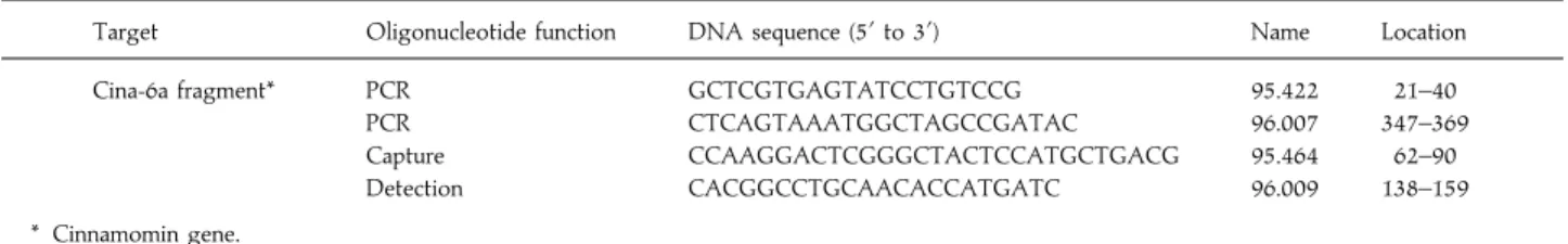
The sequence of the primers and the probes and their position inside the sequenced and cloned fragment named
Table 3.Primers and probes used in the amplification reaction and detection system
Target Oligonucleotide function DNA sequence (5« to 3«) Name Location
Cina-6a fragment* PCR GCTCGTGAGTATCCTGTCCG 95.422 21–40
PCR CTCAGTAAATGGCTAGCCGATAC 96.007 347–369
Capture CCAAGGACTCGGGCTACTCCATGCTGACG 95.464 62–90
Detection CACGGCCTGCAACACCATGATC 96.009 138–159
* Cinnamomin gene.
cina-6a, are in Table 3. The detection probe was polyaminated in a multi-fork like structure and multibiotinylated (eight biotins) at the 5« end (De Vos et al., 1994). The capture probe was covalently linked to polystyrene microplates (Polymer Laboratories, Birmingham, U.K.) by chemical condensation (De Vos et al., 1994).
Colorimetric hybridization assay
The PCR mixture (10µl) was treated with 40µl of 0±25 NaOH for 10 min. Simultaneously oligonucleotide-coated plates were incubated at room temperature with 250µl}well of a Tris-buffered solution (0±1 Tris-HCl, 1 NaCl, 2 m MgCl#, 0±05% Triton X-100, pH 7±5), containing 5% non-fat dried milk. The solution was eliminated after 10 min. Four pmol of the detection probe were mixed with the hybridization buffer composed of Tris-buffered solution, containing 0±2 acetic acid and 2±5% non-fat dried milk. 50µl of this solution and 50µl of chemically denatured PCR product were applied into a well of a 96-well plate, carrying the covalently linked capture oligonucleotide probe. This procedure was always made in duplicate. After 90 min of incubation at 37° under slight agitation the wells were washed five times with Tris-buffered solution, containing 5 % non-fat dried milk. For detection, 100µl of streptavidin-horseradish peroxidase (DAKO, Copenhagen, Denmark), diluted 1000 fold in the Tris-buffered solution containing 3 % of Bovine Serum Albumin (Sigma), were added to each well and plates were incubated at room temperature for 30 min. Then, the plates were washed five times with 250µl of Tris-buffered solution and incubated for 15 min in the dark with 200µl of 3,3 «,5,5«-tetramethylbenzidine (TMB) (Sigma). The reaction was stopped by the addition of 100µl of 10 % sulphuric acid and the optical density (OD) at 450 nm was then measured in a Novapath microplate reader (Biorad, Richmond, U.S.A.). The PCR product concerning the blank PCR (negative control of PCR reaction) was tested in five wells in each hybridization assay. The cut-off value was defined as three standard deviations above the mean of the blank PCR mixtures.
DNA sequencing
The PCR amplified fragments were cloned using A–T Cloning in the Invitrogen pCR II4 vector (Invitrogen, U.S.A.). The DNA sequences were obtained for double strands by the chain termination method, using an automated sequencer ABI 373A model. Samples were prepared following the Kit Prism4 Ready Reaction Dye Deoxy4 Terminator cycle sequencing protocol.
R E S U L T S
Development of a PCR assay for the specific identification of P. cinnamomi
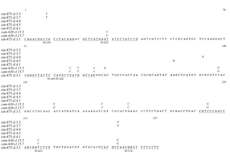
Based on the published aminoacid sequence of cinnamomin (Huet & Pernollet, 1989) and taking the Phytophthora codon usage into account, we defined a pair of degenerated oligonucleotides for the PCR amplification of a 257 bp DNA fragment, using the approach described in Materials and Methods. The nucleotide sequences of these primers (94.239, 95.374) and their corresponding aminoacid sequence are shown in Fig. 1. Two isolates of P. cinnamomi (875, cin-876) were amplified yielding DNA fragments with the expected length. These DNA fragments were cloned and sequenced in an automatic sequencer. The sequences showed slight differences between the two isolates and clones of the same isolate (Fig. 2).
5« 3« 5«TAC GCC AAC GGC TTC TCG TC3«
G C
95.374 CAG ACC GCC GCC TAC AAG
G T
94.239T
TACTATQQTAAYKTLVSILSESSFSQCSKDSGYSMLTATALPTNAQYKLMCASTACNTMIKKIVALNPPDCDLTVPTSGLVLDVYTYANGFSSKCASL
Fig. 1.Aminoacid sequence of cinnamomin. Location and sequence of degenerated primers (94.239 ; 95.374), used for PCR amplification, are shown. cin-875 cl 3.3 cin-875 cl 3.7 cin-875 cl 4.9 cin-875 cl 4.5 cin-875 cl 4.1 cam-630 cl 15.5 cam-630 cl 15.7 cin-875 cl 3.1 cin-875 cl 3.3 cin-875 cl 3.7 cin-875 cl 4.9 cin-875 cl 4.5 cin-875 cl 4.1 cam-630 cl 15.5 cam-630 cl 15.7 cin-875 cl 3.1 cin-875 cl 3.3 cin-875 cl 3.7 cin-875 cl 4.9 cin-875 cl 4.5 cin-875 cl 4.1 cam-630 cl 15.5 cam-630 cl 15.7 cin-875 cl 3.1 cin-875 cl 3.3 cin-875 cl 3.7 cin-875 cl 4.9 cin-875 cl 4.5 cin-875 cl 4.1 cam-630 cl 15.5 cam-630 cl 15.7 cin-875 cl 3.1 95.423 C C G G 95.374 G G 257 G G C C T T G G 141 211 71 94.239 C C T T A A G G G 210 140 70 C C 1 T T 95.422 95.441/95.442
Fig. 2.Nucleotide sequence of clones obtained by PCR amplification from P. cinnamomi isolates (cin-875 ; cin-876) and from P. cambivora (cam-630). Primers used for PCR amplification are underlined : degenerated primers (94.239 ; 95.374) ; primers used for the amplification of the 199 bp fragment (95.422 ; 95.423) ; and initiating primers used in the PCR-vectorette approach (95.441 ; 95.442). Consensus sequence corresponds to cin-875 cl. 3.1. Nucleotide numbering starts at 1 corresponding to the first nucleotide of the codon of aminoacid eight of cinnamomin.
From these sequences we defined and synthesized another pair of oligonucleotide primers used for the amplification of a 199 bp fragment. The nucleotide sequences of these primers (95.422, 95.423) and of the amplified fragment are shown in Fig. 2. The sensitivity of the PCR assay was assessed on DNA isolated from the strain 20.875 of P. cinnamomi. The parameters, such as magnesium concentration, number of cycles and temperature of hybridization were modified in order to achieve the highest sensitivity. This sensitivity did not go beyond 25 pg, which corresponds to about 250–450 genome equivalents. The fungal genome is from 6¬10( to 3¬10) base long, weighing from 55 to 100 fg. Our estimate for the haploid genome size of P. cinnamomi is based on the published values of the genome sizes of P. infestans – 2±5¬10) bp (Tooley & Therrie$ n, 1987) – and of P. sojae – 6±3¬10( bp (Mao & Tyler, 1991). The specificity of the pair of primers was evaluated on 17 isolates representing eight other Phytophthora
morphospecies and four isolates of P. cinnamomi. All four P.
cinnamomi isolates gave a positive result. All the other species,
with the exception of P. cambivora, gave negative (results not shown). Amplification of the DNA from P. cambivora yielded a fragment of identical size (199 bp) to that from P. cinnamomi.
Cloning and sequencing of the cinnamomin gene of
P. cinnamomi and the ‘ cinnamomin-like ’ gene of P. cambivora
In order to improve the sensitivity and the specificity of the PCR assay, we cloned and sequenced the DNA fragment of P.
cambivora obtained by amplification with the primers
94.239}95.374 and a DNA fragment extending to the 3« non-coding region of the cinnamomin gene of P. cinnamomi. By comparing these two sequences, we were able to define with accuracy specific primers capable of distinguishing these two related species. The DNA sequence of P. cambivora, appeared to be 96 % homologous to that of the clones derived from P.
cinnamomi (Fig. 2). This result explains the recognition of P.
cambivora by the primers 95.422}95.423.
We chose the vectorette PCR approach to clone the cinnamomin gene (Arnold & Hodgson, 1991). The mutually complementary oligonucleotides 95.441}95.442 (Fig. 2) within the cinnamomin gene were used for initiating primers. Amplification reactions were carried out under conditions chosen to yield DNA fragments& 3 kb long. Agarose gel electrophoresis analysis of amplification reaction products allowed us to select a 646 bp DNA fragment that is generated only in presence of the initiating primer (95.441) and of one vectorette primer. This 646 bp DNA fragment was cloned using the A–T cloning kit (Invitrogene) and sequenced. Sequence analysis showed 99±6% homology with the sequence obtained by PCR amplification with the degenerated primers 94.239}95.374 (Fig. 3). This DNA fragment extends to the 3« non-coding region of the cinnamomin gene.
Choice of specific amplification primers and of specific capture and revelation probes
The information obtained from the sequencing of the 646 bp DNA fragment allowed us to define a pair of amplification primers (95.422}96.007), capture (95.464) and detection (96.009) oligonucleotides that are specific for P. cinnamomi. Their nucleotide sequence and position in the DNA fragment are underlined in Fig. 3.
Sensitivity assessment of the detection system
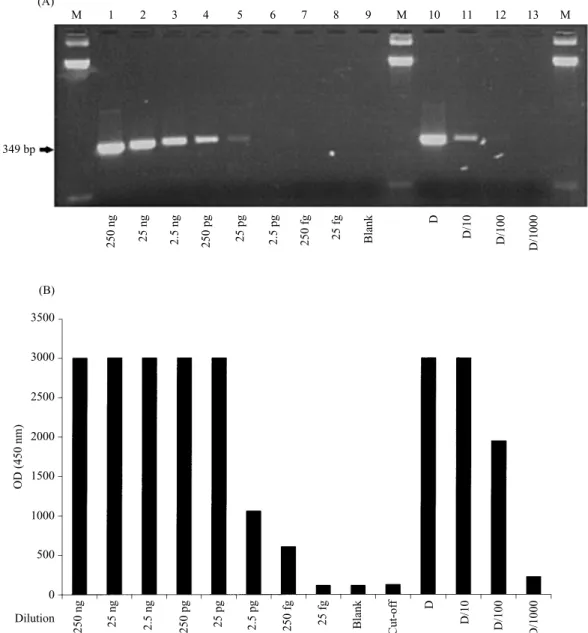
The sensitivity of the detection system using the oligo-nucleotides described above was evaluated either on products obtained from samples of varying concentrations of target DNA (from 250 ng to 25 fg, corresponding to 2±5¬10' to less than one genome copy) or on varying dilutions of PCR product obtained from 250 ng of target DNA included in the PCR assay. The sensitivity was between 25 to 250 fg of DNA (about 2–5 genome copies). This value is one hundred times higher than that achieved analysing the PCR product on ethidium bromide stained agarose gel (Fig. 4).
cina-6a 5«
Fig. 3.Nucleotide sequence of the fragment named cina-6a, obtained from the vectorette PCR amplification, initiated with primer 95.441 (see Fig. 2), extending into the 3« non-coding region. Also shown, are the first 70 nucleotides of the 257 bp fragment amplified with the degenerated primers. Capture (95.464) and detection (96.009) and PCR amplification oligonucleotides (95.422 ; 96.007), are underlined.
Specificity analysis of the detection system
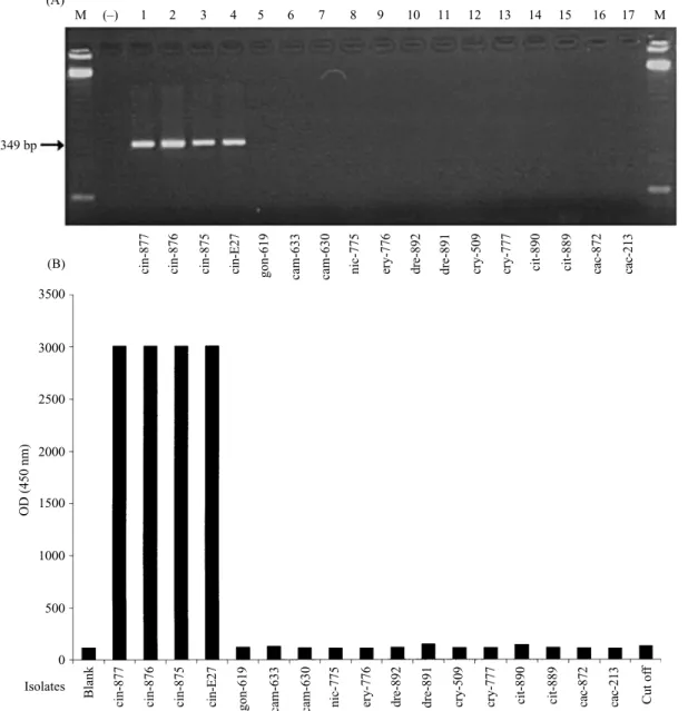
The specificity of this detection system was assessed against the isolates of the eight other reference species (Fig. 5). The analysis of the amplification reactions on agarose gel showed the presence of the expected DNA fragment for P. cinnamomi isolates and its absence from all other species. Analysis using the colorimetric assay showed a response (OD :& 3±0) clearly above the cut-off value (mean optical density of control samples, prepared from PCR in absence of DNA, plus three standard deviations) for P. cinnamomi isolates (Fig. 5). The optical densities measured for isolates from other species were well below the cut-off value, confirming the high specificity of this detection system.
Analysis of P. cinnamomi isolates from roots and soil
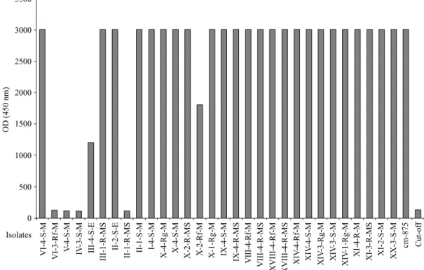
We applied this detection system to a test of 30 isolates previously identified as P. cinnamomi (Table 2) isolated from roots of Q. suber and from soils of affected areas in the Algarve region (Portugal). Twenty six samples out of thirty gave a clear positive response (Fig. 6). Four gave a clear negative response corresponding to that of the cut-off.
The four negative isolates were rechecked for affinity to P.
cinnamomi using a range of routine diagnostic tests including
colony pattern, chlamydospore production, presence of coralloid hyphae and radial growth rate (all on carrot agar at 25°); mating type, selfing in response to Trichoderma volatiles and morphological characteristics of selfed gametangia. They showed no evident differences from the other 26 P. cinnamomi
M 1 2 3 4 5 6 7 8 9 M 10 11 12 13 M 349 bp (A) (B) 250 ng 25 ng 2.5 ng 250 pg 25 pg 2.5 pg 250 fg 25 fg Blank D D/10 D/100 D/1000 250 ng 25 ng 2.5 ng 250 pg 25 pg 2.5 pg 250 fg 25 fg Blank D D/10 D/100 D/1000 Cut-of f Dilution 0 500 1000 1500 2000 2500 3000 3500 OD (450 nm)
Fig. 4.Detection of amplified P. cinnamomi DNA by agarose gel electrophoresis (A) and by colorimetric hybridization assay (B). Sensitivity assays : samples correspond to 10-fold dilution starting at 250 ng of DNA from the isolate cin-875 submitted to PCR (lanes 1–8) and to 10-fold dilution of amplified DNA (lanes 10–13). Lane M : molecular marker 123 bp DNA ladder. Lane 9 : negative control of the PCR reaction. The cut-off value was defined as 3 standard deviations above the mean for the blank PCR mixtures.
isolates and were again considered to fall within the normal range of variation for the traditional P. cinnamomi morpho-species (C. M. Brasier, personal communication).
D I S C U S S I O N
Identification of P. cinnamomi and other phytophthoras has until recently mainly relied on analysis of morphological characteristics. However, the value of these characters in delineating species units in Phytophthora is in question (Brasier, 1990, 1997). Moreover, such identification usually involves prior isolation by using baits and selective medium, which is itself time consuming and is an unreliable procedure. We have developed a method based on PCR with a fast hybridization for the specific identification of P. cinnamomi. Such methods could be easily and routinely applied to analysis of a large number of Phytophthora cultures.
The discrimination of Phytophthora taxa by PCR has been achieved previously by gel electrophoresis and, sometimes, by Southern blots (Goodwin, Kirkpatrick & Duniway, 1989 ; Goodwin, Kirkpatrick & Duniway, 1990 ; Goodwin et al., 1990 ; Lee & Taylor, 1992 ; Dobrowolski & O’Brien, 1993 ; Lee, White & Taylor, 1993 ; E; rsek, Schoelz & English, 1994). In the latter cases the sensitivity was only a minimum of 10 pg, about 400 times less than that we have achieved (see below), by using this combined PCR-hybridization method. The present method, evaluated against a limited number of traditional morphospecies (P. cactorum, P. cambivora, P.
citrophthora, P. cryptogea, P. drechsleri, P. gonapodyides, P. erythroseptica and P. nicotianae) showed an excellent specificity.
The related P. cambivora, which was shown to have highly homologous DNA sequences (Fig. 2) was also clearly discriminated.
Our approach is based on a colorimetric sandwich capture hybridization assay that has been developed for the
cr y-777 dre-892 cam-630 cin-E27 M 1 2 3 4 5 6 7 8 9 10 11 12 13 17 M 349 bp (A) (B) cin-877 Isolates 0 500 1000 1500 2000 2500 3000 3500 OD (450 nm) (–) 14 15 16
cin-876 cin-875 gon-619 cam-633 nic-775 ery-776 dre-891 cry-509 cit-890 cit-889 cac-872 cac-213
cr
y-777
dre-892
cam-630
cin-E27
cin-877 cin-876 cin-875 gon-619 cam-633 nic-775 ery-776 dre-891 cry-509 cit-890 cit-889 cac-872 cac-213
Blank Cut of
f
Fig. 5.Specificity analysis of P. cinnamomi : 2 % agarose gel electrophoresis of PCR amplified DNA (A) and colorimetric hybridization assay (B). Lane M : molecular marker 123 bp DNA ladder (Life Technology). (®): negative control of the PCR reaction. The cut-off value was defined as 3 standard deviations above the mean for the blank PCR mixtures. Isolate name corresponds to the species shown on Table 1.
identification of infectious agents in clinical specimens (Fauville-Dufaux et al., 1995 ; Lage et al., 1996 ; Mansy et al., 1996). The procedure takes advantage of an original DNA chemistry to generate multi-biotinylated probes and to immobilize oligonucleotides onto plastic support (De Vos, Van Elsen & Bollen, 1994). Analysis of the hybridization results can be carried out on an automated microplate reader allowing simplification of manipulations and rapid testing of a high number of isolates.
We chose to target the cinnamomin gene of P. cinnamomi. The cinnamomin protein is a polypeptide secreted by P.
cinnamomi which belongs to a family of proteins designated as
elicitins. It was shown to induce a hypersensitive response in tobacco (Huet & Pernollet, 1989). To target the corresponding coding DNA we had to use degenerated primers chosen on the basis of the codon usage for Phytophthora. The objective of this amplification was to make available a DNA fragment
ready for cloning and sequencing. The results obtained showed a slight variability between two isolates, cin-875 and cin-876 that could be explained by assuming discrepancies induced by the Taq DNA polymerase during the amplification. However, we can not discard the possibility of a natural variability among these isolates. This will be investigated further and was considered beyond the scope of the present work.
The establishment of the nucleotide sequence allowed us to define a non-degenerated pair of primers to amplify a consensus DNA fragment of 199 bp inside the coding region of the cinnamomin gene. In spite of several attempts to increase sensitivity and specificity using these primers under different experimental conditions we were not able to surpass the sensitivity limit of 25 pg. Furthermore, one out of the eight Phytophthora species analysed as control for the establishment of specificity to P. cinnamomi gave a positive
VI-4-S-M Isolates 0 500 1000 1500 2000 2500 3000 3500 OD (450 nm) VI-3-Rf-M V -4-S-M IV -3-S-M III-4-S-E III-1-R-MS II-2-S-E II-1-R-MS II-1-S-M I-4-S-M X-4-Rg-M X-4-S-M X-2-R-MS X-2-Rf-M X-1-Rg-M IX-4-S-M IX-4-R-MS VIII-4-Rf-M VIII-4-R-MS XVIII-4-Rf-M XVIII-4-R-MS XIV -4-Rf-M XIV -4-S-M XIV -3-Rg-M XIV -3-S-M XIV -1-Rg-M
XI-4-R-M XI-3-R-MS XI-2-S-M XX-3-S-M cm-875 Cut-of
f
Fig. 6.Test of P. cinnamomi isolates from soil and root samples by the colorimetric assay. The origin of the isolates is shown on Table 2. Cin-875 : positive control of the reaction.
response. Indeed, this pair of primers also initiated the amplification of P. cambivora DNA. P. cambivora is known to be in the same major molecular cluster as P. cinnamomi (Cooke & Duncan, 1997) and is therefore a good test for specificity. Hence the lack of specificity observed is not unexpected.
In order to achieve specificity against P. cambivora, we cloned and sequenced a DNA fragment from an isolate of this species, corresponding to the ‘ cinnamomin-like ’ gene, using the same pair of degenerated primers for PCR amplification. Not surprisingly, the comparison of the sequences showed 96 % homology between homologous fragments of the two species. It is evident that P. cambivora contains nucleotide sequences corresponding to a hypothetical ‘ cambivorin ’ gene. Whether these sequences actually code for an active polypeptide having elicitor properties remains to be demon-strated.
The sequence of the amplification primers was therefore modified in order to make them specific to P. cinnamomi and to choose divergent regions as capture and detection targets to the oligonucleotides used in the colorimetric sandwich hybridization assay. These regions need to be defined out of the cloned DNA fragment, where high homology prevents specificity. In order to look for divergent sequences we have applied the ‘ vectorette ’ system to clone the entire cinnamomin gene together with 5« and 3« coding and non-coding sequences. Several trials were carried out with different pairs of amplification primers, capture and detection oligonucleotides. Optimization was obtained targeting a region comprising 3« coding and 3« non-coding sequences (Fig. 4). The high specificity and sensitivity (up to 25 fg) achieved prompted us to apply the optimized detection system to 30 isolates of P.
cinnamomi obtained from infested soils and roots of Q. suber
from the Algarve region, identified by conventional methods.
Twenty six out of 30 isolates gave the expected positive response. The four isolates giving a negative response were re-examined, and were still considered to fall within the normal range of variation of the morphospecies P. cinnamomi. They may lack the cinnamomin gene or may have a different elicitor pattern from the others. However, it is clear that the present method is likely to identify some 80 % of P. cinnamomi isolates from cork oak areas in southern Portugal.
We have now extended our study to a larger number of isolates of P. cinnamomi including isolates from other parts of the world and isolates of the A1 mating type. We plan to carry out nucleotide sequencing analysis on the negative isolates in order to look for variations in the elicitor pattern. We also plan to further test the application of this method to the diagnosis of P. cinnamomi directly from roots and soil. We are grateful to Professor C. M. Brasier for both the initial and the reexamination identification of isolates referred in Table 2 and Fig. 6, and for critically reading the manuscript.
R E F E R E N C E S
Arnold, C. & Hodgson, I. J. (1991). Vectorette PCR : a novel approach to genomic walking. PCR Methods and Applications 1, 39–42.
Beaucage, S. L. & Caruthers, M. H. (1981). Deoxynucleoside phosphor-amidites, a new class of key intermediates for doxypolynucleotide synthesis. Tetrahedron Letters 22, 1859–1862.
Brasier, C. M. (1990). Current questions in Phytophthora systematics : the role of the population approach. In Phytophthora (ed. J. A. Lucas, R. C. Shattock, D. S. Shaw & L. R. Cooke), pp. 104–128. Cambridge University Press : Cambridge, U.K.
Brasier, C. M. (1992). Oak mortality in Iberia. Nature London 360, 539. Brasier, C. M. (1993). P. cinnamomi as a contributory factor in European oak
on oak decline. (ed. N. Luisi, P. Lerario & A. Vannini), pp. 49–57.
Dipartimento de Patologia Vegetale, Universita' degli Studi: Bari, Italy. Brasier, C. M. (1997). Fungal species in practice : identifying species units in
fungi. In Species : the units of biodiversity (ed. M. F. Claridge, H. A. Dahwah & M. R. Wilson), pp. 135–170. London and New York : Chapman & Hall. Brasier, C. M., Robredo, F. & Ferraz, J. F. P. (1993). Evidence for P. cinnamomi
involvement in Iberian oak decline. Plant Pathology 42, 140–145. Cooke, D. E. L. & Duncan, J. (1997). Phylogenetic analysis of Phytophthora
species, based on ITSI and MS II sequences of the ribosomal RNA gene repeat. Mycological Research (in press).
Crandall, B. S., Gravatt, G. F. & Rian, M. M. (1945). Root disease of Castanea species and some coniferous and broadleaf nursery stocks caused by P.
cinnamomi. Phytopathology 35, 162–180.
De Vos, M. J., Van Elsen, A. & Bollen, A. (1994). New non nucleosidic phosphoramidites for the solid phase multi-labelling of oligonucleotides : comb- and multifork-like structures. Nucleosides & Nucleotides 13, 2245–2265.
Dobrowolski, M. P. & O’Brien, P. (1993). Use of RAPD-PCR to isolate a species specific DNA probe for Phytophthora cinnamomi. FEMS Microbiology
Letters 113, 43–48.
E; rsek, J. E.-Schoelz, J. E. & English, J. T. (1994). PCR amplification of species-specific DNA sequences can distinguish among Phytophthora species.
Applied Environmental Microbiology 60, 2616–2621.
Fauville-Dufaux, M., Maes, N., Severin, E., Farin, C., Serruys, E., Struelens, M., Younes, N., Vincke, J.-P., De Vos, M.-J., Bollen, A. & Godfroid, E. (1995). Rapid identification of Mycobacterium xenopi from bacterial colonies or ‘ Bactec ’ culture by the polymerase chain reaction and a luminescent sandwich hybridization assay. Research in Microbiology 146, 349–356. Goodwin, P. H., English, J. T., Neher, D. A., Duniway, J. M. & Kirkpatrick,
B. C. (1990). Detection of Phytophthora parasitica from soil and host tissue with a species-specific DNA probe. Techniques 80, 277–281.
Goodwin, P. H., Kirkpatrick, B. C. & Duniway, J. M. (1989). Cloned DNA probes for identification of Phytophthora parasitica. Phytopathology 79, 716–721.
Goodwin, P. H., Kirkpatrick, B. C. & Duniway, J. M. (1990). Identification of
Phytophthora citrophthora with cloned DNA probes. Applied Environmental Microbiology 55, 669–674.
Huet, J.-C. & Pernollet, J.-C. (1989). Amino acid sequence of cinnamomin, a new member of the elicitin family, and its comparison to cryptogein and capsicein. FEBS Letters 257, 302–306.
Judelson, H. S. & Messenger-Routh, B. (1996). Quantitation of Phytophthora
cinnamomi in avocado roots using a species-specific DNA probe. Phytopathology 86, 763–768.
(Received 17 March 1997)
Kamoun, S., Young, M., Glascock, C. B. & Tyler, B. M. (1993). Extracellular protein elicitors from Phytophthora : host-specificity and induction of resistance to bacterial and fungal phytopathogens. Molecular Plant-Microbe
Interactions 6, 15–25.
Lage, A. P., Fauconnier, A., Burette, A., Glupczynski, Y., Bollen, A. & Godefroid, E. (1996). Rapid colorimetric hybridization assay for detecting
Helicobacter pylori DNA in gastric specimens. Journal of Clinical Microbiology
34, 530–533.
Lee, S. B. & Taylor, J. W. (1992). Phylogeny of five fungus-like protoctistan
Phytophthora species, inferred from the internal transcribed spacers of
ribosomal DNA. Molecular Biology and Evolution 9, 636–653.
Lee, S. B., White, T. J. & Taylor, J. W. (1993). Detection of Phytophthora species by oligonucleotide hybridization to amplified ribosomal DNA spacers. Phytopathology 83, 177–181.
Mansy, F., Hoyois, B., De Vos, M.-J., Van Elsen, A., Bollen, A. & Godefroid, E. (1996). A colorimetric solid phase capture hybridization assay for detection of amplified Borrelia burgdorferi DNA. BioTechniques 21, 122–125. Mao, Y. & Tyler, B. M. (1991). Genome organization of Phytophthora
megasperma f. sp. glycinea. Experimental Mycology 16, 283–291.
Matteucci, M. D. & Caruthers, M. H. (1981). Synthesis of deoxyoligo-nucleotides on a polymer support. Journal of the American Chemical Society 103, 3185–3191.
Pimentel, A. A. L. (1949). P. cinnamomi (Rands) um outro agente extremamente virulento da ‘‘ doença da tinta ’’ do castanheiro. Agronomia lusitana 9, 181–191.
Rozman, D. & Komel, R. (1994). Isolation of genomic DNA from filamentous fungi with high glucan level. BioTechniques 16, 382.
Shearer, B. L. & Tippett, J. T. (1989). Jarrah Dieback : The Dynamics and Management of P. cinnamomi in the Jarrah (Eucalyptus marginata) Forest of South-western Australia. Department of Conservation and Land Man-agement. Western Australia : Research Bulletin No. 3, 1–76.
Tooley, P. W. & Therrie$ n, C. D. (1987). Cytophotometric determination of the nuclear DNA content of 23 Mexican and non-Mexican isolates of
Phytophthora infestans. Experimental Mycology 11, 19–26.
Waterhouse, G. M., Newhook, F. J. & Stamps, D. J. (1983). Present criteria for classification of Phytophthora. In Phytophthora (ed. D. C. Erwin, S. Bartnicki-Garcia & P. H. Tsao), pp. 139–147. The American Phytopathology Society : St. Paul, Minnesota.
Wills, R. T. (1993). The ecological impact of Phytophthora cinnamomi in the Stirling Range National Park, Western Australia. Australian Journal of
Ecology 18, 145–159.
Zentmeyer, G. A. (1980). P. cinnamomi and diseases it causes. American