An Oligonucleotide Probe Derived from kDNA Minirepeats
is Specific for
Leishmania (Viannia)
Octavio Fernandes/*/
+, Marcelo Bozza**, Juan M Pascale***, Antonio B de
Miranda, Ulisses G Lopes**, Wim M Degrave
Departamento de Bioquímica e Biologia Molecular, Instituto Oswaldo Cruz, Av. Brasil 4365, 21045-900 Rio de Janeiro, RJ, Brasil *Departamento de Patologia, Faculdade de Ciências Médicas, Universidade do Estado do Rio
de Janeiro, RJ, Brasil **Departamento de Biofísica, Universidade Federal do Rio de Janeiro, RJ, Brasil ***Departamento de Parasitología, Universidad de Panama, Panama
Sequence analysis of Leishmania (Viannia) kDNA minicircles and analysis of multiple sequence alignments of the conserved region (minirepeats) of five distinct minicircles from L. (V.) braziliensis
species with corresponding sequences derived from other dermotropic leishmanias indicated the pres-ence of a sub-genus specific sequpres-ence. An oligonucleotide bearing this sequpres-ence was designed and used as a molecular probe, being able to recognize solely the sub-genus Viannia species in hybridiza-tion experiments. A dendrogram reflecting the homologies among the minirepeat sequences was con-structed. Sequence clustering was obtained corresponding to the traditional classification based on similarity of biochemical, biological and parasitological characteristics of these Leishmania species, distinguishing the Old World dermotropic leishmanias, the New World dermotropic leishmanias of the sub-genus Leishmania and of the sub-genus Viannia.
Key words: Leishmania (Viannia) identification minicircle DNA sequences oligonucleotide probe -Leishmania braziliensis - Leishmania panamensis
The parasitological diagnosis of leishmaniasis has received, in recent years, a growing attention due to the development of molecular probes, ca-pable both of detecting and typing the infecting parasite (Ramirez & Guevara 1987, Van Eys et al. 1987, 1991, Howard et al. 1991, Degrave et al. 1994a, b). Traditional methods for identification are slow and cumbersome, and suffer from a lack of sensitivity (Weigle et al. 1987). Thus, methods like culture of the protozoa from human samples, microscopic analysis of stained smears or histopathological study of biopsy material might be replaced in the future by fast, sensitive and ac-curate diagnostic assays, using molecular probes and/or the polymerase chain reaction (PCR)(Barker et al. 1986, Barker 1987, Uliana et al. 1991).
Probes derived from different sequences of the nuclear and mitochondrial genome have been used in an attempt to develop Leishmania detection and typing assays (Wirth & McMahon-Pratt 1982, Van Eys et al. 1992). However, the kinetoplast DNA
(kDNA) network, which is comprised of maxi- and minicircle molecules, has been the target of choice in many laboratories (Simpson 1987). Leishma-nia minicircles show features that make them al-most ideal as molecular probes, since they are present in the kinetoplast network in a high copy number and contain a conserved region of at least 120 bp that can be evidenced in every molecule (Rogers & Wirth 1987, 1988). Specific probes based on total kDNA, complete minicircle mol-ecules or restriction fragments, have been gener-ated and used in the identification of different leish-manias from the Old and New World (Lopes & Wirth 1986, Das Gupta et al. 1991, Laskay et al. 1991). This approach proved to be suitable for epidemiological purposes and as a diagnostic method (Wirth et al. 1986, Rogers et al. 1988, Barker 1989).
Detection systems based on PCR are very prom-ising due to their sensitivity and specificity (Saiki et al. 1985). However, secondary detection and specific typing probes for identification of PCR amplified products are highly desirable, since they offer a drastically increased specificity of the PCR assay and raise about ten-fold the sensitivity of the system, allowing for automation of the PCR scor-ing procedure, concomitantly with the typscor-ing of the infecting Leishmania species. Several PCR systems use amplification oligonucleotides complementary to the conserved sequence motifs corresponding to the origins of replication of the
Sponsored by the UNDP/World Bank/WHO Special Pro-gram for Research and Training in Tropical Diseases (TDR), the International Atomic Energy Agency, the FIOCRUZ PAPES Program and CNPq.
+Corresponding author. Fax: 55-21-590.3495
minicircle light and heavy strands (Rodgers et al. 1990).
Whole kDNA can be used as a molecular probe to distinguish between the two New World dermotropic Leishmania groups: the sub-genus
Viannia (braziliensis, guyanensis, panamensis, etc.) and Leishmania (amazonensis, mexicana, etc.) (Wirth & McMahon-Pratt 1982). However, cross hybridization with Old World leishmanias and L. chagasi occurs (Das Gupta et al. 1991). Synthetic oligonucleotides directed to specific sequences can circumvent these problems and can be used in a PCR assay.
We report here the partial nucleotide sequences of L. braziliensis and L. panamensis minicircles and the identification of an oligonucleotide se-quence specific to the subgenus Viannia, based on multiple sequence alignments of the minicircles conserved regions (minirepeats) from New World dermotropic leishmanias. Furthermore, a dendro-gram reflecting the different minirepeat sequence homologies between these dermotropic leishma-nias was constructed.
MATERIALS AND METHODS
Parasite culture and kDNA isolation - The
Leishmania strains used in this study were: L. braziliensis MHOM/BR/75/M2903, kindly pro-vided by Dr Gabriel Grimaldi, FIOCRUZ, Rio de Janeiro, and L. panamensis IPAN V (Panama Uni-versity, Panama). Promastigotes were grown at 24°C in Schneider’s medium (GIBCO, Grand Is-land, NY) supplemented with 10% heat-inactivated fetal calf serum. The taxonomic nomenclature fol-lows the recommendation of the World Health Organization.
The cells were spun down at 5000 X g for 10 min at 4°C, washed with cold saline/EDTA (0,15 M NaCl/10 mM EDTA), centrifuged and resus-pended in TE10 (10 mM Tris-HCl pH 7.4 /10 mM EDTA). Proteinase K (Boehringer Mannheim) and sarkosyl were added to final concentrations of 100
µg/ml and 0.5%, respectively. The lysate was in-cubated at 56°C for 2 hr and submitted to mechani-cal shear, followed by two ultracentrifugations at 30.000 rpm for 1 hr at 4°C. The pellet was resus-pended in 200 µl of TE buffer and extracted with phenol/chloroform (1:1). The DNA was precipi-tated with ethanol and resuspended in TE1 (10 mM Tris-HCl pH 7.4/ 1 mM EDTA).
Minicircle cloning and nucleotide sequence determination - Total L. braziliensis kDNA was digested with Msp I and minicircles were cloned in the Acc I site of M13mp18. L. panamensis was digested with Dra I and cloned in the Hinc II site of pBluescript® KS-. Nucleotide sequencing was done by the chain termination method using either
USB® Sequenase kit and α-32P-dATP labeling or the Taq Dye Primer Cycle Sequencing Kit on a 373A Automated DNA Sequencer (Applied Biosystems), according to the instructions of the manufacturer.
Computer analysis and oligonucleotide synthe-sis - Nucleotide sequences were analyzed using software from the Genetics Computer Group (GCG) on a VAX computer. Oligonucleotides were synthesized on an Applied Biosystems 381A DNA synthesizer and purified by thin layer chro-matography (TLC).
Cell dot experiments and hybridization assays
- Leishmania cells were pelleted, washed with TE10 and counted. 105 cells were applied on nylon fil-ters (Zetaprobe, Biorad) treated with 10mM Tris-HCl pH 8.0, 10 mM EDTA, 0.1% Triton X-100, 100 µg/ml proteinase K at 56°C during 1 hr. The filter was transferred to 0.4 N NaOH for 5 min and then rinsed with 2xSSC and dried. Hybridization conditions and washing stringencies are detailed below in results.
RESULTS
Minicircle cloning and sequencing - One L. panamensis, one L. amazonensis and several cloned
L. braziliensis minicircles were partially sequenced and the data were submitted to GenBank (Fig. 2, Table). The molecules apparently belong to dif-ferent minicircle classes, as judged from the ex-tensive heterogeneity of the minirepeat neighbor-ing sequences (data not shown).
Multiple sequence alignment of minirepeats -We performed a multiple alignment analysis of the conserved minirepeats from New and Old World dermotropic Leishmania minicircles (Fig. 2), us-ing the Pileup program from the GCG package. Regions with higher or lower conservation become evident when comparing the alignment with a de-duced consensus sequence. A dendrogram reflect-ing the homologies between these sequences is shown in Fig. 3. Although this dendrogram does not necessarily reflect a phylogenetic relationship between the different Leishmania species, it does cluster the Leishmania species in groups that are coherent with previous classifications based upon biological, parasitological and biochemical obser-vations (WHO 1980).
Oligonucleotide design - The multiple align-ment of the minirepeats regions has enabled us to evidence a region, adjacent to the 5' end of the ca-nonical twelvemer sequence, that seems highly specific to leishmanias of the sub-genus Viannia.
sug-gested by the sequence analysis. Using stringent hybridization conditions and washing in 0.5xSSC at 37°C, we demonstrated that this oligonucleotide is specific to the sub-genus Viannia (Fig. 1A), in contrast to hybridization with an oligonucleotide complementary to the minicircle “universal 12-mer sequence” (5’-GGGGTTGGTGTAAT/AATAGG/ TGGC-3’- OLT) (Fig. 1B) or with L. brasiliensis
total kDNA (Fig. 1C).
DISCUSSION
Leishmania species belonging to the sub-ge-nus Viannia are the main agents of cutaneous leishmaniasis in Latin America. However, there are other leishmanias from the sub-genus Leish-mania, which are also responsible for human cuta-neous infections (Grimaldi & Tesh 1993). In some areas there is an overlapping of different dermotropic or viscerotropic leishmanias, creating a complex epidemiologic pattern of transmission. Moreover, the prognosis of human leishmaniasis seems also to depend on the species involved in the infection. It is thus highly desirable to develop molecular probes that are able to identify and type the different leishmanias (Barker 1989).
Since the use of oligonucleotides as molecular probes is inexpensive, more specific, faster and more practical as compared to whole kDNA, we cloned and partially sequenced five different minicircles from L. (V.) braziliensis, searching for short sequences specific to minicircles from this relevant human pathogenic parasite group.
TABLE
Minicircle sequences used in the multiple alignment
Minicircle Leishmania strain GenBank Described by
nomenclature accession
number
L. braziliensis 1 MHOM/BR/75/2903 U19803 this article L. braziliensis 2 MHOM/BR/75/2903 U19804 this article L. braziliensis 3 MHOM/BR/75/2903 U19805 this article L. braziliensis 4 MHOM/BR/75/2903 U19806 this article L. braziliensis 5 MHOM/BR/75/2903 U19807 this article
L. braziliensis 6 MHOM/BR/75/2904 M87315 de Bruijin & Barker 1992 L. guyanensis 1 151 M X54470 Samuelson et al. 1991 L. guyanensis 2 151 M X54471 Samuelson et al. 1991 L. guyanensis 3 151 M X54472 Samuelson et al. 1991 L. guyanensis 4 UMB/BR/76/M4196 M87316 de Bruijin & Barker 1992 L. panamensis 1 MHOM/PA/75/M4037 M87314 de Bruijin & Barker 1992 L. panamensis 2 IPAN V U19811 this article
L. peruviana MHOM/PE/76/SL5 M87317 de Bruijin & Barker 1992 L. amazonensis 1 IFLA/BR/67/PH8 Rogers & Wirth 1987 L. amazonensis 2 MHOM/BR/73/M2269 U19810 this article
L. major MHOM/TN/84/ISS108 J04654 Smith et al. 1989
L. aethiopica pPK763 Barker et al. 1986
L. tarentolae Clonal strain C-1 K01979 Kidane et al. 1984
Fig. 1: cell dot hybridization of different Leishmania species with the oligonucleotide OLB (1.A), OLT (1.B) or L. braziliensis total kDNA (1.C) as molecular probes. 1) L. (V.) guyanensis (M4147); 2) L. (V.) braziliensis (M2309); 3) L. (V.) panamensis (IPAN5); 4) L. (L.) amazonensis (M2269); 5)
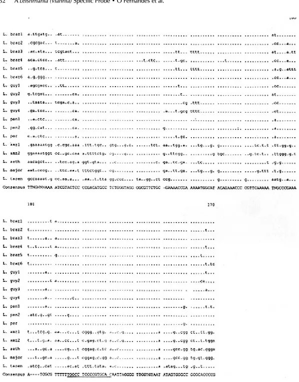
Fig. 2: multiple alignment of 170 bp of the conserved region from six different minicircle molecules of Leishmania braziliensis, four minicircles of L. guyanensis, two of L. panamensis, two of L. amazonensis, one of L. peruviana,L. aethiopica, L. major and
L. tarentolae. The leishmania strains, the minicircle nomenclature and the GenBank accession numbers are listed in Table. The conserved 12mer sequence, present in all Leishmania minicircles, is marked in bold. The Viannia specificoligonucleotide sequence is underlined. Nucleotide positions identical to the consensus sequence were marked with a dot.
Based on the multiple alignment of the obtained sequences with those of the conserved regions of other Leishmania minicircles from different spe-cies, we identified a region of 16 nucleotides (OLB, Fig. 2), that proved to be specific for the
sub-ge-nus Viannia in cell dot hybridization experiments (Fig. 1), demonstrating its use as a typing probe. This region is 5' adjacent to the conserved block of 12 nucleotides, found in all Leishmania
served region of the minicircle molecule. Species of the sub-genus Leishmania are also grouped, and separated from the Viannia cluster.
ACKNOWLEDGEMENTS
To Dr Gabriel Grimaldi for strains donations and culture facilities.
REFERENCES
Barker D 1987. DNA Diagnosis of Human Leishmaniasis. Parasitol Today 3: 177-184. Barker D 1989. Molecular approaches to DNA
diagno-sis. Parasitol99: 129-146.
Barker DC, Gibson LJ, Kennedy WPK, Nasser AAAA, Williams RH 1986. The potential of using recombi-nant DNA species-specific probes for the identifi-cation of tropical Leishmania. Parasitol 91: S139-S174.
Das Gupta S, Ghosh DK, Majumder HK 1991. A cloned kinetoplast DNA minicircle fragment from a Leish-mania sp. specific for post-kala-azar dermal leishmaniasis strains. Parasitol102: 187-191. De Bruijn M, Barker D 1992. Diagnosis of New World
leishmaniasis: specific detection of species of the Leishmania braziliensis complex by amplification of kinetoplast DNA. Acta Tropica 52: 45-48. Degrave WM, Fernandes O, Campbell D, Bozza M,
Lopes UG 1994b. Use of molecular probes and PCR for detection and typing of Leishmania - A mini-review. Mem Inst Oswaldo Cruz89: 463-469. Degrave WM, Fernandes O, Thiemann O, Wincker P,
Britto C, Cardoso A, Pereira JB, Bozza M, Lopes U, Morel C 1994a. Detection of Trypanosoma cruzi and Leishmania using the Polymerase Chain Reac-tion. Mem Inst Oswaldo Cruz89: 367-368. Grimaldi G, Tesh RB 1993. Leishmaniases of the New
World: current concepts and implications for future research. Clin Microbiol Rev6: 230-250.
Howard MK, Kelly JM, Lane RP, Miles MA 1991. A sensitive repetitive DNA probe that is specific to the Leishmania donovani complex and its use as an epi-demiological and diagnostic reagent. Mol Biochem Parasitol44: 63-72.
Kidane G, Hughes D, Simpson L 1984. Sequence het-erogeneity and anomalous electrophoretic mobility of kinetoplast minicircle DNA in Leishmania tarentolae. Gene27: 265-277.
Laskay T, Kiessling R, Dewit TFR, Wirth DF 1991. Gen-eration of species-specific DNA probes for Leish-mania aethiopica. Mol Biochem Parasitol44: 279-286.
Lopes UG, Wirth DF 1986. Identification of visceral Leishmania species with cloned sequences of kine-toplast DNA. Mol Biochem Parasitol20: 77-84. Ramirez JL, Guevara P 1987. The ribosomal gene spacer
as a tool for the taxonomy of Leishmania. Mol Biochem Parasitol22: 177-183.
Rodgers MR, Popper SJ, Wirth DF 1990. Amplifica-tion of kinetoplast DNA as a tool in the detecAmplifica-tion and diagnosis of Leishmania. Exp Parasitol 71: 267-275.
Rogers WO, Wirth DF 1987. Generation of sequence
synthesized (OLT, Fig. 1B, Fig. 2). Apart from its use as a typing probe in cell-dot experiments, the OLB oligoprobe proved to be very useful for the detection and typing of PCR products (Degrave et al. 1994a), generated using oligonucleotides complementary to the minicircle light and heavy strand origins of replication.
Moreover, using oligonucleotides directed out-wardly from the Viannia specific region and from the origin of replication of the minicircle heavy strand (12mer sequence), as primers in a PCR re-action, it is possible to amplify the whole minicircle molecule. This method may be useful in typing experiments in a schizodeme-like analysis after full length minicircle amplification (Degrave et al. 1994b). De Bruijn and Barker (1992) have previ-ously used such amplification of whole minicircles for diagnostic purposes.
The L. Viannia minirepeat sequences demon-strate extensive sequence conservation. Moreover, point mutations are clustered in a few regions and do not distinguish between the different species, as shown by the clustering produced by the com-puter analysis of the Leishmania minirepeats. All the Viannia species are grouped together, with a near homogeneity inside the sub-genus taxon in
Leishmania species when considering the
diversity in the kinetoplast DNA minicircles of Leish-mania mexicana amazonensis. Mol Biochem Parasitol30: 1-8.
Rogers WO, Burnheim PF, Wirth DF 1988. Detection of Leishmania within sand flies by kinetoplast DNA hybridization. Am J Trop Med Hyg 39: 434-439. Saiki R, Scharf SJ, Faloona F, Mullis K, Horn G, Erlich
H, Arnheim N 1985. Enzymatic amplification of beta globin genomic sequence and restriction site analy-sis for diagnoanaly-sis of sickle cell anemia. Science230: 1350-1354.
Samuelson J, Lerner E, Tesh R, Titus R 1991. A mouse model of Leishmania brasiliensis brasiliensis infec-tion produced by coinjecinfec-tion with sandfly saliva. J Exp Med173: 49-54.
Simpson L 1987. The mitochondrial genome of kinetoplastid protozoa: genomic organization, tran-scription, replication and evolution. Annu Rev Microbiol41: 363-382.
Uliana SRB, Affonso MHT, Camargo EP, Floeter-Win-ter LM 1991. Leishmania: genus identification based on a specific sequence of the 18S ribosomal RNA sequence. Exp Parasitol72: 157-163.
Van Eys GJJM, Guizani I, Ligthart GS, Dellagi K 1991. A nuclear DNA probe for the identification of strains within the Leishmania donovani complex. Exp Parasitol72: 459-463.
Van Eys GJJM, Schoone GJ, Kroon NCM, Ebeling SB 1992. Sequence analysis of small subunit ribosomal RNA genes and its use for detection and identifica-tion of Leishmania parasites. Mol Biochem Parasitol 51: 133-142.
Van Eys GJJM, Schoone GJ, Ligthart GS, Laarman JJ, Terpstra WJ 1987. Detection of Leishmania para-sites by DNA in situ hybridization with non-radio-active probes. Parasitol Res73: 199-202.
Weigle KA, Davalos M, Heredia P, Molineros R, Saravia N, D’Alessandro A 1987. Diagnosis of cutaneous and mucocutaneous leishmaniasis in Colombia: a comparison of seven methods. Am J Trop Med Hyg 36: 489-496.
WHO 1980 - World Health Organization - Biochemical characterization of Leishmania. Proceedings of a workshop held at the Pan American Health Organi-zation, Washington, D.C. ML Chance & BC Walton (eds).
Wirth DF, McMahon-Pratt D 1982. Rapid identifica-tion of Leishmania species by specific hybridiza-tion of kinetoplast DNA in cutaneous lesions. Proc Nat Acad Sci USA77: 6810-6814.