U
NIVERSIDADE DEL
ISBOAF
ACULDADE DEC
IÊNCIASD
EPARTAMENTO DEQ
UÍMICA EB
IOQUÍMICAM
OLECULAR DIAGNOSIS OF
F
AMILIAL
H
YPERCHOLESTEROLEMIA
AND
F
UNCTIONAL CHARACTERIZATION OF MISSENSE VARIANTS IN
THE
LDLR
GENE
Sílvia Valentim de Azevedo
DissertaçãoMestrado em Bioquímica
Orientação: Doutora Mafalda Bourbon 2014/2015
i
Agradecimentos
Primeiramente, quero agradecer à Doutora Mafalda Bourbon por me ter aceite, motivado e ensinado que o trabalho e o esforço compensam sempre. Graças a si e à sua paixão pela investigação sou hoje uma aluna mais capaz e dedicada. É a responsável pelo início do Estudo Português de Hipercolesterolemia Familiar, do qual fazem parte dezenas de médicos e centenas de doentes, aos quais deixo também o meu agradecimento.
À Patrícia, que tanto me ensinou e tantas brainstorms teve comigo. Foi, sem dúvida e sem sequer se aperceber, a minha maior inspiração no decorrer deste projecto. Quando for grande quero ser como tu!
À nossa grande família do GIC. Tanto aos que foram passando como aos que ainda ficam, pois tornaram todos os dias aqui únicos e animados. À Salima, Rita, Cibelle e Joana Ch., o meu obrigada. Um agradecimento especial à Ana, pela boa disposição constante, pelo seu interesse e curiosidade naturais; e outro à Catarina, por toda a ajuda e pelas desventuras vividas em Bilbau. Um gigante obrigada à minha Juca, pela partilha de histórias e sentimentos que levaram ao nascer de uma boa amizade, pelas horas de confidências, por tornar todas as horas passadas no laboratório mais felizes.
Ao César por toda a ajuda e orientação em Bilbau. Ao Asier, ao Aitor, à Rocío e à Garazi, por toda a animação e boa disposição que os dias cinzentos teimavam em contrariar. Às minhas colegas de casa Águeda e Angela. Fui super bem recebida e vocês foram, sem dúvida, os responsáveis pelos meses espetaculares que passei nesta cidade fantástica.
Ao meu núcleo de amigos, que nos últimos cinco anos me acompanharam neste percurso. Aos rapazes João, Frederico, Tomás, Gui e Lucas por todo o ânimo; ao Bustorff e ao Coelho pela paciência e por serem bons ouvintes, mesmo quanto 90% das frases que dizia eram constituídas por palavrões de fúria. Às minhas amigas do coração, Inês, Ana, Leonor, Madalena e Margarida, por todos os momentos, em especial pela mágica viagem a Berlim. Estiveram sempre presentes e contribuíram tanto para a minha formação e crescimento enquanto pessoa. Dizem que os amigos da faculdade são para sempre, e espero que sejam mesmo. Obrigada, sem vocês não teria sido o mesmo.
Às pessoas que são a presença mais constante desde há 13 anos, Joana e Andreia. O tempo tem-me trazido muitas coisas e levado outras tantas, mas vocês permanecem. Um obrigada gigante por todas as choradeiras e gargalhadas, por todas as horas a fio passadas à conversa, mesmo quando nada mais havia por dizer. À Joana por me ouvir vezes sem conta e por fazer de mim cúmplice de todos os seus esquemas... bom, nossos esquemas. À Andreia, por estar sempre presente e por todos os conselhos em alturas de, como o seu pai diria, “Oh meu deus, há dramas no mundo encantado das queens!”. Foi tão bom crescer convosco!
ii
Deixo ainda aos meus pais o maior agradecimento de todos, pois não podiam ter-me amado mais. Sinto que tenho demasiada sorte em ter-vos. Uma vez li que a maior dificuldade que os pais enfrentam é deixar um filho ser ele próprio, e vocês deixaram. Sempre. Muito obrigada. Termino com um agradecimento gigantesco aos meus queridos irmãos Patrícia e Rodrigo, simplesmente por existirem, a vida é tão melhor ao vosso lado!
Sou uma pessoa verdadeiramente feliz.
“Somos o resultado dos livros que lemos, das viagens que fazemos e das pessoas que amamos.” (Airton Ortiz)
iii
Abstract
Familial Hypercholesterolemia (FH) is an autosomal dominant disorder characterized by increased levels of plasmatic cholesterol since birth.
FH occurs due to functional variants in one of three genes: low density lipoprotein receptor (LDLR), apolipoprotein B-100 (APOB) and proprotein convertase subtilisin/kexin type 9 (PCSK9).
This disorder has an estimated prevalence of 1:500 individuals, being more than 90% of FH patients identified with a variant in the LDLR gene. The LDLR is a membrane glycoprotein, responsible for binding and uptake of low density lipoproteins (LDL), the major cholesterol transporter in blood.
Variants in LDLR gene may result in a defective LDL catabolism, leading to increased cholesterol levels in plasma, accumulating in tendons and arteries. Cholesterol accumulation in untreated FH patients leads to premature atherosclerosis and cardiovascular disease development.
Although more than 1600 variants identified in this gene are reported in databases, the majority of them remains, until now, without functional studies proving their pathogenicity. For patients carrying these variants, a definitive molecular diagnosis for FH is not possible.
The main purpose of Portuguese Study of Familial Hypercholesterolemia (EPHF) is to perform the molecular diagnosis of patients who were clinically diagnosed. Criteria used are adapted from the ones of Simon Broome. Furthermore, the performance of functional studies for variants of unknown pathogenicity is imperative, in order to better clarify the molecular basis of this disorder.
During this project, the molecular diagnosis was performed for 25 index cases, participants of Portuguese FH Study. Among all of them, 11 variants were identified in 12 patients. Among these, 2 were novel, being first described in this project. The remaining 9 have been previously reported, although only 7 have been functionally assessed. The search for large rearrangements was performed by Multiplex Ligation-dependent Probe Amplification (MLPA), but no alteration of this type was identified.
The 10 most common LDLR variants described during the Portuguese FH Study, to date without functional assessment, were selected. In silico assessment was performed using described tools, in order to predict their pathogenicity. Site-directed mutagenesis was successfully performed in a pcDNA3_LDLR plasmid for all variants, being expressed in CHO–ldlA7 cells, lacking endogenous expression of LDLR. LDLR expression, binding and uptake were independently assessed by flow cytometry.
Results suggest that among 10 functionally studied variants, 7 cause an impairment in LDLR function. Variants c.1802A>T p.(Asp601Val), c.1876G>A p.(Glu626Lys), c.631C>G p.(His211Asp), c.661G>T p.(Asp221Tyr), c.618_638del p.(Gly207_Ser213del), c.551G>A
iv
seem to be neutral, not revealing any kind of impact on the LDLR function.
Comparing functional studies with in silico tools predictions led to the conclusion that these are useful, but should not be the only source of evidence for a diagnosis associated to a pathology.
FH is a disease for which, fortunately, a genetic diagnosis and therapeutic options exist. Although it remains as a subdiagnosed disorder, the performance of a molecular diagnosis, along with functional assessment of variants with unknown pathogenicity, allows a definite diagnosis. This way, preventive measures and personalized counseling can be made in order to improve FH patients’ prognostic, providing them longer and better lives.
Key words: Familial hypercholesterolemia, LDLR, molecular diagnosis, variants, functional studies.
v
Resumo
A Hipercholesterolemia Familiar (FH) é uma patologia genética que é transmitida de forma autossómica dominante e é caracterizada por elevados níveis de colesterol no plasma desde o nascimento.
A FH ocorre devido a variantes funcionais num dos genes codificantes de três proteínas: recetor de lipoproteínas de baixa densidade (LDLR), apolipoproteína B-100 (APOB) ou pró-proteína convertase subtilisina quexina tipo 9 (PCSK9).
O LDLR é uma glicoproteína membranar que liga e internaliza colesterol associado às lipoproteínas de baixa densidade (LDL), que constituem o principal transportador de colesterol no sangue. Variantes no gene LDLR podem resultar num catabolismo deficiente das LDL, tendo como consequência o aumento do colesterol no plasma, que se acumula nos tendões e artérias. Esta acumulação pode levar ao desenvolvimento prematuro de aterosclerose e doença cardiovascular.
A FH apresenta duas formas clínicas: a forma heterozigótica, que apresenta um fenótipo menos agressivo, com valores de colesterol total entre 290 e 500 mg/dl (com LDL>190 mg/dl); e a forma homozigótica, que apresenta um fenótipo mais agressivo, com valores de colesterol total entre 600 mg/dl e 1000 mg/dl.
Estima-se que as prevalências das formas heterozigótica e homozigótica sejam de 1/500 e de 1/1000000 indivíduos, respetivamente. Sabe-se ainda que mais de 90% dos casos identificados apresentam uma variante no LDLR, fazendo deste gene o mais associado a esta doença.
Apesar de haver mais de 1600 variantes do LDLR reportadas em bases de dados, para a maior parte delas não existe registo de estudos funcionais que provem a sua patogenicidade, levando a que não seja possível atribuir um diagnóstico molecular definitivo a estes casos. Assim sendo, a necessidade de avaliar funcionalmente estas variantes, de modo a perceber em que medida afetam a função do recetor, também se torna imperativa.
Tendo em conta as prevalências acima referidas, estima-se que em Portugal existam cerca de 20000 casos de FH. O Estudo Português de Hipercolesterolemia Familiar (EPHF), implementado em 1999 no Instituto Nacional de Saúde Dr. Ricardo Jorge, tem como objectivo a determinação da prevalência e distribuição desta patologia em Portugal. A população em estudo é constituída por indivíduos de ambos os sexos e todas as idades, desde que cumpram com os critérios para o diagnóstico clínico de FH.
O diagnóstico clínico de FH é feito, em Portugal, de acordo com os critérios clínicos adaptados de “Simon Broome Heart Research Trust” e idealmente deverá ser realizado o estudo genético, com a identificação da variante, pois só desta forma é possível confirmar o diagnóstico clínico. A realização de estudos funcionais para determinar o efeito de
vi
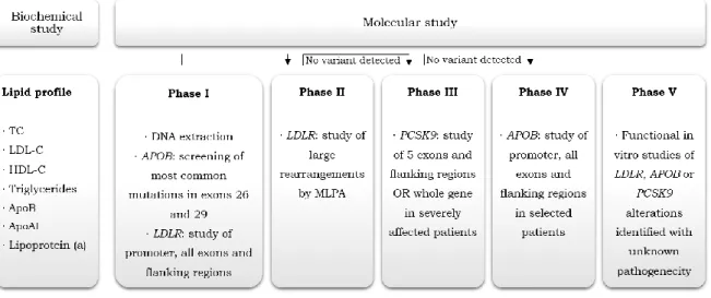
O EPHF está dividido em duas partes: o estudo bioquímico e o estudo molecular. Este último está sub-dividido em cinco fases. A fase I compreende a extração de DNA, o estudo do promotor, todos os exões e regiões adjacentes do LDLR, bem como das variantes mais comuns nos exões 26 e 29 da APOB. A fase II consiste no estudo de grandes rearranjos por Multiplex Ligation-dependent Probe Amplification (MLPA). A fase III consiste no estudo do PCSK9 e a fase IV no estudo de todo o gene da APOB. A V e última fase consiste na realização de estudos funcionais in vitro para variantes cuja patogenicidade ainda é desconhecida.
Assim sendo, este projeto está dividido em duas partes, que compreendem três das cinco fases do EPHF, nomeadamente as fases I, II e V.
A primeira parte consistiu na realização do estudo molecular em participantes do EPHF, estudando o promotor e os 18 exões e regiões adjacentes do LDLR, bem como o estudo de parte dos exões 26 e 29 da APOB por Polymerase Chain Reaction (PCR) e sequenciação de Sanger. De seguida, procedeu-se à pesquisa de grandes rearranjos por MLPA. Uma predição, utilizando as ferramentas in silico, foi também realizada, de modo a prever o impacto das alterações encontradas ao nível da proteína. Esta predição foi realizada tanto para alterações identificadas ao nível do exão, com as ferramentas Polymorphism Phenotyping (PolyPhen-2), Sorting Tolerant From Intolerant (SIFT) e Mutationtaster, como para as alterações identificadas ao nível do intrão, de modo a prever efeitos no splicing, com as ferramentas Human Splicing Finder (HSF), the Splice Site Prediction by Neutral Network (NNSSP) e FSPLICE.
A segunda parte deste projeto consistiu então no estudo funcional das 10 variantes do LDLR mais comuns na população portuguesa, até à data sem estudos funcionais. As diferentes variantes do LDLR foram obtidas por mutagénese dirigida num plasmídeo pcDNA3_LDLR sob o controlo do promotor viral SV40. Toda a região de interesse foi confirmada por sequenciação de Sanger e foi feita uma reclonagem, em que o gene LDLR, já com a variante, foi transferido para um vetor limpo. Células CHO-ldlA7, que não expressam endogenamente o recetor, foram transfetadas com os diferentes plasmídeos. A expressão do recetor foi avaliada através da deteção com anticorpos; a ligação e a internalização foram avaliados através do uso de LDL fluorescentemente marcada com FITC. O impacto de todas as variantes ao nível da expressão, ligação e internalização foi analisado por citometria de fluxo.
Em 25 casos índex estudados, 11 variantes foram identificadas em 12 doentes, embora duas destas sejam provavelmente benignas. A variante patogénica mais frequentemente encontrada na APOB foi identificada em apenas 2 doentes. De entre as alterações encontradas no LDLR (10), 2 foram aqui primeiramente reportadas e 8 já tinham sido anteriormente identificadas. De entre estas últimas, 7 já apresentavam estudos funcionais comprovando a sua patogenicidade.
vii
Aquando da pesquisa por grandes rearranjos nestes doentes por MLPA, nenhuma alteração deste tipo foi identificada no grupo em estudo.
O estudo funcional das dez alterações mais frequentemente identificadas no decorrer do EPHF, até à data sem estudos funcionais que comprovem o impacto na função do recetor, revelou que, de entre as 10 variantes estudadas, 7 são patogénicas, afetando de alguma forma a função do LDLR.
A variante c.1802A>T p.(Asp601Val) é patogénica, fazedo com que não haja sequer LDLR à superfície celular. Como não atinge a membrana (por não ser expressa ou por não se ancorar a esta) apresenta valores de ligação e de internalização igualmente baixos. As variantes c.1876G>A p.(Glu626Lys), c.631C>G p.(His211Asp), c.661G>T p.(Asp221Tyr), c.618_638del p.(Gly207_Ser213del) e c.551G>A p.(Cys184Tyr) apresentaram uma expressão normal. No entanto, foram observáveis valores muito reduzidos para a fluorescência associada à união das LDL nestes casos. Consequentemente, a internalização também é defetiva, parecendo lógico classificar estas variantes como patogénicas. A variante c.1775G>A p.(Gly592Glu) resulta num defeito, possivelmente, ao nível da reciclagem do recetor, sendo também considerada patogénica. Por outro lado, as variantes c.1816G>T p.(Ala606Ser), c.1966C>A p.(His656Asn) e c.2177C>T p.(Thr726Ile) parecem não revelar qualquer impacto no LDLR, sugerindo a sua neutralidade, pois obtiveram-se valores de fluorescência, associados à expressão e atividades de ligação e internalização, comparáveis ao wt. Estes resultados sugerem que os doentes portadores destas variantes devem apresentar outra justificação para o seu fenótipo hipercolesterolémico.
A análise destas variantes através de ferramentas de predição in silico, realizada para todas as variantes identificadas no decorrer deste projeto, permitiu concluir que estas predições nem sempre vão de encontro ao determinado através da realização de estudos funcionais. Assim sendo, estes programas deverão ser usados, mas com a devida ressalva de que são apenas preditores.
A FH é uma doença para a qual, felizmente, existe um diagnóstico definitivo (genético) e variados tratamentos farmacológicos. Apesar de ser uma doença subdiagnosticada, estão a ser realizadas diversas iniciativas para que haja uma maior divulgação da doença, nomeadamente dos benefícios do diagnóstico precoce. No âmbito da EPHF tem-se feito um esforço para que seja implementado o diagnóstico molecular como diagnóstico preferencial, juntamente com a execução de estudos funcionais para determinar a patogenicidade de variantes desconhecidas. Só assim os doentes têm a possibilidade de ter um diagnóstico definitivo para a sua patologia tornado possível instituir medidas preventivas com uma terapêutica dirigida e personalizada, melhorando o prognóstico destes doentes.
Palavras chave: Hipercolesterolemia familiar, LDLR, diagnóstico molecular, variantes, estudos funcionais.
viii
ix
Abbreviations
Aa Amino acid
ACAT Acyl-CoA:Cholesterol acyltransferase) ARH Autosomal Recessive Hypercholesterolemia Apo Apolipoprotein
bp Base pair
cDNA Complementar DNA CHD Coronary Heart Disease CHO Chinese Hamster Ovary
DMEM Dulbecco's Modified Eagle Medium DMSO Dimethyl sulfoxide
DNA Deoxyribonucleic acid
dNTP Desoxirribonucleótido trifosfatado EDTA Ethylenediamine tetraacetic acid EGF Epidermal Growth Factor
EPHF Estudo Português de Hipercolesterolemia Familiar ER Endoplasmic Reticulum
FACS Fluorescence Activated Cell Sorter FH Familial Hypercholesterolemia FITC Fluorescein isothiocyanate
g gram
g Relative centrifugal force HDL High Density Lipoprotein HDL-C HDL Cholesterol
HGVS Human Genetic Variation Society HMGCoA 3-hydroxy-3-methylglutaryl coenzyme A IC Index case
IDL Intermediate Density Lipoprotein
INSA Instituto Nacional de Saúde Dr. Ricardo Jorge
kb Kilobase
kDa Kilodalton
LDL Low Density Lipoprotein LDL-C LDL Colesterol
LDLR Low Density Lipoprotein Receptor LDLRAP 1 LDLR adaptor protein 1
LPL Lipoproteín lipase Lp(a) Lipoproteína (a) M Molar concentration
x
MLPA Multiplex ligation-dependent probe amplification mM Milimolar (10-3 M)
mRNA Messenger Ribonucleic acid ng Nanogram (10-9 g)
NGS Next Generation Sequencing
NNSSP Nearest-neighbor Secondary Structure Prediction PBS Phosphate buffered saline
PCR Polymerase Chain Reaction
PCSK9 Proprotein convertase subtilisin/kexin type 9 PTCA Percutaneous transluminal coronary angioplasty RNA Ribonucleic acid
RT Room temperature
SAP Shrimp Alkaline Phosphatase
SB Simon Broome
SDS Sodium dodecyl sulfate
SIFT Sort Intolerant From Intolerant TBE Tris-borate-EDTA
TG Triglyceride
U Enzyme unit
VLDL Very Low Density Lipoprotein VUS Variant of Uncertain Significance V/V Volume/volume WT Wild type W/V Weight/volume ºC Celsius degree μg Microgram (10-6 g) μL Microliter (10-6 L) ρmol Picomol (10-12 mol)
xi
Contents
Agradecimentos………. i Abstract……….. iii Resumo……… v Abbreviations……… ix Chapter 1. Introduction 1.1. Cholesterol and lipoproteins ... 11.1.1. The LDLR pathway ... 3
1.2. Familial Hypercholesterolemia ... 5
1.2.1. Therapeutic approaches for FH ... 5
1.2.2. Genetics behind FH ... 7
1.3. Molecular FH ... 8
1.3.1. The LDLR gene and protein ... 8
1.3.2. Classes of variants in LDLR ... 11
1.3.3. Variants ... 12
1.4. Portuguese FH Study ... 13
1.5. Aim of the project ... 14
Chapter 2. Materials and Methods 2.1. Molecular diagnosis ... 15
2.1.1. Patients Recruitment ... 15
2.1.2. Blood samples collection ... 16
2.1.3. Biochemical determination ... 16
2.1.4. Molecular biology techniques ... 17
2.1.4.1. Genomic DNA extraction ... 17
2.1.4.2. DNA amplification by Polymerase Chain Reaction (PCR) ... 17
2.1.4.3. Assessment of DNA fragments and PCR products ... 18
2.1.4.4. Automated sequencing ... 18
2.1.4.5. Multiplex Ligation-dependent Probe Amplification (MLPA) ... 19
2.1.5. In silico analysis ... 19
2.2. Production of LDLR gene variants ... 20
2.2.1. Site-directed mutagenesis ... 20
2.2.2. Bacteria transformation; Plasmid DNA extraction and purification ... 21
2.2.3. Confirmation of variant and insert of interest ... 22
2.2.4. Recloning ... 22
xii
2.3.1.1. Cell culture ... 23
2.3.1.2. Transfection ... 24
2.3.2. Flow cytometry ... 24
2.3.2.1. Lipoprotein labelling with FITC ... 24
2.3.2.2. Expression assessment by flow cytometry ... 25
2.3.2.3. Binding and uptake assessment by flow cytometry ... 25
2.3.2.4. Measurements by Flow Cytometry ... 26
Chapter 3. Results 3.1. Molecular diagnosis ... 29
3.2. Production of LDLR variants ... 35
3.3. Functional studies ... 40
3.3.1. Lipoprotein labelling with FITC ... 40
3.3.2. FACS assays ... 41 3.4. In silico vs. in vitro ... 46 Chapter 4. Discussion 4.1. Molecular diagnosis ... 49 4.2. Functional Studies ... 52 4.2.1. Neutral Variants ... 54 4.2.2. Null variants ... 55 4.2.3. Binding-defective variants ... 55 4.2.4. Recycling-defective variants ... 57 4.3. In silico vs. in vitro ... 58 Appendices Appendix I - Molecular Biology Techniques ... 69
Appendix II - Production of LDLR gene variants ... 71
Appendix III – Production of LDLR gene variants ... 73
xiii
List of Tables
Table 1.1. Characteristics and percentage content of the distinct lipoprotein
particles……….. 2
Table 2.1. FH criteria adapted from “Simon Broome Heart Research trust……… 17
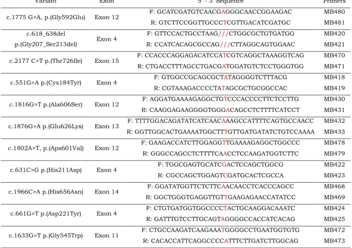
Table 2.2. LDLR variants and negative control under functional assessment and respective mutagenic primers……… 23
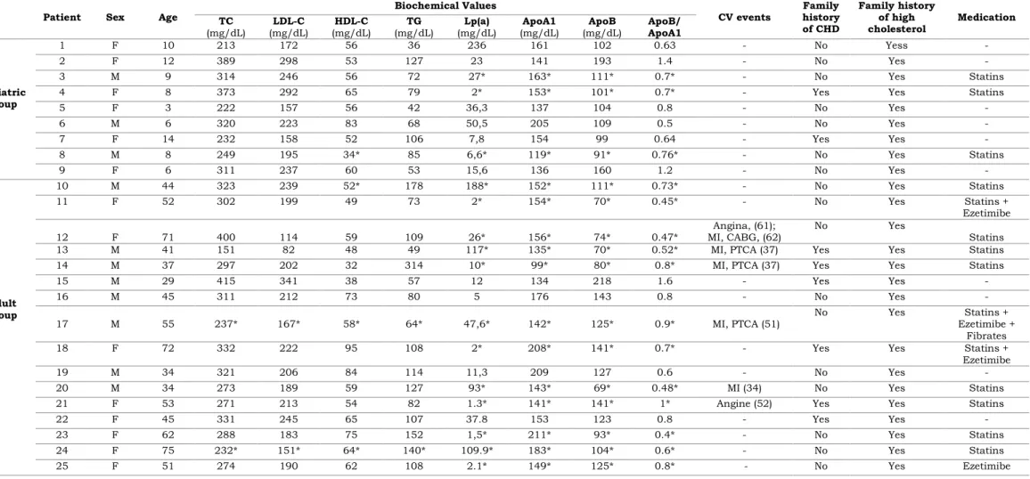
Table 3.1. Clinical and biochemical characterization of studied index cases………. 32
Table 3.2. Variants in LDLR and APOB genes identified in studied index cases…... 33
Table 3.3. Characteristics and in silico prediction of the most common variants found in Portuguese population, without functional studies to date……….. 39
Table 3.4. Comparison between in silico prediction and functional assessment………….. 48
List of Figures
Figure 1.1. LDL, a cholesterol transporter……… 3Figure 1.2. The LDLR pathway………. 4
Figure 1.3. - Pathophysiology of Familial Hypercholesterolemia………..……… 8
Figure 1.4. Schematic representation of the human LDLR gene and protein……… 10
Figure 1.5. Classes classes of variants that disrupt the structure and function of LDLR………...11
Figure 1.6. Portuguese FH Study phases……… 15
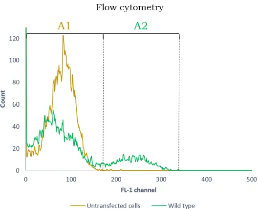
Figure 2.1. Count of events in function of fluorescence measured through the FL-1 channel………. 29
Figure 3.1. Schematic representation of Molecular diagnosis results……….. 35
Figure 3.2. MLPA results……… 36
Figure 3.3. Number of patients carrying the most common variants identified in Portuguese FH Study………..… 37
Figure 3.4. Family trees of index cases (IC) carrying variants c.2177C>T p.(Thr726Ile) and (B) c.618_638del p.(Gly207_Ser213del)………... 40
xiv
Figure 3.6. Major human plasma lipoprotein fractions after one-step salt gradient ultracentrifugation with KBr…... 42 Figure 3.7. Flow cytometry outputs – example of LDLR expression in CHO-ldlA5 cells……… 44 Figure 3.8. Functional characterization of LDLR variants in transfected CHO-ldlA7 cells……… 45 Figure 3.9. Schematic representation of characteristics assessed by flow cytometry and obtained results, for each variant under study………. 47
1
Chapter 1
Introduction
1.1. Cholesterol and lipoproteins
Cholesterol is a very important molecule in biology, which led to the awarding of several Nobel prizes to scientists who devoted part of their careers to its study [1][2][3][4]. It is an organic molecule of extreme importance in mammalian cells, being an integral component of cell membranes, due to its insolubility. Furthermore, it is essential for steroid hormone synthesis, bile acid metabolism, and as a building block for cellular platforms such as lipid rafts [5]. However, its insolubility has as much advantages as disadvantages – cholesterol is capable of creating a barrier to protect the cell through regulation of its interaction with the exterior but, when it accumulates within the wall of an artery, it cannot be readily mobilized, and its presence eventually leads to the formation of an atherosclerotic plaque [5].
Cholesterol is biosynthesized in all animal cells through the mevalonate pathway, being the mevalonate production the rate-limiting and irreversible step of its biosynthesis [5][6]. This important step is performed by the enzyme Hydroxymethylglutaryl-CoA (HMG CoA) reductase, which constitutes a target for cholesterol-lowering drugs [7]. In addition to biosynthesis, cholesterol is obtained through the diet, being withdrawn from the intestinal lumen, through the intestinal epithelial cells, and reaching the blood flow.
In mammals, the cholesterol transport is facilitated by esterifying the sterol with long-chain fatty acids and packaging these esters within the hydrophobic cores, inside plasma lipoproteins [5].
Lipoproteins can be separated by ultracentrifugation based on their densities (table 1.1.) and have been categorized into six major classes: chylomicrons, Very-Low-Density Lipoprotein (VLDL), Intermediate-Density Lipoprotein (IDL), Low-Density Lipoprotein (LDL)
2
and High-Density Lipoprotein (HDL) and Lipoprotein(a) (Lp(a)) [8]. All proteins play major roles in mammals’ organism [9][10], as they are the biological mediators of cholesterol and triglycerides transport, taking part of numerous processes in several pathways. However, LDL is the most prominent lipoprotein in plasma as well as the primary plasma carrier of cholesterol, being responsible for its delivery to all tissues [11].
Table 1.1. Characteristics and percentage content of the distinct lipoprotein particles. Adapted from [12].
Lipoprotein particle
Size (Å) Density C % TG % PL % ApoP %
Major apoproteins
Chylomicrons 800–5000 0.95 3 90 5 9 AI, AII, B, CI, CII, CIII
VLDL 300–800 0.95–1.006 10 70 10 10 BI, CI, CII, CIII, E
IDL 250–350 1.006–1.019 – – – – B, CIII, E
LDL 180–280 1.019–1.063 26 10 15 25 B
HDL 50–120 1.063–1.210 20 5 25 50 AI, All
C, cholesterol; TG, triglycerides; PL, phospholipids; ApoP, apoprotein.
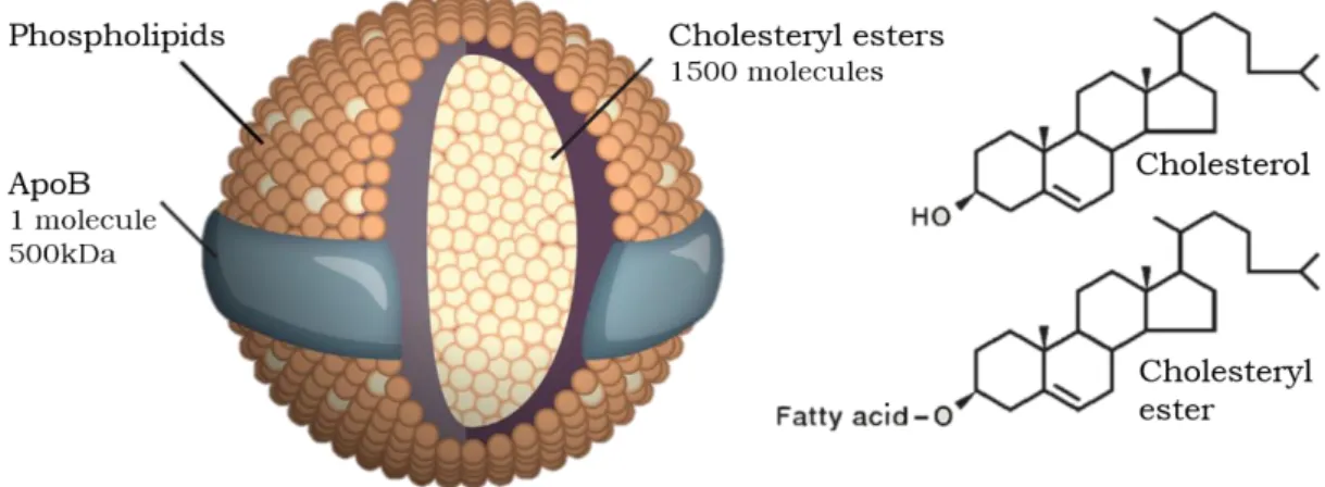
Each LDL particle has a diameter of about 22 nm and a mass of 3000 kDa, containing approximately 1500 molecules of cholesteryl ester in a hydrophobic core surrounded by a polar phospholipid coat and a single large protein called apolipoprotein B (apoB) [13] (Figure 1.1.).
LDL is not secreted directly from the liver, but rather produced in the circulation from VLDL, which is secreted by the liver and transports mainly triglycerides to adipose tissue and muscle. After the removal of these triglycerides in capillaries, it is transformed in IDL. Some of IDL particles, which have apolipoprotein E (apoE) and apoB-100 in their constitution, are rapidly taken up by the liver; others remain in the circulation, where they undergo further triglyceride hydrolysis, lose their apoE particles and are converted to LDL [14].
3
Figure 1.1. LDL, a cholesterol transporter. LDL is a spherical particle with 220 nm of diameter and a mass of 3000 kDa. Each particle contains approximately 1500 molecules of cholesteryl ester in the hydrophobic core and a hydrophilic coat composed of phospholipids, unesterified cholesterol molecules and 1 molecule of apoB. Adapted from [7].
The packaging of cholesterol into lipoproteins allows its correct transport to cells. However, as cholesteryl esters are too hydrophobic to pass through membranes, this delivery problem is solved by lipoprotein receptors, namely the LDL receptor (LDLR), through which approximately two thirds of LDL clearance is normally mediated [5].
1.1.1.
The LDLR pathway
The LDLR is a cell membrane glycoprotein which is ubiquitously expressed. Nevertheless, the largest number of LDLR is produced by the liver [5]. The LDLR binds two proteins: apoB-100, the 387 kDa glycoprotein that is the sole protein of LDL, and apoE, a 34kDa protein that is also found in multiple copies in IDL [5].
The LDLR is synthesized in the rough endoplasmic reticulum (ER) as a precursor with an apparent molecular weight of 120 kDa. Posteriorly, it migrates to the Golgi apparatus, where it undergoes extensive glycosylation, reaching the mature form of 160 kDa [15].
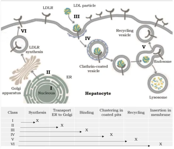
After synthesis, LDL receptors appear on the cell surface, where they gather in coated pits, ready to perform the receptor-mediated endocytosis [5] (figure 1.2.). The receptor binds a LDL particle, due to its affinity to apoB, and the coated pits invaginate to form coated endocytic vesicles [5]. Here, depending on the cell type, specific proteins (e.g. LDLR adaptor protein 1 (LDLRAP1) and Disabled homolog 2 (Dab2) [16]) (not shown in figure 1.2.) play an important role because they bind the cytoplasmic tail of LDLR and governs its clustering into clathrin-coated pits, being required for internalization of the LDL-LDLR complex and
4
for efficient binding [17]. Very quickly, the clathrin coat dissociates and multiple endocytic vesicles then fuse to create endosomes, where the LDLR separates from the LDL due to the acid pH, created by ATP-driven proton pumps [18]. A segment of the endosomal membrane forms a recycling vesicle, responsible of returning the LDLR to the cell surface. The endosome containing LDL fuses with a lysosome and its protein content is hydrolysed to amino acids and cholesteryl esters are hydrolysed to cholesterol. The liberated cholesterol is used to cellular functions as plasma membranes, bile acids and steroid hormones synthesis; or stored in the form of cytoplasmic cholesteryl ester droplets until further use by the cell. The receptor can be recycled several times, since one round trip lasts 10 minutes and LDLR has a 20-hour lifespan [5].
The LDLR also has affinity to proprotein convertase subtilisin/kexin type 9 (PCSK9) [19], secreted from hepatocytes. Similarly to the LDL, PCSK9 can also interact with LDLR, forming a tight ligation impairing, in the endosome, the recycling of the receptor to cell surface and targeting LDLR to the lysosome for degradation [20]. Thus, PCSK9 is also a modulator of the LDLR pathway.
Figure 1.2. The LDLR pathway - The LDLR is synthesized in the ER, undergoes extensive glycosylation in the Golgi apparatus and it transported to the cell surface. The LDLR specifically binds ApoB in LDL particles, internalizing them by endocytosis. Inside the endosome the complex dissociates; the receptor is recycled to the cell surface, whereas the LDL particle is degraded into the lysosome. The PCSK9 is synthesized and excreted, binding LDLR–LDL complex extracellularly under regulatory signals. PCSK9 prevents the dissociation of LDLR-LDL complex, leading to its degradation in the endosomal compartment. Adapted from [21].
The LDLR is a crucially important modulator of plasma LDL levels as it removes not only LDL but its precursors IDL from circulation due to the presence of apoE and apoB, whereby the receptor binds [1][20]. Its high affinity for LDL and ability to cycle multiple
5
times in and out of the cell allow the delivery of large amounts of cholesterol to body tissues. In addition, both cholesterol biosynthesis and LDLR pathway are regulated by negative feedback in order to keep the level of cholesterol in cell membranes constant, through regulation of HMG CoA reductase, and LDLR transcription factors [1],[20].
When a defect arises in genes codifying one of the proteins necessary to the performance of the LDLR pathway, this can lead to a lipid disorder known as Familial Hypercholesterolemia (FH), or Autosomal Recessive Hypercholesterolemia (ARH) in case of defects in LDLRAP1. It was the study of FH by several scientists which threw light on fundamental biological and regulatory mechanisms related with cholesterol.
Familial Hypercholesterolemia
The correlation between plasma cholesterol and coronary heart disease (CHD) was first postulated through the description of families in which high plasma cholesterol levels and its associated coronary problems were transmitted as an autosomal dominant trait [22]. Later, it was found that these families had a genetic disorder of lipid metabolism - Familial hypercholesterolemia (FH).
FH is a common autosomal dominant characterized by high levels of low density lipoprotein cholesterol (LDL-C) in plasma and increased risk of premature coronary heart disease (CHD) [5]. FH has an estimated prevalence of 1/500 individuals, although a prevalence of 1/200 has been observed in some populations [23], which leads to a calculated value between 14 and 34 million affected individuals worldwide, being among the commonest inherited disorders [24].
Clinically, FH exists essentially in two forms: a more common and less severe heterozygous form, and a rare (prevalence 1/1000000) and more severe homozygous form [5][15]. FH heterozygotes have a two-fold increase in the number of LDL particles in plasma from the time of birth, which predisposes to premature CHD as early as 30 years old [5]. Homozygotes patients have a much worse prognosis, presenting six to ten-fold elevations in plasma LDL levels from the time of birth, often having a myocardial infarction in childhood [5].
1.2.1.
Therapeutic approaches for FH
Although cholesterol is endogenously synthesized, the diet is also a source of cholesterol, and these are two points where therapeutic measures can be applied. FH patients should follow a strict diet, poor in fat and perform daily physical activity, but these measures are not enough to lower their increased LDL-C values. Fortunately, FH is a disorder for which effective treatment exists and current therapies revolve around
6
cholesterol-lowering drugs and in some cases LDL apheresis [25]. For FH patients, medication should be initiated as soon as possible still in childhood, being strongly considered starting between 8 and 10 years old [23][26].
Several lipid lowering drugs are currently available due to knowledge of fundamental properties of the cholesterol metabolism, as well as LDLR and its interactions with other important molecules as APOB and PCSK9. These interactions represent central implications for therapy of FH.
The first inhibitor of HMG CoA reductase, and consequently inhibitor of the endogenous cholesterol synthesis, initiated the class of cholesterol-lowering drugs known as statins [27]. This drug leads to the synthesis of more LDLR, in order to satisfy the cell demands for cholesterol. The final outcome is the reduction of LDL-C between 20-45%, depending on the dosage [28]. Nonetheless, statins do not lower LDL significantly in FH homozygotes (HoFH), who have null variants in both copies of the LDLR [29].
Statins are often coadministered with ezetimibe [23], a cholesterol absorption inhibitor that actuates at the level of small intestine, reducing the amounts of dietary cholesterol that reach the liver. This results in increased LDL withdrawn from the circulation. Ezetimibe might as well be administrated alone in adults, in case of intolerance to statins, resulting in a reduction of approximately 20% of the LDL-C levels [28][29][30][31].
For a long time, statins and ezetimibe were the only pharmacological treatments available for FH [32]. However, management of homozygous FH requires additional treatment as LDL apheresis, which provides transient reductions in LDL-C levels by 40% [33][34]. LDL apheresis is typically performed once or twice-a-week in patients with homozygous FH and is also an option for the treatment of heterozygous FH, intolerant to statins. Nonetheless, this invasive therapy is far from being the ideal, since it has several side effects associated and the treatment sessions are expensive and time consuming [21]. Advances in genetic-based pharmacology have empowered the study of new LDL-lowering agents, which are currently at advanced stages of development. These comprise the development of monoclonal antibodies targeting PCSK9, an anti-sense oligonucleotides targeting APOB and cholesteryl ester transfer protein inhibitors [23]. Studies are underway to determine the long-term safety of these therapeutic measures and their efficacy in preventing CHD [35][36]. Nevertheless, further studies on these new medications’ long-term safety and efficacy are still needed.
It is now clear that LDLR pathway is of extreme importance in regulation of cholesterol levels in blood and its activity has an impact on the response of the system to lowering the levels of cholesterol. However, although FH has a high prevalence and a clear relation with premature atherosclerosis and CHD, it remains as an extremely underdiagnosed disorder worldwide [23], which emphasises the need of investigation at the genetic level. Only the molecular diagnosis can confirm a clinical diagnosis of FH, hence
7
the genetic demonstration of a causative variant in specific genes is important for FH diagnosis, allowing a more personalised treatment [23].
1.2.2.
Genetics behind FH
Genetically, heterozygous FH is caused by variants in three genes: LDLR, APOB, and PCSK9 [5][37][38].
Variants in the LDLR gene are the most common, being the cause of more than 90% of the identified FH cases worldwide [39]. These variants can result in an impaired function of the LDLR, which will be further discussed in section 1.2. Molecular FH.
Variants in APOB sequence, affecting amino acids which are important for the binding to LDLR, can disable the recognition of LDL by the LDLR [40]. This results in a reduction of LDL withdrawn from the circulation, leading to increased cholesterol levels. Due to APOB size, only part of exons 26 and 29 is routinely studied, although disease-causing variants have been reported out of these sites [41].
PCSK9 gain-of-function variants result in disruption of the recycling mechanism, which is responsible for the LDLR return to cell surface after internalization, as the PCSK9 targets the complex LDLR-LDL to lysosomal degradation. As a result, the number of cell-surface LDLR declines and LDL rises [42][43]. PCSK9 loss-of-function variants have as well been reported, resulting in an enhancement of the recycling process, consequently reducing LDL-C levels [44][45].
Variants in APOB and PCSK9 genes are found in ~5% and ~1%, respectively, of heterozygous FH subjects with a causative variant [40]. Homozygous individuals are uncommon and present a more aggressive clinic phenotype, leading to extremely increased cholesterol values, since there is no production of normal and functional protein. Some rare subjects are “double heterozygotes”, which means they carry variants in two of the above-mentioned genes.
Recently, variants in LDLRAP1 were identified as the cause of autosomal recessive hypercholesterolemia (ARH) [46], a disorder distinct from FH as it causes a less severe phenotype and these patients are more responsive to lipid lowering therapies. The protein LDLRAP1 facilitates the internalization in clathrin coated-pits, thus variants causing its loss of function can result in reduced clearance of circulating LDL by the liver [47], leading to a phenotype between heterozygous and homozygous FH [48].
A genetic defect in one of the three FH-related genes will lead to defects in distinct steps of the LDLR pathway, depending on the affected gene, which can culminate in premature cardiovascular disease, as represented in figure 1.3.
8
Figure 1.3. - Pathophysiology of Familial Hypercholesterolemia. A heterozygous disease-causing variant in one of the three genes associated with FH leads to a situation where the liver only produces 50% of functional LDLR, resulting in elevated LDL-C. Subsequently premature atherosclerosis can urge, culminating in CHD. Adapted from [23].
In view of genetic variability and when lifelong drug treatment is under consideration, the comprehension of the origin of a LDLR defect is imperative. LDLR variants represent the most known cause of FH, thus knowledge on the LDLR gene and protein will be covered in next section, as it allows profound understanding of the molecular basis of this disorder.
Molecular FH
1.3.1.
The LDLR gene and protein
The striking feature of the LDLR pathway is that it requires a highly selective and distinct movement for each one of the components involved, characteristics that may reside in its structure. These domains are encoded in the LDLR gene, which lies on the short arm
9
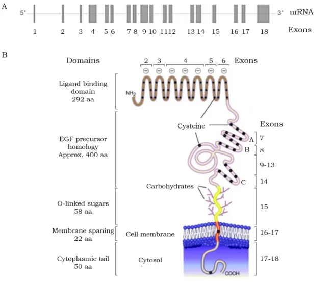
of chromosome 19 - 19p13 -, spans 45kb, and is comprised of 18 exons and 17 introns [15]. The protein coding sequence is interrupted by introns in such a way that many of the protein segments are revealed as products of individual exons [49] (Figure 1.4.).
The LDLR is synthesized as a precursor of 860 amino acids. Nevertheless, the first 21 amino acids, at the extreme NH2 terminus, constitute a typical hydrophobic signal sequence that is cleaved from the protein prior to its appearance on the cell surface. The short 5’ untranslated region plus the signal sequence of the protein is encoded by exon 1 [49]. The mature form of LDLR has 839 amino acids with five recognizable domains [49][50], described below.
The first domain of the LDLR consists of the NH2-terminal 292 amino acids and is assembled from multiple repeats of 40 residues each. Each repeat has six cysteine residues, all involved in disulphide bonds [51], which must be the cause of the extreme stability of the binding domain of the receptor. All of the charged residues that are conserved bear a negative charge, which might be responsible for the LDLR ability to bind closely spaced positively charged residues. It contains the binding site for apoB and apoE. The repeats I, III, VI and VI are encoded exons 2, 3, 5 and 6, respectively. The other three repeats (III, IV and V) are all contained in a single exon, the exon 4 [49].
The second LDLR domain, constituted by approximately 400 amino acids, is homologous to a portion of the extracellular domain of the Epidermal Growth Factor (EGF) precursor. This part of the LDLR is implicated in the release of bound lipoproteins at low pH in the endosome [18]. This EGF precursor homology domain contains three repetitive sequences of about 40 amino acids, that are designated A, B and C, containing each one six cysteine residues spaced at similar intervals. Each repeat is contained within a single exon – exons 7, 8 and 14. The exons 9 to 13 codify for the region of β-propeller, between the repeats B and C [49].
The third domain is encoded within the single exon 15 [49], consisting of a stretch of 58 amino acids that contains 18 serine or threonine residues, many of which appear to serve as attachment sites for O-linked carbohydrate chains added to serine and threonine residues during posttranslational processing events.
The fourth domain, with 22 hydrophobic amino acids, is the membrane spanning region, which is poorly conserved among species. This transmembrane domain is encoded by exons 16 and 17 [49].
10
Figure 1.4. Schematic representation of the human LDLR gene (A) and protein (B). The mature human LDLR is composed of five domains. The 5’ untranslated region, as well as the signal peptide are not represented in the scheme. Aa, amino acids. Adapted from [52].
The last protein domain is a 50 amino acid COOH-terminal cytoplasmatic tail, projected into the cytoplasm [49][50]. This domain is strongly conserved among species and plays a crucial role in clustering in coated pits. The cytoplasmic domain is encoded by exons 17 (13 amino acids of the transmembrane domain and the first 39 amino acids of the cytoplasmic domain) and 18, the largest exon in the gene. It encodes the last 11 amino acids of the LDLR and a 2.5 kb DNA sequence that represents the 3’ untranslated region of the mRNA [49].
When a change in the LDLR codifying sequence is noted, it might have serious repercussions at the protein and consequently at the LDLR pathway level, implying modifications in the cholesterol level.
According to the domain altered by these modifications at the DNA sequence level, different functions of the LDLR can be affected, which leads us to the LDLR classes of variants.
11
1.3.2.
Classes of variants in LDLR
Consistent with the first reports, there were four classes of LDLR variants [53]. However, posterior new findings brought the necessity to subdivide some of these classes and to create a new one [1][15], existing currently five classes of LDLR variants (figure 1.5.).
Figure 1.5. Classes of variants that disrupt the structure and function of LDLR. Each variant affects a different region in the gene resulting in defects in distinct parts of the cycle. Adapted from [5][21].
Class I Variants: Null Alleles. The variants responsible for this class lead to the production of no LDLR or only trace amounts of it. The absence of receptor protein in these cells may be due to a rapid turnover of the mRNA or to accelerated degradation of the receptor protein [15].
Class II Variants: Transport-defective Alleles. These alleles encode LDLR which is either completely (Class II A) or partially (Class II B) blocked in transport between the endoplasmic reticulum and the Golgi apparatus, not reaching the cell surface. Class II variants comprehend the most common at the LDLR locus [15].
12
Class III Variants: Binding-defective Alleles. These LDLR are normally synthesized, matured and reach the cell membrane. However, their affinity to apoB in LDL or apoE in IDL can be affected, resulting in an impaired binding to LDL [50].
Class IV Variants: Internalization-defective Alleles. Here the receptors move to the cell surface and bind LDL normally. The problem resides in the clustering in clathrin-coated pits, preventing the LDL internalization. These variants have been classified into two groups: variants that alter the cytoplasmic domain alone (Class IV A) and variants that involve the cytoplasmic domain together with the adjacent membrane-spanning region (Class IV B) [15]. This last one produce truncated receptors that lack the membrane-spanning domain as well as the cytoplasmic tail. Most of these molecules are secreted from the cell, but approximately 10% remain adherent to the cell membrane where they bind LDL but do not internalize it.
Class V variants: Recycling-deficient Alleles. The LDLR encoded by these variants perform all steps of the LDLR cycle until the recycling of the receptor, as they fail to release the ligands in the endosome. This results in the degradation of the complex LDLR-LDL, not being recycled to the cell surface [15].
Recently, a sixth class (class VI) has been defended as affecting LDLR insertion in cell membrane [54]. Here should be included variants in the cytoplasmic domain, which makes the anchoring of the receptor to the cell membrane impossible.
1.3.3.
Variants
A mutation has been primarily defined as a heritable change in a specific DNA sequence when compared with the reference sequence [55]. However, the mutation within this definition can have an effect at the phenotype level or be a neutral variation without an observable effect. If it is a rare variation, it is called mutation. However, if it occurs in the population at a frequency above 1%, it might be defined as polymorphism [55][56].
Therefore, in order to end these ambiguities, every change in DNA is called variant [56]. Thus, if a dominant phenotype, as in FH, segregates with the variant and does not segregate in its absence, then the variant is compatible with a genotype that can be called disease-causing [55]. These are variants that are likely to have greater functional importance, affecting polypeptide structure and function [55].
The impact of a variant at the protein function level can be assessed with in silico tools, allowing a bioinformatics analysis that can be helpful in characterizing new variants found, on which pathogenicity is unknown. However, only the in vitro study of the effect of a variant in a protein (functional assay) can correctly determine its pathogenicity.
13
Portuguese FH Study
Universal screening for FH was recommended from the World Health Organization (WHO) in 1998, which led to the implementation, at National Institute of Health (INSA) Doutor Ricardo Jorge, of the Portuguese FH Study [57]. The Portuguese FH Study has the purpose of determining the prevalence and distribution of FH in Portugal. The population under study consisted of individuals of both sexes and all ages with a clinical diagnosis of FH (Simon Broome criteria). The implementation of the molecular study of this disorder in Portugal, promotes its early identification in patients and respective relatives, leading to a correct counselling as soon as possible, decreasing their CV risk.
Since 1999, a total of 2122 individuals were enrolled due to the participation of numerous clinicians from several clinics and hospitals in all country. Among these, 623 heterozygous patients had putative pathogenic variants in LDLR, 33 in APOB, and 4 in PCSK9 [58].
This study implies a biochemical and a molecular assessment, being the last subdivided into 5 phases, as schematically represented in figure 1.6.
Figure 1.6. Portuguese FH Study phases, comprehending the biochemical and the several phases of the molecular study. Adapted from [58].
When a putative disease-causing variant is identified in an index case, a cascade screening is performed, when samples of relatives are available, as recommended by international guidelines [59]. The cascade screening has been proved as the most cost-effective method of identification and has a special importance for children [60], since a genetic defect can be identified before the atherosclerotic process, leading to the implementation of preventive measures and correct treatment.
Cascade screening might be life-saving, especially in young patients, since FH is a serious disease that requires early intervention, lifelong treatment and regular follow-up.
14
Aim of the project
Although the Portuguese FH Study has been implemented in Portugal in 1999 and more than 800 families have been enrolled [58], FH remains as an underdiagnosed disorder.
A clinical diagnosis is not enough to identify a FH patient, and only with molecular diagnosis is possible to identify the disease-causing variants. However, not all of identified variants are functionally assessed in order to understand their involvement in LDLR function. The performance of functional studies is imperative, providing a definite FH diagnosis.
In order to clarify the relationship between genotype and phenotype in FH, the aim of the present work is to perform the molecular diagnosis for 25 index cases with clinic criteria of FH.
Moreover, functional characterization of the 10 most common LDLR variants, which remain to date without functional studies, identified in Portuguese FH patients, also constitutes an aim, being the objective to determine the effect of these 10 LDLR variants in the cell surface expression, binding and uptake of the LDLR.
These results will allow the increase of knowledge about the functionality of LDLR variants, contributing to the elucidation of molecular basis of FH not only in Portugal, but worldwide, as some of the alterations under study were reported in several other countries. The knowledge about genetic causes of FH and the relation between patients’ genotype and phenotype, allows an accurate and definite diagnosis along with early personalized counselling and treatment, improving FH patients’ prognosis and effectively reducing their cardiovascular risk.
15
Chapter 2
Materials and Methods
Molecular diagnosis
2.1.1.
Patients Recruitment
All the patients were recruited for the Portuguese Familial Hypercholesterolemia Study, which protocol and database have been approved by the National Institute of Health Ethics Committee and the National Data Protection Commission, respectively.
During these past 15 years, patients with a clinical diagnosis of FH (Criteria in Table 2.1., adapted from those of the Simon Broome Heart Research Trust [61]) have been recruited all over the country by clinicians from several specialties [58]. When a pathogenic variant is identified in a patient, the clinician is notified and asked to perform cascade screening in other relatives with and without a clinical diagnosis of FH for co-segregation analysis. Written informed consent was obtained from all participants before their inclusion in the study.
Table 2.1. – FH criteria adapted from “Simon Broome Heart Research trust”
Confirmed familial hypercholesterolemia is defined as:
Index case: Child under 16 with total cholesterol over 260 mg/dL (6.7 mmol/L) or LDL cholesterol over 155 mg/dl (4 mmol/L);
Index case: Adult with total cholesterol over 290 mg/dl (7.5 mmol/L) or LDL cholesterol over 190 mg/dl (4.9 mmol/L),
and
Tendon xanthoma in the index case or relative (parents, children, grandparents, siblings, aunts or uncles),
16 or
Genetic evidence of a variant in the LDLR, APOB or PCSK9 genes.
Possible familial hypercholesterolemia is defined as:
Index case: Child under 16 with total cholesterol over 260 mg/dl (6.7 mmol/L) or LDL cholesterol over 155 mg/dl (4 mmol/L);
Index case: Adult with total cholesterol over 290 mg/dl (7.5 mmol/L) or LDL cholesterol over 190 mg/dl (4.9 mmol/L),
and
Family history of myocardial infarction before the age of 50 in grandparents or aunts or uncles, or before the age of 60 in parents, siblings or children, and/or family history of elevated cholesterol levels (>290 mg/dL) in parents, siblings or children;
or
Total cholesterol (TC) over 290 mg/dl (7.5 mmol/L) in grandparents and/or aunts or uncles.
2.1.2.
Blood samples collection
For each index case and respective relatives, fasting blood samples (7.5 mL in serum tube and 3 x 2.7 mL in EDTA tubes for adults; 5 mL in serum tube and 2 x 2.7 mL in EDTA tubes for children) were collected in order to perform DNA extraction. Moreover, 5 mL were collected in serum tubes to obtain serum, used in biochemical determination. For each sample a confidential identification number was assigned and all the information concerning the patients was registered in a confidential database, according to legal requirements. At maximum 48 h after the blood collection, the biochemical determination was performed.
2.1.3.
Biochemical determination
The biochemical determination was executed by technicians of the Unidade Laboratorial Integrada at INSA. It included measurement of TC, HDL-C, LDL-C, triglycerides (TG), apoAI, apoB and Lp(a), performed in an autoanalyser Cobas Integra 400 Plus (Roche) by enzymatic, colorimetric and immunoturbidimetric methods.
17
2.1.4.
Molecular biology techniques
The molecular analysis comprises 5 phases, as described in Introduction, section 1.4. Portuguese FH Study [58].
2.1.4.1.
Genomic DNA extraction
Genomic DNA was extracted from leucocytes in ~5 mL samples of peripheral blood, collected in EDTA tubes. For index cases, 10 mL were collected and DNA extraction was performed for both 5 mL tubes, in independent days, providing two different DNA samples for diagnosis confirmation. This extraction was performed as an adaption of the protocol described in [62]. The proportion of each reagent to mL of blood is disclosed in Appendix I, Table A I.1.
The blood was well homogenised and transferred to a 15mL falcon tube; it was added equal volume of TKM X-100 (low salt buffer containing 10 mM Tris-HCl pH 7.6, 10 mM KCl, 10 mM MgCl2, 2 mM EDTA and 25 mL Triton-X 100/L) and mixed several times by inversion. Then IGEPAL was added, mixed until total solubilisation. The tubes were centrifuged – 10 min 2200 rpm at room temperature (RT) (centrifuge 5810 R, Eppendorf) and the supernatant was despised; the pellet was washed in TKM1 (TKM-X100 without the Triton-X 100) and centrifuged again for 10 min 1600 rpm at RT. The wash was repeated at least once, or twice when the pellet was still red. The pellet was resuspended with TKM2 (high salt buffer containing 10 mM Tris-HCl pH 7.6, 10 mM KCl, 10 mM MgCl2, 0.4 M NaCl and 2 mM EDTA) and then SDS 10% was added, followed by a 10 min incubation at 55ºC, for protein denaturation. All the content was transferred to a 2 mL Eppendorf tube, NaCl 5 M was added and mixed by inversion, for protein precipitation. The tubes were centrifuged for 20 min, 13200 rpm at RT, forming a dark protein pellet. All the supernatant was transferred to a clean falcon tube and absolute ethanol at RT was added and gently mixed by inversion, in order to denature and precipitate DNA. The DNA fibrils were removed with a loop and washed in a 70% ethanol solution at 4 ºC. When DNA was completely dry, it was resuspended in TE. DNA content was quantified in a spectrophotometer (NanoDrop 1000, Thermo Scientific), assessed by electrophoresis gel (2.1.4.3. Assessment of DNA fragments and PCR products) and stored in 1.5 mL tubes.
2.1.4.2.
DNA amplification by Polymerase Chain Reaction (PCR)
Fragments containing parts of the APOB exons 26 and 29 of APOB, and the LDLR promoter and 18 exons, with respective flanking regions, were amplified by PCR. All the primers used, as well as distinct annealing temperatures for each one are written in Appendix I, table A I.2.
The PCR was performed with BIOTAQ™ DNA Polymerase kit (Bioline), according to the manufacturer’s instructions: to a reaction tube 1 l of dNTPs (100 mM dNTP Mix), 2.5
18
and reverse) (10 ρmol/l, Invitrogen), 1.25 U of BioTaq polymerase (Bioline) and bidistilled water was added up to a final volume of 24 µL. At last, 1 l (100-200 ng) of genomic DNA was added. It was performed a replicate without DNA as a control for each exon amplification reaction.
The PCR reaction was held in a thermocycler (model 2720, Applied Biosystems), as follows: initial denaturation for 3 min at 95 ºC; 35 cycles of three steps: denaturation for 45 sec at 94 ºC, annealing for 30 sec at 57 ºC to 62ºC depending on the primer (Appendix I, Table A.2.), and elongation for 1 min at 72 ºC; and final extension for 30 min at 72 ºC. All PCR products were assessed by an agarose gel electrophoresis.
2.1.4.3.
Assessment of DNA fragments and PCR products
An agarose gel was prepared in 100 mL of TBE buffer 1x (TBE 10x ultrapure, pH 8.4, 1.0 M Tris, 0.9 M boric acid, 0.01 M EDTA, Invitrogen). It was used a concentration (weight/volume) of 0.8% (w/v) for DNA qualitative assessment and of 1.5% (w/v) for PCR products visualization. SYBR safe (10000x concentrate in DMSO, Invitrogen) was added to the gel before polymerization. When polymerized (20 to 30 min), the samples (5 µL PCR product or DNA, gel loading die (Orange 6x, New Englands Biolabs) and bidistilled water to a final volume of 10 µL) were loaded, as well as a molecular weight marker (1 kB DNA Ladder, Boehringer Mannheim). The electrophoresis was performed for 40 min at 90 volt (Bio-Rad Power Pac 3000 equipment), in TBE 1x. The gel was visualized in a Safe Imager™ blue light transilluminator (Invitrogen).
2.1.4.4.
Automated sequencing
Before Sanger sequencing, PCR products must be purified to remove the excess of primers and dNTPs. This was made by an enzymatic digestion using two hydrolytic enzymes; Exonuclease I and Shrimp Alkaline Phosphatase (SAP), combined in a commercial product named ExoStar (IllustraTM ExoStarTM, GE lifesciences). To 2.5 µL of PCR product, 1 µL of ExoStar was added in a reaction tube and incubated for 15 min at 37 ºC (enzyme optimum temperature) and for 15 min at 80 ºC for enzyme inactivation. Purification products were stored at 4ºC until further use.
The sequencing reaction was prepared as follows: to a reaction tube, 2 ρmol of primer (Invitrogen), 1 µL of BigDye (Terminator Cycle Sequencing Ready reaction kit, Applied Biosystems) and bidistilled water was added up to a final volume of 9 µL. At last, 1 µL of purified DNA was added. The sequencing reaction was performed as follows: initial denaturation for 30 sec at 96 ºC; 25 cycles of three steps: denaturation for 10 sec at 96 ºC, annealing for 5 sec at 50 ºC and elongation for 4 min at 60 ºC. Sequencing products were stored at 4 ºC. Resulting products were sequenced by Unidade de Tecnologia e Informação (UTI – INSA) (3130xl Genetic Analyser, Applied Biosystems). The subsequent .AB sequence files were analyzed with Staden Package software (version 2.0). The reference sequence
19
NM_000527.4 for LDLR and NM_000384.2 for APOB was used and the novel variants were numbered according to the Human Genetic Variation Society (HGVS) guidelines, where +1 is the A of the ATG translation initiation codon of the coding DNA [63].
Whenever an alteration was found, all PCR, assessment and sequencing protocols were repeated in the second aliquot of index case, as well as made in its relatives, when samples were available, for confirmation and co-segregation studies.
2.1.4.5.
Multiplex Ligation-dependent Probe Amplification (MLPA)
The search for large rearrangements (duplications or deletions) was performed by MLPA. This was made with SALSA® MLPA® kit (probemix P062-C2 LDLR, MRC-Holland, The Netherlands) according to the manufacturer’s instructions. The MLPA reaction can be divided in five major steps: 1) DNA denaturation and hybridization of MLPA probes; 2) ligation reaction; 3) PCR reaction; 4) separation of amplification fragments by electrophoresis, which was performed by Unidade de Tecnologia e Informação (UTI – INSA) (3130xl Genetic Analyser, Applied Biosystems); and 5) data analysis, which was performed using the Coffalyser – MLPA analysis tool (developed at MRC-Holland, The Netherlands).2.1.5.
In silico analysis
Whenever a LDLR or APOB variant was found, the predicted effects of alterations were assessed using the following open access software: Polymorphism Phenotyping (PolyPhen-2) [64], Sorting Tolerant From Intolerant (SIFT) [65] and Mutationtaster [66] for prediction of single nucleotide substitutions. Briefly, SIFT [65] takes into account evolutionary conservation through the use of sequence alignments, while MutationTaster [66] and PolyPhen-2 [64] base their predictions in protein structure/function and evolutionary conservation, being that PolyPhen-2 also uses a prebuilt sequence alignment.
Human Splicing Finder (HSF) (http://www.umd.be/HSF3/HSF.html; [67]), the
Splice Site Prediction by Neutral Network (NNSSP)
(http://www.fruitfly.org/seq_tools/splice.html; [68]) and the FSPLICE (http://linux1.softberry.com/) tools were used for prediction of splicing defects. HSF [67] uses position-dependent logic, identifying exonic and intronic motifs, NNSSP [68] is based in neural networks combined a sequence similarity matrix with a local structural environment scoring scheme for predicting protein secondary structure and FSPLICE (http://linux1.softberry.com/) bases its predictions on weight matrices model, which consider the importance of the presence of a determinate nucleotide in a specific position. Mutation Taster also predicts a phyloP score. PhyloP score is a measurement of evolutionary conservation, thus the higher its score is, the stronger is the evolutionary conservation for a specific nucleotide.
20
A variant was considered probably pathogenic when all three software tools had a prediction of pathogenic (probably damaging, deleterious or disease causing), variant of unknown significance (VUS) if predictions were contradictory and neutral when tools predicted it to be benign (benign, tolerated or polymorphism). Regarding splice-site analysis tools, a variant was considered pathogenic if there was a deletion of the actual splice-site or the addition of a new one.
Production of LDLR gene variants
2.2.1.
Site-directed mutagenesis
In order to express all 10 different alterations in the LDLR gene, individual point mutations were introduced into the human LDLR cDNA, previously subcloned in the mammalian expression vector pcDNA3, under the control of a SV40 promoter (pcDNA3_LDLR, kindly offered by César Martín) (Appendix 2, Figure A II.1.). This was performed by oligonucleotide site-directed mutagenesis with the Quik-Change XL mutagenesis kit (Agilent Technologies) according to the manufacturer’s instructions. This procedure is based on a PCR reaction that uses specific complementary mutagenic primers of the fragment of the interest (insert – complete LDLR cDNA) (Table 2.2.).
Each mutagenesis reaction tube was composed of 5 µL of reaction buffer (10x, Agilent Technologies), 125 ng of each pair of mutagenic primers (forward and reverse), 1 µL of dNTPs, 1 µL of PfuUltra High Fidelity DNA polymerase (2.5 U/µl), 50-100 ng of double-stranded plasmid DNA (pcDNA3_LDLR) and bidistilled water up to 50 µL. The PCR program was performed as follows: initial denaturation for 10 min at 95 ºC; 12 cycles of three steps: denaturation for 30 sec at 90 ºC, annealing for 1 min at 55 ºC and extension for 12 min at 68 ºC. Completed the program, 10 µL of the products were visualized in an 1% agarose gel (see Annex III, figure A III.1.), as well as 50 ng of the original plasmid (the same amount of DNA of the mutagenesis reaction) properly mixed with DNA loading dye and bidistilled water up to a final volume of 20 µL. Next, the mutagenesis products were treated with the restriction enzyme DpnI (provided with the kit) for 1 h at 37 ºC, in order to digest the parental DNA that does not contain the desired alteration. This strategy is based in the fact that plasmids grown in almost all E. coli strains are dam methylated and therefore are susceptible to DpnI specific digestion of methylated and hemimethylated DNA.