Universidade de Aveiro Ano 2019
Departamento de Ciências Médicas
INÊS FILIPA PEREIRA
ABRUNHOSA AMARAL
Análise da expressão de lncRNAs em monócitos e
osteoclastos de pacientes com artrite reumatóide
Analysis of lncRNAs expression in monocytes and
osteoclasts of rheumatoid arthritis patients
Universidade de Aveiro Ano 2019
Departamento de Ciências Médicas
INÊS FILIPA PEREIRA
ABRUNHOSA AMARAL
Análise da expressão de lncRNAs em monócitos e
osteoclastos de pacientes com artrite reumatóide
Analysis of lncRNAs expression in monocytes and
osteoclasts of rheumatoid arthritis patients
Dissertação apresentada à Universidade de Aveiro para cumprimento dos requisitos necessários à obtenção do grau de Mestre em Biomedicina Molecular, realizada sob a orientação da Doutora Ana Gabriela Henriques, Investigadora Auxiliar do Departamento de Ciências Médicas da Universidade de Aveiro, e co-orientação científica do Doutor Ângelo Miguel Silva Calado, Professor Auxiliar da Faculdade de Medicina da Universidade de Lisboa.
o júri
presidente Doutora Ana Margarida Domingos Tavares de Sousa
Professora Auxiliar Convidada do Departamento de Ciências Médicas da Universidade de Aveiro
Doutora Anita Raquel Quintal Gomes
Professora Adjunta da Escola Superior de Tecnologia de Saúde de Lisboa
Doutor Ângelo Miguel Silva Calado
Professor Auxiliar da Faculdade de Medicina da Universidade de Lisboa
agradecimentos Em primeiro lugar gostaria de agradecer à Doutora Vânia Glória, que me aceitou em primeira instância e me proporcionou a oportunidade de realizar a dissertação de mestrado na Unidade de Investigação em Reumatologia do Instituto de Medicina Molecular João Lobo Antunes.
Ao Doutor Ângelo Calado por me acolher, pela orientação, e pelo apoio no laboratório, sem dúvida fundamental.
À Doutora Ana Gabriela Henriques, pelo acompanhamento e orientação.
Ao Doutor João Eurico Fonseca, pela liderança e supervisão do projeto. A toda a equipa da Unidade de Investigação em Reumatologia agradeço o companheirismo e amizade, as ajudas no trabalho de laboratório e também os momentos de conversa e descontração que me proporcionaram.
Ao Instituto de Medicina Molecular João Lobo Antunes e a todos os funcionários e colegas de outros laboratórios, um sentido agradecimento por proporcionarem um ambiente de trabalho e amizade sem igual.
A ambas as equipas do Serviço de Reumatologia e do Centro de Investigação Clínica do Hospital de Santa Maria, cuja colaboração foi fundamental para a concretização deste trabalho.
A todos os doentes e dadores de sangue, o maior agradecimento pela disponibilidade em participarem no estudo.
Um agradecimento muito importante e especial aos meus amigos mais próximos, que me acompanharam ao longo de todos os altos e baixos característicos desta etapa. Sem o vosso apoio não conseguia. Obrigada pela amizade.
Às amigas que o mestrado me deu, obrigada por todos os momentos de diversão, de horas infinitas de trabalho, e também por todo o apoio no nosso grupo. Sem vocês não era igual. Que continuemos assim, sempre.
Ao José Miguel, por me apoiar em mais uma das minhas aventuras.
E por último, e sem dúvida mais importante, agradeço profundamente aos meus pais. Obrigada por todo o esforço e sacrifício. Obrigada por acreditarem sempre em mim, por serem o meu suporte, e por todo o apoio incondicional. Sem vocês, nada disto seria possível. É a vós que dedico todas as minhas conquistas académicas e pessoais. Obrigada por fazerem de mim a pessoa que sou hoje. Obrigada por me fazerem querer ser sempre melhor neste caminho cheio de lutas e incertezas, mas também de muitas esperanças e sonhos.
palavras-chave Artrite reumatóide, erosão óssea, monócitos, osteoclastos, osteoclastogénese, RNAs longos não-codificantes (lncRNAs).
resumo A artrite reumatóide (AR) é uma doença reumática crónica, inflamatória e imuno-mediada. A erosão óssea é uma das características da fisiopatologia da AR e está associada à gravidade da doença. As erosões ósseas em pacientes com AR resultam de um metabolismo ósseo alterado devido, em parte, a osteoclastogénese (o processo pelo qual as células precursoras da linhagem de monócitos se diferenciam em osteoclastos) e a atividade osteoclástica excessivas. Num estudo recente, observou-se uma expressão variável de RNAs longos não codificantes (lncRNAs) durante a osteoclastogénese no ratinho, sugerindo um papel fisiológico dos lncRNAs neste processo. Em pacientes com AR, vários estudos observaram até à data uma expressão alterada de lncRNAs em tipos celulares críticos da fisiopatologia da doença, como leucócitos mononucleares do sangue periférico e sinoviócitos ativados tipo-fibroblastos. No entanto, nenhum estudo examinou, até ao momento, a expressão de lncRNAs em monócitos e osteoclastos de pacientes com AR. Este trabalho visou abordar esta questão analisando a expressão de um painel de 8 lncRNAs (GAS5, NEAT1, Meg3, DANCR, HOTAIR, Meg9, H19 e Sox2OT) em monócitos e osteoclastos de pacientes com AR, artrite inicial e controlos saudáveis. Os doentes foram recrutados no Serviço de Reumatologia do Hospital de Santa Maria, Centro Académico de Medicina de Lisboa. Dadores emparelhados por idade e sexo foram usados como controlos saudáveis. Foi colhido sangue heparinizado, a partir do qual, se isolaram células mononucleadas de sangue periférico (PBMCs) por centrifugação em gradiente de densidade. Os monócitos aderentes foram depois diferenciados in vitro em osteoclastos por um protocolo de diferenciação de 21 dias. RNA total foi extraído de monócitos e osteoclastos, para posterior análise da expressão de lncRNAs por PCR quantitativo em tempo real (RT-qPCR). Estudou-se a expressão de lncRNAs para dezassete indivíduos, incluindo 7 pacientes com artrite reumatóide estabelecida, 3 pacientes com artrite inicial e 7 dadores saudáveis. Observou-se um número substancialmente superior de osteoclastos em pacientes com artrite inicial face ao obtido para controlos saudáveis. Observámos um aumento da expressão de NEAT1 e GAS5, e uma diminuição da expressão de DANCR, em monócitos de pacientes com AR estabelecida. Por outro lado, observou-se uma expressão aumentada de NEAT1 e diminuída de GAS5 e DANCR em osteoclastos de pacientes com artrite inicial. Não foram encontradas diferenças estatisticamente significativas. Os resultados obtidos são consistentes com um potencial osteoclastogénico aumentado em monócitos periféricos de pacientes com artrite inicial e, portanto, com um metabolismo ósseo alterado. Globalmente, os resultados de expressão de lncRNAs sugerem que os lncRNAs testados possam ter um papel na osteoclastogénese na AR, atendendo aos seus níveis de expressão alterados em monócitos de pacientes com artrite inicial e com AR.
keywords Rheumatoid arthritis, bone erosion, monocytes, osteoclasts, osteoclastogenesis, long non-coding RNAs (lncRNAs).
abstract Rheumatoid arthritis (RA) is a chronic inflammatory immune-mediated rheumatic disease. Bone erosion is one of the hallmarks of RA pathophysiology and is associated with disease severity. Bone erosions in RA patients result from an imbalanced bone metabolism due, in part, to excessive osteoclastogenesis (the process by which precursor cells of the monocyte lineage differentiate into osteoclasts) and osteoclastic activity. Variable expression of long non-coding RNAs (lncRNAs) has been observed during mouse osteoclastogenesis, suggesting a physiologic role for lncRNAs in the process. In RA patients, lncRNA expression has been further shown to be altered in cellular types critical for its pathophysiology, like peripheral blood mononuclear leukocytes and activated fibroblast-like synoviocytes, in comparison to healthy controls. However, no study has yet examined lncRNA expression in monocytes and osteoclasts of RA patients. This work aims to address this question by analyzing the expression of a panel of 8 lncRNAs (GAS5, NEAT1, Meg3, DANCR, HOTAIR, Meg9, H19, and Sox2OT) in monocytes and osteoclasts of early arthritis patients, established RA patients and healthy controls. Both groups of patients were recruited at the Rheumatology Department, Hospital de Santa Maria, Lisbon Academic Medical Centre, Portugal. Age and sex matched donors were used as healthy controls. Heparinized blood was collected from each participant. Peripheral blood mononuclear cells (PBMCs) were isolated from whole blood samples by density gradient centrifugation. Adherent monocytes were in vitro differentiated into osteoclasts by a 21-day differentiation protocol. Total RNA was extracted from both monocytes and osteoclasts, for further analysis of lncRNA expression by real-time quantitative PCR (RT-qPCR). LncRNA expression analysis was performed for seventeen subjects, including 7 established rheumatoid arthritis patients, 3 early arthritis patients and 7 healthy donors. In vitro osteoclastogenesis produced a substantially higher number of osteoclasts in early arthritis patients, when compared to healthy controls. From our lncRNA panel, only GAS5, NEAT1, and DANCR presented a measurable expression in all tested samples. Our results showed an increased expression of NEAT1 and GAS5 along with a decreased expression of DANCR in monocytes of established RA patients, in comparison to those of healthy controls. An increased expression of NEAT1 along with a decreased expression of GAS5 and DANCR was observed in osteoclasts of early arthritis patients, when compared to those of healthy controls. No statistically significant differences were found for both analyses. Our data are consistent with an increased osteoclastogenic potential of peripheral monocytes of early arthritis patients, and thus, with an imbalanced bone metabolism in this pathological condition. Overall, our results prompted us to suggest that the lncRNAs here analyzed may in fact play a role in osteoclastogenesis in RA, as an altered lncRNA expression was observed in monocytes of early arthritis and established RA patients, when compared to healthy controls.
xv
POSTER PRESENTATIONS UNDER THE SCOPE OF THIS THESIS
“Probing the expression of lncRNAs in monocytes and osteoclasts of rheumatoid arthritis patients”, Inês Abrunhosa Amaral, Ângelo Calado, Vânia G. da Glória, Rui L. Teixeira, Sofia Barreira, João E. Fonseca. Poster presented at XXI Congresso Português de Reumatologia, p123, 2019, Vilamoura, Portugal.
“Analyzing the expression of lncRNAs in monocytes and osteoclasts of rheumatoid arthritis patients”, Inês Abrunhosa Amaral, Ângelo Calado, Vânia G. da Glória, Rui L. Teixeira, Sofia Barreira, João E. Fonseca. Poster presented at V Post-graduate Symposium in Biomedicine, p22, 2019, Aveiro, Portugal.
xvii
LIST OF ABBREVIATIONS
ACPA Anti-citrullinated protein antibody
ACR American College of Rheumatology
ADA Adalimumab
anti-CCP Anti-cyclic citrullinated peptide
APC Antigen-presenting cell
BM Bone marrow
BMU Basic multicellular unit BMU Basic multicellular unit
CCP Cyclic citrullinated peptide
CCR6 C-C chemokine receptor type 6
COX Cyclooxygenase
CRP C-reactive protein
CTLA4 Cytotoxic T-lymphocyte–associated antigen 4
CZP Certolizumab pegol
DANCR Differentiation antagonizing non-protein coding RNA DAS28 Disease activity score based on 28 joints
DCs Dendritic cells
DMARD Disease-modifying antirheumatic drug DMEM Dulbecco's modified eagle medium DNA Deoxyribonucleic acid
ENCODE Encyclopedia of DNA Elements ESR Erythrocyte sedimentation rate
ETC Etanercept
EULAR European league against rheumatism
FBS Fetal bovine serum
FLS Fibroblast-like synoviocyte
GAPDH Glyceraldehyde-3-phosphate dehydrogenase GAS5 Growth arrest specific 5
xix
LIST OF ABBREVIATIONS (continuation)
GRE Glucocorticoid response element GWAS Genome-wide association studies
H19 H19 imprinted maternally expressed transcript
HLA Human leukocyte antigen
HOX Transcript antisense RNA HSCs Hematopoietic stem cells
IFN Interferon γ
IFX Infliximab
IGF Insulin-like growth factor
IL Interleukin
IL23R Interleukin 23 receptor IRF5 Interferon regulatory factor 5 lncRNA Long non-coding RNA
LPS Lipopolysaccharides
MCSF Macrophage colony stimulating factor MEG3 Maternally expressed 3
MEG9 Maternally expressed 9
MHC Major histocompatibility complex
miRNA Microrna
MMP Matrix metalloproteinase
MO Monocyte
MTX Methotrexate
ncRNA Non-coding RNA
NEAT1 Nuclear paraspeckle assembly transcript 1 NSAID Non-steroidal anti-inflammatory drug
OC Osteoclast
OPG Osteoprotegerin
xxi
LIST OF ABBREVIATIONS (continuation)
PADI4 Peptidyl arginine deiminase type IV PBMCs Peripheral blood mononuclear cells PBS Phosphate buffer saline
PGs Prostaglandins
PTPN22 Protein tyrosine phosphatase, non-receptor type 22
RA Rheumatoid arthritis
RAF1 Raf-1 proto-oncogene, serine/threonine kinase RANK Receptor activator of nuclear factor kappa-Β RANKL Receptor activator of nuclear factor kappa-Β ligand
RF Rheumatoid factor
RF Rheumatoid factor
RNA Ribonucleic acid
sncRNA Small non-coding RNA
SNPs Single-nucleotide polymorphisms snRNA Small nuclear RNA
Sox2ot SOX2 overlapping transcript (non-protein coding) STAT4 Signal transducer and activator of transcription 4
Th1 T helper 1
Th17 T helper 17
TNF Tumor necrosis factor alpha TRAP Tartrate-resistant acid phosphatase
xxiii
LIST OF FIGURES
Figure 1 - Cartoon representation of a normal joint (left) and a rheumatoid arthritis joint (right). Adapted from Strand et al., 2007 [265]. ... 33
Figure 2 - Cartoon representation of the role of monocytes and osteoclasts in RA pathogenesis. ... 41
Figure 3 - PBMCs isolation with Lymphocyte Separation Medium (https://bioscience.lonza.com) ... 54
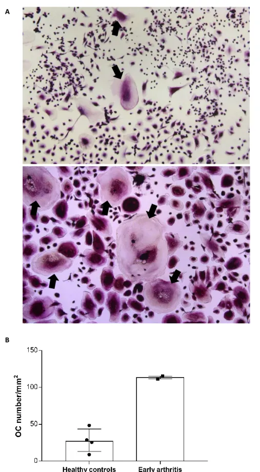
Figure 4 - TRAP staining of in vitro differentiated osteoclasts. A: representative image of OCs of healthy controls (top) and early arthritis patients (bottom) (10× magnification) at culture day 21, stained for TRAP. Black arrows – osteoclasts. B: OC number were increased in early arthritis patients (n=2), when compared to healthy controls (n=4). Each dot represents a sample. Data presented as median with interquartile range. 65
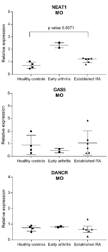
Figure 5 - Relative expression of NEAT1, GAS5 and DANCR in monocytes from healthy controls (n=4), early arthritis patients (n=2) and established rheumatoid arthritis patients (n=6). Data was normalized for housekeeping gene (GADPH) expression levels. No statistically significant differences were found between healthy controls and established RA patients. Each dot represents a sample. Data presented as median with interquartile range. RA – rheumatoid arthritis; MO – monocytes. ... 68
Figure 6 - Relative expression of NEAT1, GAS5 and DANCR in osteoclasts from healthy controls (n=3), and early arthritis patients (n=2). Data was normalized for housekeeping gene (GADPH) expression levels. No statistical analysis was performed due to insufficient number of samples. Each dot represents a sample. Data presented as median with interquartile range. OC – osteoclasts. ... 69
Figure 7 - Relative expression of NEAT1, GAS5 and DANCR in monocytes (n=4) and osteoclasts (n=3) from healthy controls. Data was normalized for housekeeping gene (GADPH) expression levels. No statistically significant differences were found between monocytes and osteoclasts of healthy controls. Each dot represents a sample. Data presented as median with interquartile range. HC – healthy control; MO – monocytes; OC – osteoclasts. ... 70
Figure 8 - Relative expression of NEAT1, GAS5 and DANCR in monocytes (n=2) and osteoclasts (n=2) from early arthritis patients. Data was normalized for housekeeping gene (GADPH) expression levels. No statistical analysis was performed due to insufficient number of samples. Each dot represents a sample. Data presented as median with interquartile range. EA – early arthritis; MO – monocytes; OC – osteoclasts. ... 71
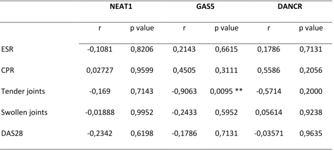
Figure 9 - Correlation analysis between GAS5 expression and the number of tender joints in RA patients. Line represents linear regression obtained for these variables: Y = -0,5045*X + 2,004; R2 = 0,3319. ... 72
xxv
LIST OF TABLES
Table 1 - Summary of the inclusion criteria for the study groups. ... 53
Table 2 - Primers used for lncRNAs expression analysis. ... 58
Table 3 - Summary of the patients and healthy controls' characteristics. ... 63
Table 4 - Summary of the obtained RNA samples from both monocytes and osteoclasts of all study groups. ... 64
Table 5 - Relative expression values for NEAT1, GAS5, and DANCR. ... 67
xxvii
LIST OF CONTENTS
INTRODUCTION ... 31 1. Rheumatoid arthritis ... 31 1.1. Epidemiology ... 31 1.2 Characteristics and Pathophysiology ... 32 1.3 Diagnostic and Treatment ... 35 1.4 Risk Factors ... 37 2. Monocytes ... 38 2.1. Role of monocytes in the pathophysiology of rheumatoid arthritis ... 38 3. Osteoclasts ... 39 3.1. Role of osteoclasts in the pathophysiology of rheumatoid arthritis ... 40 4. Bone metabolism ... 41 4.1. Normal bone metabolism: modeling and remodeling ... 42 4.2. Altered bone metabolism in rheumatoid arthritis: bone erosion and osteoclasts ... 45 5. Long non-coding RNAs (lncRNAs) ... 46 5.1 lncRNAs in rheumatoid arthritis ... 47 6. Aim of the study ... 49 PATIENTS AND METHODS ... 53 1. Patients and healthy controls ... 53 2. Peripheral blood mononuclear cells (PBMCs) isolation ... 53 3. Cell culture and osteoclastogenesis ... 54 4. Osteoclasts functional assay (TRAP staining) ... 55 5. Total RNA extraction ... 56 6. Complementary DNA (cDNA) synthesis ... 57 7. Real-time quantitative PCR (RT-qPCR) ... 57 8. Statistical analysis ... 59 RESULTS ... 63 1. Patients and healthy controls ... 63 2. Cell culture, osteoclastogenesis, and total RNA extraction ... 64 3. Osteoclasts functional assay (TRAP staining) ... 64 4. lncRNAs expression analysis by RT-qPCR ... 66 5. Correlation of lncRNAs expression and clinical variables ... 72 DISCUSSION... 75 FINAL REMARKS ... 85 REFERENCES ... 89
Analysis of lncRNAs expression in monocytes and osteoclasts of rheumatoid arthritis patients Inês Abrunhosa Amaral
31
INTRODUCTION
1. Rheumatoid arthritis
Rheumatoid arthritis (RA) is a chronic inflammatory and immune-mediated disease [1,2]. It is an idiopathic disease (of unknown cause), that can be defined as a clinical syndrome spanning several disease subsets, with multiple pathogenic mechanisms [1–3]. The hallmark of RA is systemic inflammation, with persistent symmetric peripheral polyarthritis and structural damage of cartilage and bone tissue, leading to joint swelling, loss of function and stiffness [2]. The hypersensitivity reaction associated with disease pathogenesis in RA is mediated by immune complexes (type III reaction), and the main autoantibodies involved are rheumatoid factor (RF) and anti-citrullinated protein antibody (anti-ACPA) [4,5]. RA patients can be subdivided into two subtypes designated “seropositive” and “seronegative”, with seropositivity being defined as the presence of serum elevations of the autoantibodies RF and/or ACPAs [6].
The pain and progressive deterioration of joint function, along with increased comorbidity, have a high impact on health and quality of life of RA patients. In severe cases, the ability to work can be compromised [7]. Different criteria can be applied to assess disease severity in RA: disease activity, extraarticular manifestations and joint destruction [8]. Concerning this, the Disease Activity Score based on 28 joints (DAS28) is one of the most widely used tools to assess disease activity in RA patients [9].
1.1. Epidemiology
Worldwide prevalence of RA is of around 1%, with a significant variation of the disease incidence and prevalence among different populations [10]. In Portugal, the total prevalence of RA is of 0.7%, with a four-fold higher prevalence in women than in men (1.2% and 0.3% respectively) [11]. It is the most common form of chronic inflammatory arthritis [12]. A systematic review by Minichiello et al., described a trend toward a decline in the incidence of RA [8]. However, while some studies support such decrement [13,14], others report an increasing trend in the incidence of RA in the general population [15,16].
Patients with rheumatoid arthritis present a life expectancy decreased by 3 to 10 years when compared with the general population, along with a 50%-54% increased risk of premature mortality [7]. Higher severity of the disease is associated with a higher risk of death [7]. Cardiovascular
Analysis of lncRNAs expression in monocytes and osteoclasts of rheumatoid arthritis patients Inês Abrunhosa Amaral
32
diseases, respiratory diseases, and infections are among the primary causes of death in RA patients [7,17]. The increased risk for infection in RA patients may be attributed to the impairedimmune function in RA or an effect of immunosuppressive therapy. Death by an infection in RA patients is often due to respiratory infection or pneumonia [17]. Recent trends show no significant reduction in mortality in RA patients worldwide [7]. However, information about the mortality rates of observational studies differ. Some studies show higher mortality in RA patients [18,19], while others report that mortality in patients with RA was not different from that of the general population [20,21].
Several studies show a decrement in disease activity over time, based on a decline of DAS28 values, suggesting a decrease in RA severity in most recent cohorts [22–24]. Regarding overall extraarticular manifestations, there is no consensus. Some authors report a decrease [25], while others describe no significant changes over time [26]. A study from the UK, in a stable well-defined population, monitored for 15 years, reported a decrease in the incidence of systemic rheumatoid vasculitis [27]. On the other hand, the need for joint surgery reflects the degree of joint destruction [8]. Increased severity of disease leads to an increased need for joint surgery. Some studies report adecline in the severity of joint destruction, in RA patients, over time [28,29]. Regarding the need for joint surgery, several authors indicate a significant reduction in both the risk and the incidence of joint surgery [30–32].
These varying results might be explained by differences in treatment or different types of cohorts and follow-up time [33]. Nevertheless, evidence indicates a decrease in severity over time, with fewer extraarticular manifestations and lower disease activity, along with a diminished need for surgery to treat joint destruction, and less severe radiological changes [8]. Van Nies et al.,reported an improvement in the outcome of early RA patients over the last decade, based on lower rates of joint destruction and higher remission rates [20]. The authors hypothesized that current treatment strategies, namely the use of disease modifying anti-rheumatic drugs (DMARDs), combination therapies and tight control of disease activity, have been having a positive impact on the survival of RA patients, thus reflected in a reduction of the mortality risk in RA [20].
1.2 Characteristics and Pathophysiology
Rheumatoid arthritis comprises different disease subsets that involve several immuno-inflammatory cascades, with predominant features varying from patient to patient [2]. Nevertheless, all the involved cascades in RA lead towards a final common pathway characterized
Analysis of lncRNAs expression in monocytes and osteoclasts of rheumatoid arthritis patients Inês Abrunhosa Amaral
33
by persistent synovial inflammation (hyperplasia) and associated damage to articular cartilage and underlying bone tissue [2]. The characteristic hyperplastic joint present in RA, with several inflammatory cellular infiltrations, is represented in Figure 1.
Even though joint involvement is the hallmark of this disease, RA is an organ nonspecific (systemic) immune-mediated disease since extraarticular pathologic lesions or manifestations can be found, including fatigue, subcutaneous nodules, lung involvement, pericarditis, peripheral neuropathy, vasculitis, and hematologic abnormalities [12].
The pathophysiological mechanisms that have been proposed for the development of RA involve a complex interplay between different cells, genetic and environmental factors, triggers, and chance [34]. Both T and B cells and the action of pro-inflammatory cytokines play key roles in the pathophysiology of RA [35]. Tumor necrosis factor alpha (TNF-) and interleukin (IL-6) are regarded as the cytokines most directly implicated in RA, although IL-1 and IL-17 may also play an important role in the disease process [35–37]. Prominent T-cell infiltrate in RA patients suggests these cells
Figure 1 - Cartoon representation of a normal joint (left) and a rheumatoid arthritis joint (right). Adapted from Strand et al., 2007 [265].
Analysis of lncRNAs expression in monocytes and osteoclasts of rheumatoid arthritis patients Inês Abrunhosa Amaral
34
are key participants in the acute inflammatory features of RA [1].Nevertheless, increasing evidence suggests that a variety of innate effector cells, such as monocytes and macrophages, are also closely involved in the development of synovial inflammation in RA. In fact, massive infiltration of activated monocytes/macrophages are frequently observed in synovial membranes of RA patients. These are a major source of cytokines (such as TNF-, IL-1β, IL-8, and GM-CSF) in the inflamed joints [38]. The initial milestone for RA pathogenesis onset is thought to be the activation of the innate immune response, including the activation of dendritic cells by exogenous material and autologous antigens [35]. Antigen-presenting cells, including dendritic cells, macrophages and activated B cells, present arthritis-associated antigens to T cells [35,39]. These T cells are upregulated by various lymphokines, including interleukin 2 (IL-2) and interferon γ (IFNγ). Upon stimulation, T cells induce activation of macrophages, B cells, fibroblasts, and osteoclasts [38,39]. In turn, B lymphocytes express various cell-surface molecules, including antigen receptor immunoglobulin and differentiation antigens, such as CD20 and CD22. Upon differentiation, they turn into plasma cells that secrete antibodies, including autoantibodies to IgG (rheumatoid factor), to citrullinated peptides such as vimentin, fibrinogen, or cyclic citrullinated peptide (CCP) and to rheumatoid arthritis antigen (RA33) [42,43]. On the other hand, the formation of immune complexes by autoantibodies can increase the production of proinflammatory cytokines such as TNF-. In fact, the occurrence of autoantibodies RF and anti-CCP are associated with severe rheumatoid arthritis and can be used as an effective strategy for identifying patients with RA at high risk for poor outcome [44]. When activated, B cells also serve as APCs, inducing T cell activation, and potentially leading to the perpetuation of the immune-mediated proinflammatory response [42]. Besides cells of the innate and adaptive immune system, several other inflammatory cell populations infiltrate the synovial membrane in rheumatoid arthritis patients [34,35,45].
The in situ pathological occurrences within the synovial inflammation include activation of endothelial cells, along with neovascularization and increased presence of fibroblast-like synoviocytes (FLSs). These synoviocytes are highly activated and secrete inflammatory mediators, cytokines and matrix metalloproteinases (MMPs) [46,47]. MMPs are enzymes that can degrade the components of the extracellular matrix of the joint [48]. By secreting MMPs into the synovial fluid, and probably by direct invasion, fibroblast-like synoviocytes can destroy cartilage and play a role in bone destruction [46,49]. Nevertheless, bone erosions occur mainly via activated osteoclasts. Bone erosions in RA patients result from an imbalanced bone metabolism due, in part, to excessive osteoclastogenesis and osteoclastic activity, that leads to increased bone resorption by osteoclasts, and inadequate bone formation by osteoblasts [50–52]. Besides being responsible for bone
Analysis of lncRNAs expression in monocytes and osteoclasts of rheumatoid arthritis patients Inês Abrunhosa Amaral
35
resorption, osteoclasts are key players in the complex crosstalk between bone and immune cells, that leads to the perpetuation of the inflammatory environment in joints of RA patients [53–55]. The role of osteoclasts in physiological bone metabolism and in RA pathogenesis will be discussed further ahead.
All the above-mentioned events and complex cross-talk of immune and inflammatory modulators promote influx, expansion, and activation of cells in the synovium, ultimately leading to the immunoinflammatory and destructive response of rheumatoid arthritis, clinically presented as joint swelling and joint and bone destruction [56,57]. However, the precise hierarchy of events remains enigmatic. It is nowadays understood that multiple and redundant pathophysiological mechanisms contribute to RA [56,57].
1.3 Diagnostic and Treatment
Diagnostic of RA is mostly clinical, along with a combination of laboratory tests, and imaging methods. The most recent criteria to the diagnosis of rheumatoid arthritis are the 2010 American College of Rheumatology (ACR) and the European League Against Rheumatism (EULAR) criteria [58], and their application is due to be made by the physician regarding each patient’s individual circumstances. These guidelines are aimed at identifying rheumatoid arthritis among patients that recently present synovitis (inflammation of a synovial membrane) in at least one joint, in the absence of an alternative diagnosis that better explains the synovitis. For patients to be diagnosed with RA, they need to achieve a total score of at least 6 (of a possible 10) within 4 evaluation domains [58,59]. These domains include the following: (1) the number and site of involved joints, (2) serologic abnormalities (presence of rheumatoid factor or anti-citrullinated peptide/protein antibody), (3) elevations of inflammatory markers (erythrocyte sedimentation rate and/or C-reactive protein [CRP]), and (4) the duration of symptoms [58]. The criteria for the evaluation of disease progression are the 2012 ACR Disease Activity Measures [60].
To treat RA patients, there are multiple drugs currently available for use. These can be non-steroidal anti-inflammatory drugs (NSAIDs), disease-modifying antirheumatic drugs (DMARDs) or adjunctive agents such as corticosteroids, and analgesics [61–63]. Most of the NSAIDs are designed to target and suppress prostaglandins (PGs), inflammatory mediators, through inhibition of the cyclooxygenase (COX) enzymes [61]. These can lead to renal, hepatic, and cardiovascular toxicity with long-term use in RA patients [61]. Within the NSAIDs, there are many different drugs, such as Ibuprofen, Aspirin, and Naproxone [61].
Analysis of lncRNAs expression in monocytes and osteoclasts of rheumatoid arthritis patients Inês Abrunhosa Amaral
36
DMARDs are a class of antirheumatic drugs which can be divided into two sub-categories: non-biological (synthetic) and non-biological [62]. Among non-non-biological DMARDs, methotrexate (MTX) has been the gold standard for the past few decades in the treatment of RA patients [62,63]. Although its exact mechanism has not been fully defined, MTX can block cytokine production and modulate specific matrix metalloproteinases (MMPs) levels [64,65].
The introduction of biological DMARDs (bDMARDs) dramatically changed the treatment and outcome of RA, as they are significantly more effective than non-biologic treatments in improving physical function in RA patients [66]. These drugs include monoclonal antibodies targeting tumor necrosis factor (TNF)-α (anti-TNF-α), IL-6 receptor (anti-IL6R), IL-1 receptor (anti-IL1R) and other inhibitors of cytokines and immune cells that play a major role in promoting RA pathogenesis [67]. As previously described, cytokines play a fundamental role in the pro-inflammatory milieu occurring within the synovium of affected joints in RA patients [36,68]. Among these, TNF-α and IL-6 are considered the most relevant and constitute the therapeutic targets of several drugs for rheumatoid arthritis [69]. These drugs can directly inhibit these cytokines, or interfere with receptor binding, or both. Up to date, a total of five anti-TNF drugs have entered clinical use, namely infliximab (IFX), adalimumab (ADA), etanercept (ETC), golimumab (GLM), and certolizumab pegol (CZP) [70]. Infliximab (IFX) was the first TNF-α inhibitor to be administered in RA patients. Both infliximab, adalimumab, and golimumab are monoclonal antibodies anti-TNF-α.Certolizumab pegol is a humanized monovalent Fab antibody fragment linked to polyethylene glycol (PEG) [70]. Etanercept was the first biological DMARD to be approved for treating RA and is a recombinant fusion protein in which the extracellular domain of TNF receptor II is fused to the Fc-portion of IgG1 [70,71]. IL-6 has also been the focus of great interest in RA. This cytokine in presumed to play key roles in the context of the pathogenesis of rheumatoid arthritis: activation of T cells, macrophages and osteoclasts, and induction of antibody production by B cells [37,72–74]. Tocilizumab is a monoclonal antibody to the IL- 6 receptor, that targets the actions of this cytokine [75,76]. However, despite the range of available treatments, several unmet needs remain. There is a lack of predictive biomarkers of prognosis, therapeutic response, and toxicity. Importantly, current synthetic and biological DMARDs therapies sometimes fail or produce only partial responses. In addition, sustained remission in RA patients is rarely achieved and requires ongoing pharmacologic therapy [77].
Analysis of lncRNAs expression in monocytes and osteoclasts of rheumatoid arthritis patients Inês Abrunhosa Amaral
37
1.4 Risk Factors
Multiple genetic and environmental factors have been associated with an increased risk of developing rheumatoid arthritis [78]. In RA, altered gene expression is present in both circulating immune cells and synovial tissues [79].
The genetic factors can include susceptibility genes such as HLA-DRB1, which shows the strongest association with RA, in particular with ACPA positive RA [80]. Most HLA DRB1 alleles that predispose to RA (HLA DRB1*01, *04, and *10 alleles) share a specific amino acid sequence in their peptide-binding region. This sequence similarity might implicate the involvement of these shared epitope alleles in presenting arthritis-related peptides. Some studies have reported that these alleles may preferably present citrullinated peptides, which could explain the strong association with ACPA positive RA [81,82]. RA patients expressing two HLA-DRB1*04 alleles are at elevated risk for nodular disease, major organ involvement and increased need for surgery related to joint destruction [83]. Furthermore, HLA and non-HLA genes may discriminate anti-cyclic citrullinated peptide (anti-CCP) antibody–positive and anti–CCP–negative RA groups [84].
Genome-wide association studies (GWAS) have expanded the number of validated RA risk loci, identifying additional alleles related to RA risk beyond HLA-DRB1 shared epitope alleles [84]. Single-nucleotide polymorphism genotyping across the MHC has identified additional alleles related to RA risk, including those found on the conserved A1-B8-DR3 (8.1) haplotype and those near the HLA-DRB1 gene. Some of the relevant single-nucleotide polymorphisms (SNPs) linked to RA pathogenesis include RAF1, STAT4, CTLA4, IRF5, CCR6, PTPN22, IL23R, and PADI4 [85–87]. Among these, some of the most relevant include PTPN22 and PADI4 [86,87]. Like HLA-DRB1, PTPN22 is mostly associated with ACPA positive RA. It encodes a tyrosine phosphatase involved in T and B cell signaling [87]. PADI4 encodes for an arginine deiminase responsible for the posttranslational conversion of arginine into citrulline residues. Besides being associated with RA susceptibility, the PADI4 haplotype is also mainly associated with ACPA positive RA [86].
Among the multiple environmental, lifestyle, and behavioral risk factors already studied, cigarette smoking is regarded as the most strongly and consistently associated with the development of RA, presenting a clear dose-response [88–90]. As well as the susceptibility genes, environmental factors such as smoking are most strongly associated with the seropositive RA phenotype [91]. It is estimated that smoking is responsible for around 35% ACPA positive RA cases. Furthermore, 55% of ACPA positive RA can be attributable to smoking, in individuals carrying two copies of the HLA-DRB1 shared epitope [91].
Analysis of lncRNAs expression in monocytes and osteoclasts of rheumatoid arthritis patients Inês Abrunhosa Amaral
38
2. Monocytes
Human monocytes represent 5 % – 10% of the blood leucocytes and their half-life in the vascular compartment is around 1 – 3 days. Monocytes in blood have some typical morphological features such as the irregular shape of the cell and its nucleus, high cytoplasm-to-nucleus ratio, and light blue cytoplasm [92–94]. These cells originate in the bone marrow from hematopoietic stem cells (HSCs). They are then released into the peripheral blood, where they circulate. These circulating monocytes give rise to tissue-resident macrophages and specialized cells, such as dendritic cells (DCs) and osteoclasts [93]. Monocytes have important roles in both innate and adaptive immunity, primarily functioning in immune defense, inflammation, and tissue remodeling, in both homeostasis and inflammation. During inflammation, monocytes circulate through the blood and extravasate into inflamed tissues, where they differentiate into macrophages, generally maintaining the same inflammatory phenotype [95].
Monocytes are categorized into three major subsets,according to the expression of CD14 (the LPS co-receptor) and CD16 (the low-affinity receptor for the Fc region of IgG): classical CD14++CD16−
that constitute about 90% of circulating monocytes, and intermediate CD14++CD16+ and
non-classical CD14+CD16+ that together constitute the remaining 10% [96,97]. The first two populations
of monocytes share many features,including a high capacity for phagocytosis and proinflammatory cytokine production. Classical monocytes, the dominant blood population, express CCR2, a receptor involved in mobilization from the bone marrow (BM) and recruitment to inflammatory sites, which is the main function of classical monocytes [98,99]. In contrast, non-classical monocytes present a reduced CCR2 but elevated CX3CR1 surface expression, which is needed for patrolling blood vessels and migrating into resting tissues [96,100]. The population of intermediate monocytes expresses both CCR2 and CX3CR1 on intermediate level and shows the highest HLA-DR expression [101].
2.1. Role of monocytes in the pathophysiology of rheumatoid arthritis
Infiltration of monocytes along with T and B cells and the exacerbated presence and activity of macrophages and osteoclasts into the synovial tissue and production of inflammatory mediators typify the immunopathology of RA [102]. These accumulating activated monocytes massively infiltrate inflammatory sites (synovial membranes) of RA patients and present a significantly increased expression of CD16 on their surface and produce TNF- in response to TLR stimulation [103–107].The impact of the monocytic lineage in determining the immune response is substantial and these cells interfere with both the innate and adaptive immunity [108]. For example, in RA,
Analysis of lncRNAs expression in monocytes and osteoclasts of rheumatoid arthritis patients Inês Abrunhosa Amaral
39
monocytes present in inflamed joints have been shown to induce the development of Th17 cells, thus contributing to the amplification of inflammation. In fact, T cell - monocyte contact is involved in cartilage destruction and cytokine production [109,110]. Therefore, targeting monocyte-derived cytokines such as tumour necrosis factor (TNF), IL-1, IL-6 or even monocyte - T cell interaction might be a therapeutic strategy for controlling inflammation in RA patients non-responsive to classical DMARDs [111]. Approaches to depletion of monocytes using specific antibodies could prevent their presence in the synovial tissue and thus, attenuate inflammation [112,113].
In RA patients, the frequencies of classical, intermediate and non-classical blood monocytes are altered, when compared to healthy controls [114–116]. CD14+CD16+ monocytes (non-classical) are
increased in the blood of RA patients, which might contribute to the persistent joint inflammation of RA [117]. These monocytes display properties for antigen presentation and inflammatory characteristics upon activation [118]. Increased expression of CD16 in monocytes was observed in patients with active disease and increased counts of tender and swollen joints. CD16 expression on RA blood monocytes was augmented in vitro by IL-10, M-CSF, and TGF-β1 [117]. These pieces of
evidence suggest that monocytes are key players in RA pathogenesis, contributing the perpetuation of the inflammatory milieu observed in the joints of RA patients. However, it is still unknown whether and how the altered frequency of monocytes subsets in RA patients affects the role of monocytes and/or macrophages in the pathogenesis of RA.
In addition to their central role in modeling inflammation, monocytes can be pinpointed as the origin of pathological bone erosion in RA, due to their excessive differentiation into osteoclasts [119]. As such, monocytes may thus constitute ideal targets to modulate osteoclastogenesis in inflammation and RA.
3. Osteoclasts
An osteoclast can be described as a tissue-specific macrophage of the bone, able to degrade bone tissue by polarized secretion of proteolytic enzymes such as cathepsin K and acid, which hydrolyzes and solubilize the organic and inorganic components of bone [50,120]. In fact, in physiological conditions, the osteoclast is the only cell type capable of resorbing bone [120]. Mature osteoclasts are fused polykaryons (multinuclear cells) arising from multiple (up to 10–20) individual cells [50]. Osteoclastogenesis is the process by which hematopoietic precursor cells of the monocyte/macrophage lineage differentiate into osteoclasts [50,120]. In particular, peripheral blood monocytes (PBMs) constitute the main source of precursors of osteoclasts, like as with other
Analysis of lncRNAs expression in monocytes and osteoclasts of rheumatoid arthritis patients Inês Abrunhosa Amaral
40
tissue macrophages [121,122]. PBMs leave the peripheral circulation by margination in capillaries and migration into the extravascular pool. Osteoclasts arise from these migrating monocytes by in situ proliferation of macrophage precursors and further differentiation in tissues [93]. For such processes, two major cytokines are required: the macrophage colony stimulating factor (MCSF) and the receptor activator of NF-kB ligand (RANKL) supplied mostly by osteoblasts and/or osteocytes [50,120]. M-CSF binds to its receptor c-FMS, (cellular-feline McDonough strain sarcoma virus oncogene homologue, or CSF-1 receptor, or CD115), which in turn induces the expression of RANK on monocytes.RANKL expression by synovial fibroblasts is induced by pro-inflammatory cytokines such as TNF-a, IL-1, IL-6, and IL-17 [123]. The RANK/RANKL ligand-receptor complex is a crucial intervenient in osteoclast differentiation and activation. RANKL is a necessary factor for the differentiation of osteoclasts and functions as both survival factor and activator [124]. Its physiological inhibitor is osteoprotegerin (OPG) [124]. Osteoblasts are the main source of RANKL. However, it can be expressed by synovial cells, activated T cells, mature B cells and NK cells. RANKL is a mediator of differentiation, survival, and activation of osteoclasts and its expression is upregulated by TNF, IL-1, IL-6, and IL-17 [124]. Overall, osteoclastogenesis and osteoclast activation are regulated, both positively and negatively, by at least 24 gene or loci [50]. Among these are as well cytokines, produced by monocytes, with important effects on osteoclast differentiation, activation, and apoptosis [121].
3.1. Role of osteoclasts in the pathophysiology of rheumatoid arthritis
Osteoclasts play a critical role in skeletal homeostasis by regulating bone resorption. However, in RA, they excessively populate chronically inflamed joints, resorbing the bone in an exacerbated way and creating localized skeletal defects, the so-called bone erosions [125]. Chronic inflammation appears to be a key mediator for both local and systemic bone loss in RA patients [124]. In fact, inflammation itself might trigger bone resorption by osteoclasts [126]. It has been reported that the high levels of pro-inflammatory cytokines prevalent in the synovial fluid of affected joints in RA patients are associated with osteoclast-mediated focal bone erosion at the margins of these joints [127]. Pro-inflammatory cytokines are potent mediators of bone loss and correlate with greater disease activity in RA patients. They can enhance osteoclastogenesis in inflamed joint and bone, acting on osteoclasts, osteoblasts, and their precursors, by inducing RANKL expression [124,128,129].
Analysis of lncRNAs expression in monocytes and osteoclasts of rheumatoid arthritis patients Inês Abrunhosa Amaral
41
RA patients present increased numbers of osteoclast progenitors in the peripheral blood, exhibiting an enhanced ability to differentiate into osteoclasts when compared to those of matched healthy controls [130]. The extensive infiltration of monocytes in the synovial tissue of inflamed joints in RA patients leads to an increased number of osteoclasts, as these function as osteoclast precursors and provide specific molecular signals that drive osteoclast formation, as previously stated [131]. Overall, osteoclasts play a critical role in bone destruction in RA and could represent a therapeutic target for RA [132].
The role of monocytes and osteoclasts in the pathophysiology of rheumatoid arthritis is represented in Figure 2.
4. Bone metabolism
Bone homeostasis is modulated by both inflammation and immunological events. The concept of osteoimmunology was coined a few years ago to account for the interplay between the bone and immune systems [123,133]. During life, bone undergoes modeling and remodeling to grow or change shape. The main cells involved in bone remodeling are osteoblasts and osteoclasts, whose
Analysis of lncRNAs expression in monocytes and osteoclasts of rheumatoid arthritis patients Inês Abrunhosa Amaral
42
functions are tightly coupled. Additional cells involved in bone metabolism are osteocytes and the bone lining cells [134].
The osteoblast is a specialized bone-forming cell derived from mesenchymal stem cells (MSCs). Osteoblasts produce bone by synthesis and secretion of type I collagen, that makes up the major bone matrix protein. They also participate in the mineralization of the newly formed bone [135]. As previously stated, osteoclasts are the cells capable of resorbing bone. For osteoclasts to be activated and bone resorption to commence, non-polarized, mature osteoclasts must bind to the bone matrix. When osteoclasts bind to bone, they become polarized [120]. Osteocytes are the most abundant and long-lived cells in bone. They comprise 90-95% of the total bone cells and have a lifespan of up to 25 years. Osteocytes originate from osteoblasts that have undergone terminal differentiation during bone formation and subsequently have been engulfed by the unmineralized osteoid. Following mineralization, these entombed cells become osteocytes [136]. Its functions include monitoring bone quality and stress, as well as coordinate remodeling through membrane-bound and secreted factors [120,137]. Bone lining cells are quiescent flat-shaped osteoblasts that cover the bone surfaces, where neither bone resorption or bone formation occurs [138].
4.1. Normal bone metabolism: modeling and remodeling
Bone is a complex tissue, undergoing continuous remodeling to heal damage, grow and maintain calcium and phosphate homeostasis [120,139,140]. The human bone is composed of trabecular bone, the fine bony network hosting the bone marrow, and cortical bone, the dense bony shell that provides structural support in weight-bearing regions [51]. A healthy bone metabolism relies on a delicate balance between bone formation by osteoblasts and bone resorption by osteoclasts [120,139,140]. The concerted and sequential actions of both osteoclasts and osteoblasts in the process of bone remodeling has been termed the basic multicellular unit (BMU) [139–141]. It is important to note that bone remodeling differs from bone modeling. Bone modeling is the process by which the generation and maintenance of the shape of bone during skeletal growth occur, allowing bones to change shape or size in accordance to physiologic influences or mechanical forces [142,143]. This process lasts from the beginning of skeletal development in fetal life until the end of the second decade of life, which is when the longitudinal growth of the skeleton is completed [140]. In turn, bone remodeling is a continuous process, occurring throughout life, with the purpose of maintaining strength and mineral homeostasis in the bone tissue [144]. Osteoclastic resorption
Analysis of lncRNAs expression in monocytes and osteoclasts of rheumatoid arthritis patients Inês Abrunhosa Amaral
43
is tightly coupled to the osteoblastic bone formation during the bone remodeling cycle [139,145,146].
The remodeling occurs within the BMU and is a process defined by five phases: activation, resorption, reversal, formation, and termination [140,146]. The bone lining cells separate from the underlying bone and form a raised cover that encases each BMU, exposing the bone surface at the site to be remodeled and creating a unique environment for coupled resorption–formation [147]. Each active BMU consists of bone-resorbing osteoclasts covering the newly exposed bone surface, resorbing the old bone and preparing the surface for the deposition of replacement bone. Following immediately behind, the osteoblasts secrete and deposit unmineralized bone osteoid [148,149]. The orderly arrangement of the cells within the BMU is critical for correct remodeling, ensuring the correct sequence of the phases of the process [148,149].
The first phase of the bone remodeling cycle is the activation phase, which is characterized by the recruitment of osteoclasts [140,146]. The initiating signal for remodeling can be either hormonal or mechanic and is supposed to be detected by the osteocytes. In fact, osteocytes are key players in the bone remodeling cycle modulation [137,150,151]. Succeeding the detection of the signal occurs the retraction of the bone lining cells and the digestion of the endosteal membrane by collagenase. Alongside with this, occurs the recruitment of precursor cells from the monocyte/macrophage hematopoietic lineage, which are recruited from circulation and are activated at the site [152–154]. Multiple mononuclear cells fuse to form multinucleated preosteoclasts that migrate and attach to the bone surface. These develop and terminally differentiate in activated osteoclasts after binding to the bone matrix and thus form bone-resorbing compartments, secreting acid and lytic enzymes to break down the bone matrix [50,155].
The second phase of the bone remodeling cycle is the resorption phase when the osteoclasts resorb bone. This phase lasts approximately two to four weeks during each remodeling cycle [140,146]. Osteoclasts generate an acidic microenvironment in the attachment zone, secreting hydrogen ions that drop the pH as low as 4.5 within the bone-resorbing compartment [156]. These acidic conditions might help mobilize bone mineral and could favor the action of secreted lysosomal proteinases in the degradation of collagen [156–158]. The degradation of the collagen-rich bone matrix by osteoclasts occurs through the secretion of specialized proteinases, such as matrix metalloproteinases (MMPs) and cathepsin K [157,159]. At last, to ensure that excess resorption does not occur, the resorption phase is terminated by osteoclasts programmed cell death [160]. The third phase of the bone remodeling cycle is the reversal phase, where the osteoclasts undergo apoptosis and the osteoblasts are recruited. This phase lasts approximately four to five weeks in
Analysis of lncRNAs expression in monocytes and osteoclasts of rheumatoid arthritis patients Inês Abrunhosa Amaral
44
each bone remodeling cycle [140,146]. Reversal phase is characterized by a shift from bone resorption to bone formation when osteoclasts are replaced by osteoblast-lineage cells which initiate bone formation [139,145]. However, the multitude of coupling signals between osteoclasts and osteoblasts that lead to this phase is not yet fully understood. Current understanding suggests that the release of osteogenic signals from the osteoclasts might initiate the coupling activity of the reversal phase [161,162]. There is evidence that osteoclasts (OCs) can produce many different molecules that affect the osteoblast lineage, both positively and negatively. These include OC-derived cell surface signals, OC-OC-derived soluble signals and resorption-OC-derived soluble signals [162]. Cells of the osteoblast lineage also appear to play an essential role in the concerted effort in bone remodeling, regulating bone-resorbing activity by osteoclasts and coupling resorption with the formation during the reversal phase [163]. These regulatory signals and coupling factors, produced by either osteoclasts or cells from the osteoblast lineage, can be cytokines and/or regulatory receptors on the cells surface, such as IL-6, BMP-2, TGF-β, insulin-like growth factor (IGF), prostaglandin E2 (PGE2), receptor activator of NF-kB (RANK)/RANK ligand (RANKL) and
osteoprotegerin (OPG)/OPG ligand (OPGL), functioning for instance in the ephrin-Eph bidirectional signaling between osteoblasts and osteoclasts [163–167]. Summarizing, the recently resorbed bone surface is prepared for the deposition of new bone matrix, and complex signaling occurs to couple resorption to bone formation, leading to the replacement of osteoclasts by osteoblast-lineage cells which initiate bone formation [161].
The fourth phase of the bone remodeling cycle is the formation phase when new bone formation occurs. This phase can last approximately up to four to six months [140,146]. Osteoblasts synthesize new bone matrix (osteoid), rich in type I collagen, filling the cavities left behind by osteoclastic bone resorption [168]. As the new bone matrix is gradually mineralized it gives origin to new bone. This process is called bone mineralization, whereby hydroxyapatite crystals are deposited among collagen fibrils. This process is regulated by controlling systemic levels of calcium and phosphate concentrations, along with the local concentration of calcium and phosphate within extracellular matrix vesicles [169–172].
The fifth and final phase of the bone remodeling cycle is the termination phase when mineralization of the bone matrix is fully completed. The bone enters in a quiescent state, and the amount of bone formed equals the amount resorbed. At this phase, osteoblasts undergo apoptosis, change into bone-lining cells or become buried within the newly formed bone matrix and terminally differentiate into osteocytes [140,146].
Analysis of lncRNAs expression in monocytes and osteoclasts of rheumatoid arthritis patients Inês Abrunhosa Amaral
45
Bone remodeling is a crucial mechanism for normal bone metabolism and to maintain overall bone homeostasis. It involves a great number of cells and complex regulatory mechanisms [148,149,152– 154]. Deregulation of this mechanism and involved cells is associated with numerous skeletal disorders such as osteoporosis and most importantly, RA [173,174]. Regarding this latter disease, an increased osteoclastogenesis and deregulated osteoclastic activity at bone resorption phase gives origin to bone erosions [51,175]. Therefore, osteoclasts activity, differentiation, and its regulatory mechanisms present as a key field to be studied in RA. The following topic will focus on this matter.
4.2. Altered bone metabolism in rheumatoid arthritis: bone erosion and osteoclasts
Bone erosion is one of the hallmarks of RA pathophysiology and is associated with disease severity, leading to a poor outcome and reduced quality of life [51,176]. It represents an independent risk factor for generalized osteopenia and osteoporosis [51]. The typical feature observed on plain radiographs of RA patients is the erosion of periarticular cortical bone. However, both types of bone (trabecular and cortical) are targeted for erosion in RA. Resulting from an imbalance in bone metabolism, bone erosions in RA patients are due to an excessive osteoclastic activity that leads to increased bone resorption by osteoclasts, and inadequate bone formation by osteoblasts [50,51]. In RA, bone damage and degradation are executed by osteoclasts while fibroblast-like synoviocytes are responsible for cartilage degradation [176]. Initially involving the cortical bone, the articular bone erosion leads to the destruction of the natural barrier between the surrounding tissue and the intertrabecular spaces of the bone marrow cavity [51]. Increased bone resorption activity in RA patients has been reported with osteoclasts originating from peripheral blood monocytes [130]. This means that the osteoclasts responsible for bone erosions in RA derive mainly from monocyte progenitors that infiltrate the synovial membrane [177]. In fact, the rheumatoid pannus has been shown to contain mononuclear cells exhibiting tartrate-resistant acid phosphatase (TRAP) activity, one of the markers for osteoclastic differentiation and activation [177]. The enhanced osteoclast activity in RA is due to a multiplicity of reasons that range from cellular effects to pro-inflammatory cytokines to autoantibodies [176]. MCSF, RANKL, TNF, IL-1, and IL-17 are pro-inflammatory cytokines that may play dominant roles in the pathogenesis of rheumatoid arthritis by participating in osteoclast differentiation [124].
There is evidence that joint damage and bone erosion in RA patients begins at a very early stage of the disease, progressing throughout its course. In fact, the first bone erosions perceived by
Analysis of lncRNAs expression in monocytes and osteoclasts of rheumatoid arthritis patients Inês Abrunhosa Amaral
46
radiographic studies appear to occur during the first 2 years of the disease [178]. Both the development of erosions and the degree of radiological progression can be predicted by the rheumatoid factor (RF) and/or anti-cyclic citrullinated peptide (anti-CCP) levels at the first presentation in RA patients. This indicates that RF and anti-CCP positive patients present a higher risk for developing bone erosions with increased severity, within the first years of disease onset [179].
Many factors contribute to control the activity of osteoclasts within normal bone tissue and normal bone remodeling metabolism [148]. The complex crosstalk between the osteoclasts and neighboring cells (such as osteoblasts, osteocytes, and hematopoietic cells), along with the biochemical microenvironment, is crucial to normal osteoclastogenesis. There is evidence that certain immune complexes found in the joint of RA patients can induce macrophage activation and participate in synovial inflammation, possibly playing a role in the control of osteoclastogenesis [180,181]. Immune complexes containing anticitrullinated peptide antibodies (ACPA) may have a greater ability to stimulate osteoclastogenesis, consequently inducing an increased bone loss in RA patients [182,183]. Recent data suggest that besides being activated by immune cells, osteoclasts can play an active role in inducing or perpetuating the autoimmune feedback loops and inflammatory response in RA. They are capable of antigen presentation, which results in T cell activation [184].
5. Long non-coding RNAs (lncRNAs)
Due to the central dogma of “DNA → RNA → protein”, the research of the last few decades have mainly been focused on the role of protein-coding genes in the pathogenesis of the disease. As proteins are counted as key players of cellular functions, RNA is regarded to be a mere intermediary between genes and proteins [185]. However, in recent years this paradigm has been changed. Due to the developing of new technologies, including genome and transcriptome sequencing, evidence arises suggesting that the noncoding portions of the genome may regulate the complexity of an organism [186]. In fact, the so-called “junk DNA”, that comprises the noncoding portions of the genome, might significantly contribute to the higher eukaryotic sophistication, regulating gene expression, structural and functional mechanisms in cells and disease pathogenesis [187]. This concept resulted in the establishment of the Encyclopedia of DNA Elements (ENCODE) Consortium in 2003, that aimed to identify all the functional elements in the human genome [188,189]. The
Analysis of lncRNAs expression in monocytes and osteoclasts of rheumatoid arthritis patients Inês Abrunhosa Amaral
47
ENCODE project reported that about 90% of the genome is transcribed as nonprotein-coding RNAs (ncRNAs), indicating that this might not be junk hereafter [190–192].
These transcripts that do not hold any protein-coding capacity are called non-coding RNA (ncRNA). Depending on their length, these can be classified as small non-coding RNAs (sncRNAs) and long non-coding RNAs (lncRNAs). Small non-coding RNAs are shorter than 200 nucleotides and consist mainly of microRNAs (miRNA) and small nuclear RNAs (snRNA) [193]. Long non-coding RNAs (lncRNAs) are RNAs longer than 200 nucleotides, that can be subclassified based on their intersection with protein-coding genes as intergenic or genic lncRNAs [194]. Intergenic lncRNAs do not intersect with any protein-coding loci. Genic lncRNAs comprise three biotypes: exonic lncRNA (at least one of its exons intersects a protein-coding exon by at least 1 bp), intronic lncRNAs (contained completely within a sense or antisense protein-coding introns) and overlapping lncRNAs (contain a sense or antisense coding gene within an intron) [194]. According to GENCODE v7, the lncRNAs are the most common however least well-understood RNA species [194].
The number of lncRNAs is larger than the number of protein-coding RNAs, but they are generally expressed at lower levels compared to protein-coding genes, and appear to be tissue or cell-type specific [193–195]. These RNAs can modulate gene expression through multiple mechanisms, such as epigenetics, miRNA sponging, alternative splicing, and transcriptional and translational regulation [193,196–199]. Additionally, lncRNAs appear to participate in several biological processes, such as cell proliferation, morphogenesis, pluripotency, development, neuronal processes, and gametogenesis [200]. Furthermore, the dysregulation of lncRNAs has increasingly been linked to many human diseases, especially in cancers [185,186,190,191,196,201,202].
5.1 lncRNAs in rheumatoid arthritis
Long noncoding RNAs (lncRNAs) are emerging as critical regulators of gene expression in the immune system and inflammatory responses [203,204]. These RNAs can regulate both the innate and adaptative immune responses [205–207]. Recent reports demonstrated that lncRNAs play a key role in the pathogenesis of a variety of autoimmune diseases, such as multiple sclerosis, systemic lupus erythematosus, type I diabetes and RA [208–211].However, current knowledge of RA-related lncRNAs remains limited. The long non-coding RNA expression profile in rheumatoid arthritis patients has been mainly studied in fibroblast-like synoviocytes (FLSs) and peripheral blood mononuclear cells (PBMCs) [212–214].
Analysis of lncRNAs expression in monocytes and osteoclasts of rheumatoid arthritis patients Inês Abrunhosa Amaral
48
An interesting analysis of the lncRNA–miRNA–mRNA functional network by Jiang et al., revealed functional lncRNAs in rheumatoid arthritis, indicating that these might play important roles in the development of RA [215]. Although the biological functions and molecular mechanisms of these lncRNAs are not yet elucidated, some appear to be highly related to RA. The authors suggest that specific lncRNAs are associated with the development of RA, and highlight three lncRNAs (S5645.1, XR_006437.1, J01878) that could be used as potential diagnostic biomarkers and therapeutic targets [215]. A comprehensive analysis of lncRNA and mRNA expression profiles in rheumatoid arthritis patients was performed by Luo et al., using human lncRNA microarray to analyze the PBMCs of RA patients and healthy controls [212]. A total of 5,045 lncRNA (2,410 upregulated and 2,635 downregulated) were differentially expressed in patients with RA [212].
The expression of lncRNAs and mRNAs in the PBMCs of RA patients and their role in RA pathogenesis was described by Yuan et al. [213]. Song et al., reported several lncRNAs with altered expression in PBMCs of RA patients, including Sox2OT, H19, Meg3, GAS5, NEAT1, Meg9, DANCR and HOTAIR [223]. Yang et al., reported that the lncRNA NTT is expressed in human monocytes/macrophages and its expression is highly upregulated (around 100- to 1000-fold of normal control) in the PBMCs of early-untreated RA patients, showing a positive correlation with the initial disease activity score (DAS28) [216].
Zhang et al., described 62 upregulated lncRNAs and 73 downregulated lncRNAs in fibroblast-like synoviocytes from RA patients [214]. ZFAS1 is a recently described lncRNA known to be upregulated and to promote cell migration and invasion is several cancers [217–219]. Ye and its co-authors hypothesize that given these effects on cancer cells, ZFAS1 could also influence the migratory and invasive phenotypes in FLSs of rheumatoid arthritis patients [220]. In fact, they found that ZFAS1 expression is increased in synovial tissue and FLS from RA patients, and showed that silencing ZFAS1 suppressed FLS migration and invasion, while overexpression of ZFAS1 showed the opposite effect [220].
Li et al., recently described a marked down-regulation of the lncRNA GAS5 in FLS of RA patients, suggesting that GAS5 down-regulation could be a pathological feature of RA [221]. GAS5 was also found to be down-regulated in both B-cells and CD4 T-cells of RA patients, although its mechanism and role in the pathophysiology of RA has not been yet elucidated [222]. One possibility is that GAS5 might participate in the regulation immune functions and pathogenesis of autoimmune and inflammatory diseases by modulating the glucocorticoid receptor (GR) transcriptional activity through its decoy RNA glucocorticoid response element (GRE) [222]. It was also reported that GAS5 expression is upregulated in PBMCs of RA patients [223].