DOI: http://dx.doi.org/10.18363/rbo.v76.2019.e1373 Original Article/Oral Biology
Topographic contribution of the foramen and
mandibular lingula in mandibular ramus surgeries
Álvaro Cavalheiro Soares,1 Carolina da Costa Lima Sampaio,2 Francisco Daniel Lima Sampaio,2 Ramon Oliveira de Jesus,3 Mariana Caires Sobral deAguiar,3 Albino Fonseca Junior,2 Thiago da Silva Torres,4 Bernardo Lucena Neto,4 Marcelo Daniel Brito Faria,3,5 Luciana Freitas Bastos,5,6 Célio Fernando
Sousa-Rodrigues4
1Posgraduate Program in Dentistry, School of Dentistry, Fluminense Federal University (UFF), Nova Friburgo, RJ, Brazil 2Biomedical Institute, Department of Morphology, Fluminense Federal University (UFF), Niterói, RJ, Brazil
3Department of Diagnosis and Therapeutics, School of Dentistry, Rio de Janeiro State University, Rio de Janeiro, RJ, Brazil 4Institute of Health and Biological Sciences, Federal University of Alagoas, Maceió, AL, Brazil
5Center of Dental Radiology and Care of Patients with Special Needs, Piquet Carneiro Polyclinic, State University of Rio de Janeiro, Rio de Janeiro, RJ, Brazil 6Department of Preventive and Community Dentistry, School of Dentistry, State University of Rio de Janeiro, RJ, Brazil
• Conflicts of interest: none declared.
AbstrAct
Objective: to analyze the topography and biometrics of the mandibular foramen and mandibular lingula in dry skulls to verify their variability and to determine their
topographic positions. Material and method: to analyze the distances, measurements were made using a properly calibrated Mitutoyo® digital caliper to collect the
distance in millimeters, from the center of the foramen to the four margins (anterior, posterior, superior and inferior) of the mandibular ramus (N=176). We performed the measurements of the mandibular foramen in millimeters to determine its external geometry. The mandibular lingula (N=76) was studied regarding its topographical location and shape when its structure was preserved. All data were documented in a data collection protocol aimed at drawing schematic diagrams and archiving the photographs of the cases and the analysis of the results. Results: in relation to the frequency, the foramen was present in all cases (100%), on both sides on the medial surface of the mandibular ramus. The number of foramen on each side is unique: 175 cases (99.5%) were observed. Only one foramen (0.5%) was double on the right side. Regarding its position in relation to the margins of the jaw, in most of them, it is located near the inferior margin, having varied between 9.6mm and 39.1mm, with an average of 26.0mm. The posterior margin of the jaw varied between 6.8mm and 24.0mm, with an average of 12.30mm. The mandibular lingula of the jaws were analyzed in 32 jaws (64 cases), having triangular shape (55%), present in most cases (86%) and in anterosuperior position in 43% of them. Conclusions: the mandibular foramen is an important anatomical element for the success of the inferior alveolar nerve block technique. Its accessory structure, the mandibular lingula, is a reference position in orthognathic surgery; due to its location and aspect, it serves as a protective shield for the inferior alveolar neurovascular bundle.
Keywords: Mandibular foramen; mandibular lingula, anesthesiology; orthognathic surgery.
Introduction
T
o be able to block the inferior alveolar nerve, the anesthetic solution should be injected at the entrance of the mandibular foramen. However, the location of this foramen is very diverse, a fact contributing significantly to the failure of the anesthetic technique.1,2The inferior alveolar nerve block technique is widely used in all fields of Dentistry, such as oral surgery, endodontics, and periodontics. Unfortunately, this block has a high rate of failure, reaching the scale from 15 to 20%, according to Sloman and Donnelly 1951, Apinhasmit, Chompoopong et
al. 2015.3,4
Therefore, studying the mandibular foramen and mandibular lingula regarding their morphological and topographical aspects is shown to be very important and a great addition to dental specialties, mainly for those using the mandibular foramen and mandibular lingula as a reference for pain control through the inferior alveolar nerve block technique or for surgical purposes, such as in the sagittal osteotomy in orthognathic surgery.5
The mandibular foramen is also an anatomical reference position in orthognathic surgeries. Bearing the various
aspects that may induce the success or failure of anesthetic techniques and orthognathic surgeries in mind, the importance of knowing the anatomical characteristics of the mandibular foramen is evidenced.6-8
In this study, we analyzed the anatomic bases of the mandibular foramen and the mandibular lingula and related their anatomical and surgical importance to the several specialties of Dentistry. Thus, the following objective was substantiated: to analyze the topography and biometrics of the mandibular foramen and mandibular lingula in dry skulls to verify their variability and to determine their topo-graphic positions, thus contributing to a better application of the anesthetic technique, relating the importance of these findings to the clinical-surgical application in Dentistry.
Material and Method
To measure the quantity, location and diameters of the mandibular foramen, we used 176 jaws of male and female adult specimens, from the collection of the Department of Training in Basic Sciences - Anatomy Sector - Nova Fribur-go Health Institute, School of Dentistry, Fluminense Federal University (UFF) and Institute of Biological and Health
2
Rev. Bras. Odontol. 2019;76:e1373Sciences of Federal University of Alagoas. They were disjointed, dry and showed no significant wear of their structure.
A Mitutoyo® caliper was used to gauge the distance,
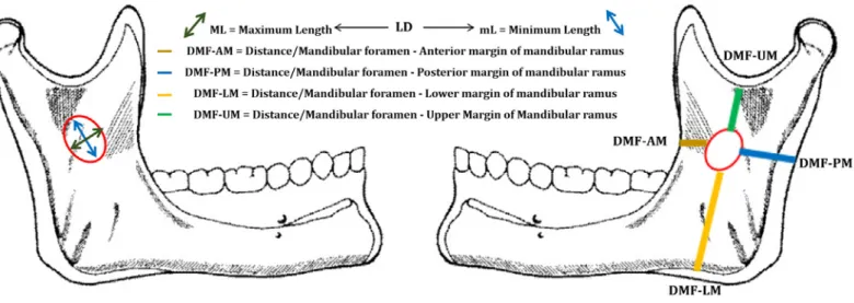
in millimeters, from the center of the foramen to the four margins of the mandibular ramus (Figure 1). This measurement was performed as follows: The opening of the mandibular foramen was modeled using pink wax 7 to indicate the midpoint of the center of the foramen and a colored head pin was put in it; then the center of the foramen was connected to each edge of the mandibular ramus (superior, inferior, anterior and posterior), using the caliper to perform these measurements and the diameter of the foramen.
The presential, morphological and topographical aspects of the mandibular lingula on both sides of the 76 disjointed and dry jaws of the same collection were also analyzed. According to their morphology, they were classified as tri-angular, trapezoidal or absent (Figure 2); and according to their positioning in relation to the mandibular foramen (Figure 1), into: anterior, anterosuperior, posteroinferior, su-perior and superoposterior (Figures 1 and 3).
All data were documented in a data collection protocol, and schematic diagrams, graphs and photographs of the cases were attached for subsequent analysis of the results. The study was approved by the Research Ethics Committee (CAEE: 142792314.1.0000.2318) of the institutions involved.
Results
In all cases studied, the mandibular foramen was present on both sides, on the medial surface of the mandibular ra-mus. One observed that the 176 jaws had a single foramen. The number of foramens on each side is unique: there were 175 cases (99.5%). Only one foramen (0.5%) was double on
the right side (Figure 3).
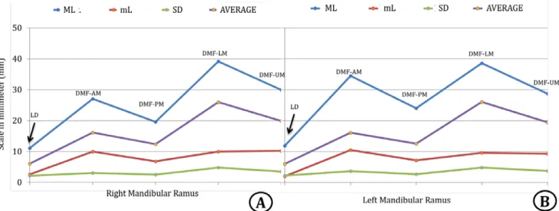
Regarding the distance of the foramen in relation to the four margins of the jaws, the average distance and standard deviation were analyzed between: 1) the foramen and the anterior margin of the jaw, which was 15.92 ± 5.63mm for the right side and 16.16 ± 3.65mm for the left side; 2) the foramen and the posterior margin of the jaw, 12.36 ± 2.54mm for the right side and 12.53 ± 2.70mm for the left side; 3) the foramen and the inferior margin of the jaw were 26.00 ± 4.74mm for the right side and 20.80 ± 4.79mm for the left side; 4) The foramen and the superior margin of the jaw were 19.87 ± 3.52mm for the right side and 19.49 ± 3, 73mm for the left side (Figures 4 and 5).
Concerning the diameter of the foramen, a mean and standard deviation of 6.04 ± 2.19mm was found, for the right and left sides, 6.04 ± 2.17mm and 6.04 ± 2.21mm, respectively (Figure 4).
In most of the cases studied (61%), the mandibular foramen was present near the posterior and superior margins of the mandibular ramus (Classification: Posterosuperior). Then, the foramen was present near the posterior and inferior margins of the mandibular ramus, but fewer in number, (Classification: Inferoposterior) 4% of the cases. In 1.29% of the cases, the foramen was present near the posterior margin and the mean height (FD-IM = FD-SM) of the mandibular ramus (Classification: Posterome-dial). In 24% of the cases, the foramen was present near the anterior and the superior margin of the mandibular ramus (Classification: Anterosuperior). In seven cases (9.09%), the foramen was at an equal distance between the anterior and posterior margins (FD-AM = FD-PM) and near the superior margin of the mandibular ramus (Classification: Superior Medial), both cases on the right side; and in only one case (1.29%), the foramen was near the anterior and
Figure 1. Scheme of the studied distances of the mandibular foramen in relation to the ramus, of the morphology and diameter of the
inferior margin of the mandibular ramus (Classification: Anteroinferior), in Figures 3, 4 and 5.
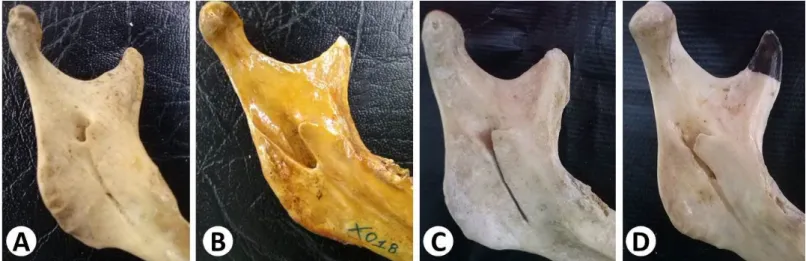
The mandibular lingula was studied in 77 cases, and the cases in which some fragment was destroyed were discarded, so we determined that the mandibular lingula can be divided into three classifications according to its shape: triangular shape, which was found in 35 cases (45%); trapezoidal shape, found in 33 cases (42.85%); and absent mandibular lingula, found in 9 cases (11.68%) in Figures 3 and 6.
Regarding its location in relation to the margins of the mandibular foramen, out of the 68 cases in which the mandibular lingula was present, 10 (12.98%) were in anterosuperior position; among these cases, 6 had a triangu-lar shape and 4 had a trapezoidal shape. In 2 cases (2.59%), the mandibular lingula was in anterior position, and both cases
Discussion
Gremigni, Moore, Miloro et al, Park et al, do not mention the location of the foramen in relation to the four margins of the jaw.6,9,10
However, we found the foramen halfway to the margins of the mandibular ramus, on its inner surface, in seven cases; this statement can be found in the descriptions by Avila Fredes and Bascunan 1965, Zografos, Gritzalis et al. 1988, Afkhami, Haraji et al. 2013.1,11,12
We believe that the observations by Menke and Gowgiel 1979, Halpern, Kaban et al. 2004, Farina, Bravo et al. 2017 are categorical when they affirm that the foramen may have a variable position depending on the individual’s biotype and age.13-15
Other assertions, described by Sekiguchi and Hikima
Figure 2. Scheme of morphological and topographic study of the
mandibular lingula
Figure 3. Morphological aspects of the mandibular foramen. In “A” the superior posterior located foramen; double presence of the mandibular
foramen in “B”; and in “C” the triangular shape of the mandible lingula, in “D” the trapezoidal shape of the lingula, in “E” absence of the mandible lingula
1973, Zografos, Kapnidou et al. 1990, Al-Shayyab 2018, defining that the foramen is near the center or almost exactly in the center become inaccurate and difficult to be identified in relation to the four margins of the foramen.16-18
We disagree with Westmoreland and Blanton 1982, Trost, Salignon et al. 2010, Thangavelu, Kannan et al. 2012 that the foramen is at the same distance between the anterior and posterior margin of the mandibular ramus, since it differs from our results, in which we did not find this description.19-21
One must highlight that, Lotric 1951, Sloman and Donnelly 1951, Mlynarczyk 1954, Avila Fredes and Bascunan 1965 refer to the location of the foramen in relation to the occlusal plane of the lower teeth, whether in the act of chewing or not. This relationship was not investigated in
4
Rev. Bras. Odontol. 2019;76:e1373Figure 4. Mean measurements, in millimeters, relative to the diameter and the distances of the mandibular foramen in relation to the anterior,
posterior, inferior and superior margins of the mandibular ramus
Abbreviations: LD = Larger Diameter, DMF-AM = Distance/Mandibular foramen - Anterior margin of mandibular ramus, DMF-PM = Distance/ Mandibular foramen - Posterior margin of mandibular ramus, DMF-LM = Distance/Mandibular foramen - Lower margin of mandibular ramus, DMF-UM = Distance/Mandibular foramen - Upper Margin of Mandibular Ramus
Figure 5. Mean measurements, in millimeters, relative to the diameter and the distances of the mandibular foramen in relation to the anterior,
posterior, inferior and superior margins of the mandibular ramus. Realize that there is no statistically significant difference (NS) in relation to the sides of the ramus in relation to the position of the mandibular foramen. In “A” LD = Larger Diameter, “B” DMF-AM = Distance/Mandibular foramen - Anterior margin of mandibular ramus, “C” DMF-PM = Distance/Mandibular foramen - Posterior margin of mandibular ramus, “D” DMF-LM = Distance/Mandibular foramen - Lower margin of mandibular ramus, “E”DMF-UM = Distance/Mandibular foramen - Upper Margin of Mandibular Ramus
this study, because we considered it to be very variable.1,4,22,23
Few authors cite the morphological aspect of the mandibular lingula, among which we can cite: Kimura 1982,24 who describes it in a triangular shape, which was
the shape found in most of our results. Tamse, Littner et al. 1988,25 in their turn, characterize it as having a prominent
crest shape, a term that is not used in our study, in which we preferred the terms trapezoidal, triangular or absent.
The shape of the mandibular lingula is important, as it may serve as a protective shield for the inferior alveolar neurovascular bundle present in the foramen content.26
Regarding the location of the mandibular lingula in relation to the margins of the foramen, Yu and Wong 2008,27
affirm it is in anterosuperior position in relation to the mandibular foramen. We did not find the same result, for it was only found in 12.98% of our cases.
We only found the mandibular lingula in anterior position at a median distance between the upper edge and the bottom edge in 2.59% of the cases. In our study, we found their absence in 12% of the cases.
Chen, Davidson et al. 2015 do not describe the presence of the mandibular lingula in the margin of the foramen, leading us to believe that the jaws did not present this structure, which can be confirmed in our results. Studying this structure is necessary because it is used as a reference for sagittal osteotomies of the mandibular ramus, when bone withdrawal should go up to the mandibular lingula, thus avoiding vascular lesions, paresthesias, or paralysis caused by rupture in the neurovascular bundle that transits through the mandibular foramen. Although we have not found statistics of these lesions in the literature, reports on them are frequent in congresses and in dental practice.28
Sekiguchi and Hikima 1973, in their turn, affirm that the distance from the foramen to the anterior margin is about 8 to 14 mm.16 This assertion was found in our findings, in
which we found an average of 14.43mm for this distance.
We agree with Senel, Ozkan et al. 2015 because, according to our casuistry, the estimate is that the ideal needle for nerve block should be long and the distance traveled from 20 to 25 mm.29 However, Chen, Davidson et al. 2015 describes
the same situation, but exceeds the distance traveled by the needle with the value of 31 mm; therefore, we understand that this distance is ideal when we use the closed-mouth block technique.30 We agree partially with Shapiro’s citation,
which informs correctly that the distance may vary in cases in which the ramus is present in a variable way. We disagree with his assertion that the approximate length of the needle to reach the foramen is 38.1 mm.31
Recent studies analyze the topography of the mandibu-lar foramen using panoramic radiographs, such as that of Akcay, Kalabalik et al. 2019, Aldosimani, Aljarbou et al. 2019, who conducted a study with children with mandibular retrognathism comparing them with children with normal skeletal occlusion at the mixed dentition stage by analyzing 120 panoramic radiographs from patients at mixed dentition period undergoing orthodontic treatment.32,33
Another study is that of Osaka 1989, Ouchi, Abe et al. 1998, analyzing the reliability of the panoramic radiographs in the location of the mandibular foramen in twenty-five dry adult jaws. They concluded that panoramic radiographs can serve as a guide to locate the mandibular foramen.34,35
Conclusion
Finally, the mandibular foramen is an important anatomical element for the success of the inferior alveolar nerve block technique. Its accessory structure, the mandibular lingula, is a reference position in ortho- gnathic surgeries. Thus, the conclusion is that the mandibular foramen was present in all cases and that it does not have an exact location and may vary in the same indivi- dual and in different individuals. One can also affirm that the mandibular lingula usually has a triangular shape and is
Figure 6. Morphological aspects of the mandibular foramen. In “A” the superior posterior located foramen; location of the posterior inferior
6
Rev. Bras. Odontol. 2019;76:e1373tion of the greater palatine foramen in the adult human skull. Anat Rec. 1982;204(4):383-8.
20. Trost O, Salignon V, Cheynel N, Malka G, Trouilloud P. A simple meth-od to locate mandibular foramen: preliminary radiological study. Surg Ra-diol Anat. 2010;32(10):927-31.
21. Thangavelu K, Kannan R, Kumar NS, Rethish E, Sabitha S, Sayeeganesh N. Significance of localization of mandibular foramen in an inferior alveo-lar nerve block. J Nat Sci Biol Med. 2012;3(2):156-60.
22. Lotric N. [Morphologic and topographic investigation on the mandibu-lar foramen]. Acta Med Iugosl. 1951;5(1-2):36-54.
23. Mlynarczyk L. [Localization of the inferior alveolar artery and of the inferior alveolar nerve in the mandibular foramen]. Folia Morphol (Warsz). 1954;5(2):81-92.
24. Kimura K. The inferior alveolar artery of the rabbit--from its origin to the mandibular foramen. Okajimas Folia Anat Jpn. 1982;59(1):25-44. 25. Tamse A, Kaffe I, Littner MM, Moskona D, Gavish A. Morphological and radiographic study of the apical foramen in distal roots of mandibular molars. Part II. The distance between the foramen and the root end. Int Endod J. 1988;21(3):211-7.
26. Pataky L, Jr. [The use of panoramic images in daily routine nerve block analgesia]. Fogorv Sz. 1998;91(5):137-41.
27. Yu IH, Wong YK. Evaluation of mandibular anatomy related to sagittal split ramus osteotomy using 3-dimensional computed tomography scan im-ages. Int J Oral Maxillofac Surg. 2008;37(6):521-8.
28. Chen WL, Wang WJ, Huang ZQ, Zhang DM. Osteotomy in the vertical ramus outside the mandibular foramen for tumours in the parapharyngeal space. J Craniomaxillofac Surg. 2014;42(3):e29-32.
29. Senel B, Ozkan A, Altug HA. Morphological evaluation of the mandibu-lar lingula using cone-beam computed tomography. Folia Morphol (Warsz). 2015;74(4):497-502.
30. Chen W, Davidson EH, MacIsaac ZM, Kumar A. Mapping the Mandibu-lar Lingula in Pierre Robin Sequence: A Guide to the Inverted-L Osteotomy. J Craniofac Surg. 2015;26(6):1847-52.
31. Shiozaki H, Abe S, Tsumori N, Shiozaki K, Kaneko Y, Ichinohe T. Mac-roscopic anatomy of the sphenomandibular ligament related to the inferior alveolar nerve block. Cranio. 2007;25(3):160-5.
32. Akcay H, Kalabalik F, Tatar B, Ulu M. Location of the mandibular lin-gula: Comparison of skeletal Class I and Class III patients in relation to ramus osteotomy using cone-beam computed tomography. J Stomatol Oral Maxillofac Surg. 2019.
33. Aldosimani MA, Aljarbou FA, Althumairy RI, Alhezam AA, Aldawsari AI. Analysis of mandibular premolar root position in relation to adjacent cortical plates and mental foramen using cone beam computed tomography in the Saudi population. Saudi Med J. 2019;40(3):298-301.
34. Osaka N. [Studies on the position of the mandibular foramen]. Shoni Shikagaku Zasshi. 1989;27(1):9-20.
35. Ouchi Y, Abe S, Sun-Ki R, Agematsu H, Watanabe H, Ide Y. Attachment of the sphenomandibular ligament to bone during intrauterine embryo de-velopment for the control of mandibular movement. Bull Tokyo Dent Coll. 1998;39(2):91-4.
References
1. Avila Fredes W, Bascunan G. [Anatomic study of the anesthesia technic of Saint-Martin-Anwandter through the mandibular foramen]. Rev Dent Chile. 1965;55(3):141-7.
2. Monnazzi MS, Passeri LA, Gabrielli MF, Bolini PD, de Carvalho WR, da Costa Machado H. Anatomic study of the mandibular foramen, lingula and antilingula in dry mandibles, and its statistical relationship between the true lingula and the antilingula. Int J Oral Maxillofac Surg. 2012;41(1):74-8. 3. Apinhasmit W, Chompoopong S, Jansisyanont P. Alternative Landmarks of the Mandibular Foramen to Prevent Nerve Injury during Ramus Surgery. J Med Assoc Thai. 2015;98(6):574-81.
4. Sloman EG, Donnelly HP. Blocking the mandibular nerve at the foramen ovale. J Oral Surg (Chic). 1951;9(4):283-91.
5. Fernandes AC, Cardoso PM, Fernandes IS, de Moraes M. Anatomic study for the horizontal cut of the sagittal split ramus osteotomy. J Oral Maxillo-fac Surg. 2013;71(7):1239-44.
6. Park JH, Jung HD, Kim HJ, Jung YS. Anatomical study of the location of the antilingula, lingula, and mandibular foramen for vertical ramus osteot-omy. Maxillofac Plast Reconstr Surg. 2018;40(1):15.
7. Aps JKM, Gazdeck LY, Nelson T, Slayton RL, Scott JM. Assessment of the Location of the Mandibular Lingula in Pediatric Patients Using Cone Beam Computed Tomography Images. J Dent Child (Chic). 2018;85(2):58-65. 8. Cvetko E. Bilateral anomalous high position of the mandibular foramen: a case report. Surg Radiol Anat. 2014;36(6):613-6.
9. Gremigni D. [On the morphology of the mandibular lingula in various cranial types]. Riv Ital Stomatol. 1966;21(3):309-23.
10. Miloro M, Halkias LE, Slone HW, Chakeres DW. Assessment of the lin-gual nerve in the third molar region using magnetic resonance imaging. J Oral Maxillofac Surg. 1997;55(2):134-7.
11. Zografos J, Gritzalis P, Martis C. [Mandibular foramen during sagit-tal split osteotomy]. Hell Period Stomat Gnathopathoprosopike Cheir. 1988;3(2):63-4.
12. Afkhami F, Haraji A, Boostani HR. Radiographic localization of the mental foramen and mandibular canal. J Dent (Tehran). 2013;10(5):436-42. 13. Menke RA, Gowgiel JM. Short-needle block anesthesia at the mandibu-lar foramen. J Am Dent Assoc. 1979;99(1):27-30.
14. Halpern LR, Kaban LB, Dodson TB. Perioperative neurosensory changes associated with treatment of mandibular fractures. J Oral Maxillofac Surg. 2004;62(5):576-81.
15. Farina R, Bravo R, Villanueva R, Valladares S, Hinojosa A, Martinez B. Measuring the condylar unit in condylar hyperplasia: from the sig-moid notch or from the mandibular lingula? Int J Oral Maxillofac Surg. 2017;46(7):857-60.
16. Sekiguchi Y, Hikima I. [The small orifice located above the mandibular foramen]. Shigaku. 1973;61(3):497-9.
17. Zografos J, Kapnidou E, Mutzuri A, Artekian L. [Position of the mandib-ular foramen in relation to the width of the ramus and the gonial angle of the mandible]. Stomatologia (Athenai). 1990;47(1):26-31.
18. Al-Shayyab MH. A simple method to locate mandibular foramen with cone-beam computed tomography and its relevance to oral and maxillofa-cial surgery: a radio-anatomical study. Surg Radiol Anat. 2018;40(6):625-34. 19. Westmoreland EE, Blanton PL. An analysis of the variations in
posi-located in superior position to the mandibular foramen, leading to the belief that the neurovascular bundle is the first to be approached in surgical interventions in this region. In relation to the mandibular foramen, it is located on the inner surface of the mandibular ramus and predominan- tly in its posterosuperior region. The dentist, knowing the
anatomical variation regarding the position of the mandibular foramen and of the mandibular lingula, should be aware that the place of deposition of the anesthetic solution is variable, both in height and in anteroposterior direction, between different individuals and between the individual characteristics of each person.
Submitted: 05/05/2019 / Accepted for publication: 09/08/2019
Corresponding author Álvaro Cavalheiro Soares
E-mail: alvarosoares90@gmail.com
Mini Curriculum and Author’s Contribution
1. Álvaro Cavalheiro Soares – DDS; MSc Student. Contribution: supervised the work and performed the data collection, manuscript writing, manuscript review. ORCID: 0000-0002-1242-5028
2. Carolina da Costa Lima Sampaio – DDS. Contribution: performed the data collection, manuscript writing. ORCID: 0000-0003-2080-0040 3. Francisco Daniel Lima Sampaio – DDS. Contribution: performed the data collection and manuscript writing. ORCID: 0000-0003-1958-7449 4. Ramon Oliveira de Jesus - DDS. Contribution: performed the data collection, manuscript writing. ORCID:0000-0002-6441-1488
5. Mariana Caires Sobral de Aguiar – DDS; MSc. Contribution: performed the data collection, manuscript writing, manuscript review. OR-CID:0000-0002-7553-0706
6. Albino Fonseca Junior – MD; PhD. Contribution: performed the data collection, manuscript writing, manuscript review. ORCID: 0000-0001-6770-4529 7. Thiago da Silva Torres – DDS; Phd. Contribution: supervised the work and performed the data collection, manuscript writing, manuscript review. ORCID: 0000-0001-8853-9948
8. Bernardo Lucena Neto – DDS; MsC. Contribution: supervised the work and performed the data collection, manuscript writing, manuscript review. ORCID: 0000-0002-2867-6008
9. Marcelo Daniel Brito Faria – DDS; PhD. Contribution: performed the data collection, manuscript writing, manuscript review, work supervisor. ORCID: 0000-0001-8853-9948
10. Luciana Freitas Bastos – DDS; PhD. Contribution: performed the data collection, manuscript writing, manuscript review and work supervisor. ORCID: 0000-0002-0788-4211
11. Célio Fernando Sousa-Rodrigues – MD; PhD. Contribution: supervised the work and performed the data collection, manuscript writing, manuscript re-view. ORCID:0000-0002-1361-8139