CIÊNCIAS AGRÁRIAS, ALIMENTARES E VETERINÁRIAS AGRICULTURAL SCIENCES, FOOD AND VETERINARY
CIENCIAS AGRÍCOLAS, ALIMENTOS Y VETERINARIA
mill
e
nium
95
Millenium, 2(ed espec nº2), 95-106.
APLICAÇÃO DE ESTRATÉGIAS PARA MINIMIZAR O ERRO EM ANATOMIA PATOLÓGICA
APPLICATION OF STRATEGIES TO MINIMIZE THE ERROR IN PATHOLOGICAL ANATOMY
IMPLEMENTACIÓN DE ESTRATEGIAS PARA MINIMIZAR EL ERROR EN ANATOMÍA PATOLÓGICA
Helena Vala
1,2,3Sónia Bernardo
1Fernando Esteves
1Carla Garcia
11
Escola Superior Agrária de Viseu, IPV, Estrada de Nelas, Quinta da Alagoa, Ranhados, 3500-606 Viseu, Portugal.
2
Centro de Estudos em Educação, Tecnologias e Saúde (CI&DETS), Portugal;
3Centro de Investigação e de Tecnologias Agro-Ambientais e Biológicas (CITAB), Portugal;
Helena Vala - [email protected] | Sónia Bernardo - [email protected] | Fernando Esteves - [email protected] | Carla Garcia - [email protected]
Autor Correspondente
Helena Vala
Escola Superior Agrária de Viseu, IPV, Estrada de Nelas, Quinta da Alagoa, Ranhados, 3500-606 Viseu, Portugal
RECEBIDO: 26 de maio de 2016 ACEITE: 12 de janeiro de 2017
Vala, H., Bernardo, S., Esteves, F & Garcia, C. (2016). Application of strategies to minimize the error in pathological anatomy. Millenium, 2(ed espec nº2), 95-106.
e
2
96
RESUMO
Introdução: cada vez mais um diagnóstico clínico correto em Medicina Veterinária pressupõe a realização de exames complementares de diagnóstico, inclusivamente da especialidade de Anatomia Patológica, devendo os riscos potenciadores de colocar em causa a qualidade e confiança do diagnóstico laboratorial ser devidamente identificados para adotar procedimentos com vista à sua minimização.
Objetivos: os objetivos deste trabalho incluem a categorização dos erros relacionados com a técnica de diagnóstico que decorre no Laboratório de Anatomia Patológica Veterinária e a enumeração de estratégias de minimização de erros frequentes.
Material e Métodos: acompanhamento da técnica de rotina de histopatologia, desde o momento da entrada e receção das amostras, para identificação e registo dos erros mais frequentemente observados.
Resultados: os erros mais frequentemente identificados neste trabalho incluíram o incompleto preenchimento da ficha de requisição, assim como a ilegibilidade da mesma, seguindo-se o incorreto acondicionamento da amostra, a inadequada quantidade de formaldeído para fixação e transporte da amostra, e a ausência de marcação e orientação da peça cirúrgica.
Conclusão: a adoção de estratégias simples, detalhadas no presente trabalho, permite resolver a maioria dos erros pré-analíticos detetados e a adoção de um sistema de siglas para a identificação das amostras pode garantir a qualidade analítica e pós-analítica no diagnóstico histopatológico, minimizando os erros.
Palavras-chave: erros pré-analíticos; erros no envio de amostras; estratégias para minimização do erro, qualidade do diagnóstico laboratorial.
ABSTRACT
Introduction: increasingly, a correct clinical diagnosis in Veterinary Medicine presupposes an accomplishment of complementary diagnostic exams, including those from Pathological Anatomy specialty and the potential risks of jeopardizing the quality and reliability of the laboratory diagnosis should be appropriately identified to adopt procedures, aiming their minimization.
Objetives: the objectives of this work include the categorization of errors related to the diagnostic technique that takes place in the Laboratory of Pathological Anatomy Veterinary and enumeration of strategies for minimizing frequent errors.
Material and Methods: monitoring the routine histological technique, from the moment of entry and receipt of samples, to identify and record the most frequently observed errors.
Results: the most frequently identified errors in this work included the incomplete filling or illegibility of the analysis request, followed by incorrect packaging of the sample, the inadequate amount of formaldehyde for fixation and transport of the sample, and the absence of indications to the correct orientation of the surgical specimen.
Conclusion: the adoption of simple strategies, detailed in the present work, solves most of the detected pre-analytical errors and the adoption of an identification system for the samples can guarantee the analytical and post-analytical quality in the histopathological diagnosis, minimizing the errors.
Keywords: pre-analytical errors; error in sending samples; strategies for minimizing error; quality of laboratory diagnosis.
RESUMEN
Introducción: cada vez más un diagnóstico clínico correcto en Medicina Veterinaria presupone la realización de exámenes complementarios de diagnóstico, incluso de la especialidad de Anatomía Patológica, debiendo los riesgos potenciadores de comprometer la calidad y la fiabilidad del diagnóstico de laboratorio ser debidamente identificados para adoptar procedimientos para su minimización
Objetivos: los objetivos de este trabajo incluyen la categorización de los errores relacionados con la técnica de diagnóstico del Laboratorio de Anatomía Patológica Veterinaria y la enumeración de estrategias para su minimización.
Material y Métodos: seguimiento de la técnica de rutina de histopatología, desde el momento de la entrada y recepción de las muestras, para identificación y registro de los errores más frecuentemente observados.
Resultados: los errores más frecuentemente identificados en este trabajo incluyeron la falta de información en los respectivos formularios de solicitud de análisis, así como su ilegibilidad, siguiendo el incorrecto acondicionamiento de la muestra, la inadecuada cantidad de formaldehído y la ausencia de indicaciones para la correcta orientación de las piezas quirúrgicas.
Conclusión: la adopción de estrategias simples, detalladas en el presente trabajo, permite resolver la mayoría de los errores preanalíticos detectados y la adopción de un sistema de siglas para la identificación de las muestras puede garantizar la calidad analítica y post-analítica en el diagnóstico histopatológico.
Palabras-clave: errores preanalíticos; errores encontrados en el envío de muestras; estrategias para minimizar el error; calidad del diagnóstico de laboratorio.
Vala, H., Bernardo, S., Esteves, F & Garcia, C. (2016).
Application of strategies to minimize the error in pathological anatomy. Millenium, (ed espec nº2), 95-106.
97
e
2
INTRODUCTION
Nowadays, an accurate diagnosis often requires the performance of complementary tests (Timens et al., 2014). To the laboratory service of Pathological Anatomy, should be forwarded the corpses for necropsy, the specimens, obtained by surgical excision or incisional biopsies, obtained by various methods, being its correct identification a key point for the diagnosis to be issued with quality and on time (Tomé & Vala, 2012; Cree et al., 2014; Stratman, 2016).
In laboratory diagnosis, the most common errors are classified into three main categories: pre-analytical, analytical and post-analytical errors (Raab et al., 2008; Dilworth et al., 2014), being the incorrect identification and orientation of the sample sent to the Laboratory of Pathological Anatomy (LPA), one of the most common pre-analytical errors (Zarbo et al., 2005; Cree et al., 2014; Dilworth et al., 2014; Vala & Pires, 2016). In this specialty we can also define other error categories (Table 1).
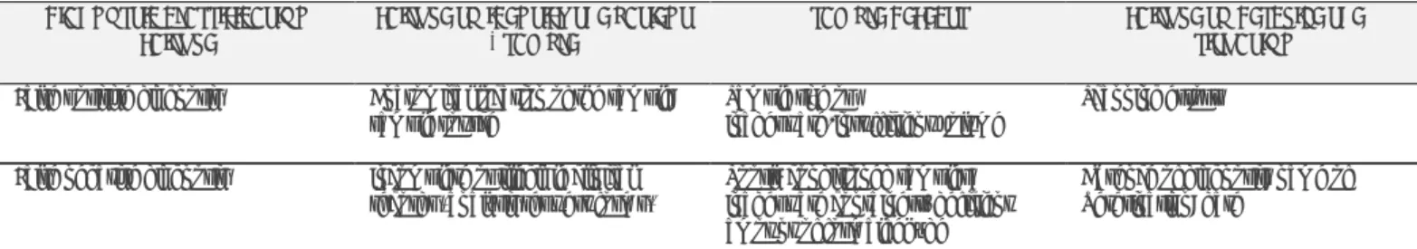
Table 1. Classification of errors in Pathological Anatomy speciality (Removed from Zarbo et al., 2005; Dilworth et al. 2014).
DIAGNOSTIC CLASSIFICATION FAILURE
FAILURE TO IDENTIFY THE PATIENT / SAMPLE
SAMPLE DEFECTS FAILURE TO DESCRIBE THE SITUATION
False positive diagnosis Anatomical location of the sample; sample source
Sample size or
inadequate/insufficient volume
Scanning errors False negative diagnosis Incomplete or illegible clinical
records (analysis request forms)
Poorly conditioned samples: inadequate containers, deficient amount of formaldehyde
Absence of diagnosis; name of Veterinarian; date
The recognition of categorized errors and the standardization of procedures is surely the best way to minimize them (Padley, 2012; Elston et al., 2016; Stratman, 2016). Thus, in this work, we will categorize the errors related to the steps of the routine histological technique, diagnostic technique that takes place in the Laboratory of Pathological Anatomy Veterinary (LPAV), particularly the errors related to the incorrect identification or orientation of the sample.
1. THEORETICAL FRAMEWORK
1.1. Orientation and marking of the surgical specimens
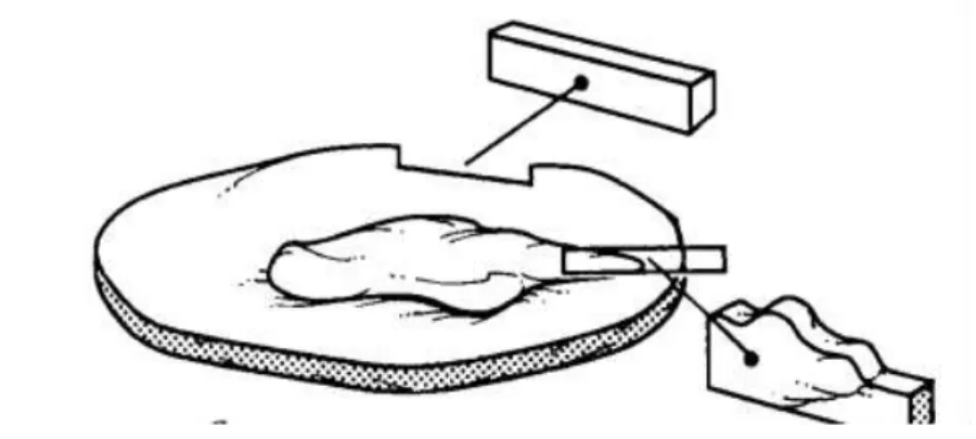
The implementation, by Veterinary Medical Centers (VMC), of measures that aim to ensure the correct orientation of excised surgical specimens, becomes imperative. These samples should be sent to the laboratory with clear marks (Figure 1), that can properly guide the anatomical position and its processing. Likewise, it is recommended that parts of the same patient are appropriately enumerated and identified with their name, organ and place of excision (Marques et al., 2007; Elston et al., 2016; Elston et al., 2016; Stratman, 2016).
The orientation of the sample to be sent, can be considered a "a path to reach the final destination", therefore, the tissue must be placed in its appropriate (clinical and anatomical) contexts, and appreciated as a structural unit, in a first stage (Galosi et al., 2011; Elston et al., 2016; Stratman, 2016; Vala & Pires, 2016).
The removal of a sample of tissue by the Veterinarian Surgeon, leads to the loss of its anatomical orientation, being essential that this professional previously examines the tissue in loco, relating it to its original anatomical location. This is crucial, given the fact that, the professional who will follow him (the pathologist in the laboratory), cannot have a full perception of the anatomical context of the isolated sample. Taking this into account, we can conclude that some measures must be taken in order to overcome this obstacle, like the placing of anatomical marks (Figure 1) and the communication between the Veterinarian Surgeon of the VMC and the Pathologist of the LPAV (Galosi et al., 2011; Elston et al., 2016; Stratman, 2016).
Vala, H., Bernardo, S., Esteves, F & Garcia, C. (2016). Application of strategies to minimize the error in pathological anatomy. Millenium, 2(ed espec nº2), 95-106.
e
2
98
Anatomical marks should be thought of as consistent features (shape, contour, structure) and should indicate a specific structure or designate a position (Figure 2) (Westra et al., 2003).
Some organs can be properly guided by their connections to adjacent structures that are easily identifiable; it is the case of the uterus, which can be correctly oriented by the relative positions of its peritoneal reflections, or even the eye, which can be guided by the insertion of a specific extraocular muscle. However, in the absence of easy-to-recognize connections, VMC professionals may use paint, sutures and coloured needles (Westra et al., 2003; Vala & Pires, 2016).
Figure 2. Use of suture at 12:00 to mark the orientation of the piece (removed from Westra et al., 2003).
The communication between the VMC and the LPAV should be brief, and should include the clinical history, clinical suspicions, description of the lesions observed, among other pertinent information, for which the guiding fields will be present in a specific analysis request form - clinical record - that is to be filled in the VMC and that must accompany each surgical specimen (Galosi et
al., 2011; Tomé & Vala, 2012; Vala & Pires, 2016).
1.2. Sample margin collection
The margin corresponds to the edge or boundary of the sample, representing the surface in which the Veterinarian Surgeon must section the tissues in order to remove the patient's lesion. It may be free of disease or, on the contrary, contain the injury, which implies that it has not been removed with safety margins. Thus, the evaluation of the margins is an important indicator of the invasive potential of the lesion, signalling the need to resort to additional treatments (e.g: chemotherapy in case of oncologic disease) (Westra et al., 2003).
It is considered a golden rule, the surgical inclusion of (excised tissue-free) the margins in the excised tissue, even in lesions with a benign clinical appearance (Figure 3) (Westra et al., 2003).
Figure 3. Cutting the margin showing the distance of the lesion to the excision limit (removed from Westra et al., 2003).
1
2
6
Vala, H., Bernardo, S., Esteves, F & Garcia, C. (2016).
Application of strategies to minimize the error in pathological anatomy. Millenium, (ed espec nº2), 95-106.
99
e
2
1.3. Sample conditioning and preparation of sample processing
After surgical excision or biopsy, the biological material obtained should be placed in a suitable container with abundant fixative liquid and sent to the laboratory (Vala & Pires, 2016).
After checking in the LPAV, the sample is appropriately identified and forwarded to processing. However, at an early stage and once the sample is fixed, the pathologist should seek to locate the lesion before proceeding to the next step: the section of the sample, in smaller and representative portions. Once the lesion is localized, the sample can be sectioned in the surface that best represents the included pathology, respecting the integrity of the organ/structure in question, as well as maintaining the connection to the surrounding anatomical structures. For nodules involving solid organs, the sample should be cut along its major axis, in order to demonstrate the total area of the nodule’s surface. However, the dissection should not only focus on the previously identified lesion but cover the entire sample, as incomplete dissections represent missed opportunities to fully disclose the extent of the lesion and to discover the suspected pathological processes (Westra et al., 2003).
A macroscopic evaluation of the sample is then carried out, which should be as comprehensive as possible, logical, factual and succinct. A logical description is one that follows a methodical sequence, which means that, in a first phase, should identify the sample and the present structures. The factual description is the one that records the objective characteristics of the sample, in particular, the size, weight, colour, shape and consistency of the respective lesion. Within these characteristics, size is particularly important, for example, in neoplasias, as well as in determining the distance between the border of the tumour and the surgical margin (Marques et al., 2007).
After dissection, the remaining tissue must be stored in order to ensure easy recovery and reconstruction of the sample. Storage tissues should be maintained in a tightly closed container with sufficient fixative volume to cover the sample (Marques et al., 2007).
2. METHODS
The routine histopathology technique, that allows the study of the tissues and constitutes the basis of the histopathological diagnosis, during a period of 6 months, was followed in the LPAV of the Agrarian School of Viseu (ASV).
The following of the technique was carried out from the moment of entrance and receipt of the samples in the laboratory, after which an input serial number has been assigned to the container of the sample, as well as to the accompanying analysis request form, and all the information has been recorded in the LPAV-ASV computer database, GestLabASV.
2.1. Sample and criteria for inclusion
During the period of this study, 118 samples were received. All the samples from the category of incisional or excisional biopsy or surgical specimen from VMC, were considered inclusion criteria. Exclusion criteria were cadavers for necropsy exam, scrapings, cytologies and cavity liquids for microscopic observation, as well as all samples from OPPs, associations, livestock farms or individual owners.
After the application of the inclusion and exclusion criteria, a total of 57 samples were analysed to identify and record the most frequently observed errors in sending, identification, conditioning of the samples, as well as in the course of processing within the laboratory.
2.2. Procedures
After the samples fixation were concluded, they were observed and described, allowing the filling of the macroscopic exam field in the diagnostic report. Afterwards, they were sectioned in a representative slice of the lesion, placed in histological cassettes, where the entry number was registered, and then they proceeded to the steps of impregnation and cutting in the microtome. The 3 μm thick sections obtained by the microtome, were placed on slides properly identified with the same input serial number, which proceeded to staining and mounting. This originated the final preparations that, when observed under the microscope, allowed the filling of the microscopic exam field, as well as the result field, in the diagnostic report, being this now ready to send to the applicant.
3. RESULTS
The main errors potentially capable of interfering with the rigor and reliability of the diagnosis in Pathological Anatomy were mainly related to pre-analytical errors. These errors included the scarcity of information in the respective analysis request form (47; 83%), with the packing and sending of samples to the laboratory (44; 77%), with the absence of marks capable of allowing the quick and efficient orientation of the surgical pieces (41; 72%) and in one case (1; 1.7%) the identification of the sample was lost during laboratory processing, thereby recording an analytical category error. In 32 samples (56%) there was a coincidence of more than one of these categories of identified errors.
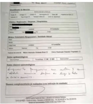
In most cases, the analysis request forms provided little information regarding clinical data, lesion’s characteristics data and intended exams (44; 93.6%) (Figures 4 and 5). The illegibility of writing was also pointed out as an observed fact (3; 6.4%).
Vala, H., Bernardo, S., Esteves, F & Garcia, C. (2016). Application of strategies to minimize the error in pathological anatomy. Millenium, 2(ed espec nº2), 95-106.
e
2
100
Figure 4. Analysis request form without information Figure 5. Analysis request form with illegible writing.
The following strategy to minimize this error consists in the correct fulfilment of all the fields of the analysis request form with legible writing, and its protection in a spill-proof casing. It should also be assured a better waterproofing of the sample container, using well-sealing containers, reinforcing the thread location with adhesive tape and placing them in a second impermeable receptacle.
The identification of a second category of errors was associated with poor packaging of samples (44; 77%), being particularly common, situations of incorrect hermeticity of the container, allowing the extravasation of the fixative liquid (5; 11.4%) (Figures 6 and 7), recipients unsuitable for the volume of the piece and insufficient amount of formaldehyde (39; 88.6%) (Figures 8 and 9), compromising the conservation of sent samples and increasing the overall processing time and delay in the emission of the final diagnosis report.
The strategy to minimize this category of errors consists on the use of wide mouth bottles, resistant, easy to seal and with a volume of fixative liquid ten to fifteen times greater than the volume of the sample, allowing a good fixation.
Figure 6. Poorly sealed bottle with visible
extravasation.
Figure 7. Analysis request form deteriorated by
Vala, H., Bernardo, S., Esteves, F & Garcia, C. (2016).
Application of strategies to minimize the error in pathological anatomy. Millenium, (ed espec nº2), 95-106.
10
1
e
2
Figure 11. Three surgical specimens from the same
container without any indication of the corresponding excision site (oriented from greater to smaler).
Figure 10. Mammary chain, without indication of the exact
location of the lesion.
The third category of errors was the absence of marking that would allow for the pathologist, to realize the correct orientation of the surgical specimens (41; 72%), commiting the macroscopic description and elaboration of the final report, with regard to the exact location of the lesions. This was particularly notorious in the orientation of the right and left sides of the uterus (5; 12.2%) (Figure 9) and in the mammary chains (20; 48.8%), making it difficult to identify the correct location of lesions in the corresponding mammary gland (Figure 10).
Every time that more than one sample was found in the same recipient, the absence of marks was also verified, as well as a description, in the analysis request form, of the exact location of each sample (16; 39%) (Figure 11).
Figure 9. Unmarked follicular cyst in one of the ovaries.
Figure 8. Inappropriate, narrow-mouth bottle. Figure 9. Insufficient volume of formaldehyde,
Vala, H., Bernardo, S., Esteves, F & Garcia, C. (2016). Application of strategies to minimize the error in pathological anatomy. Millenium, 2(ed espec nº2), 95-106.
e
2
102
To minimize errors regarding the lack of identification and orientation of the sample, the authors suggest that samples of different animals should always be sent in separate containers and that different samples of the same animal are identified by means of a sign, indication of an anatomical mark, suture point, indelible ink, coloured pins or coloured syringe needles (Figure 12) and its correct explanation and subtitling in the analysis request form.
Figure 12. Color syringe needles which can be introduced into samples for Identification and orientation,
indicating one side (removed from https://www. www.esslinger.com).
Although the loss of identification of a sample within the laboratory represents a low percentage of errors (1; 1.7%), which was later recovered, it brought as a consequence, the delay of the issuing of the final diagnosis. This may, in some more serious diseases, compromise the adoption of effective and early therapeutic measures aimed to recover the animal´s health, being this error often faced with such seriousness that it must be completely avoided, in order not to compromise the quality and speed of the diagnosis.
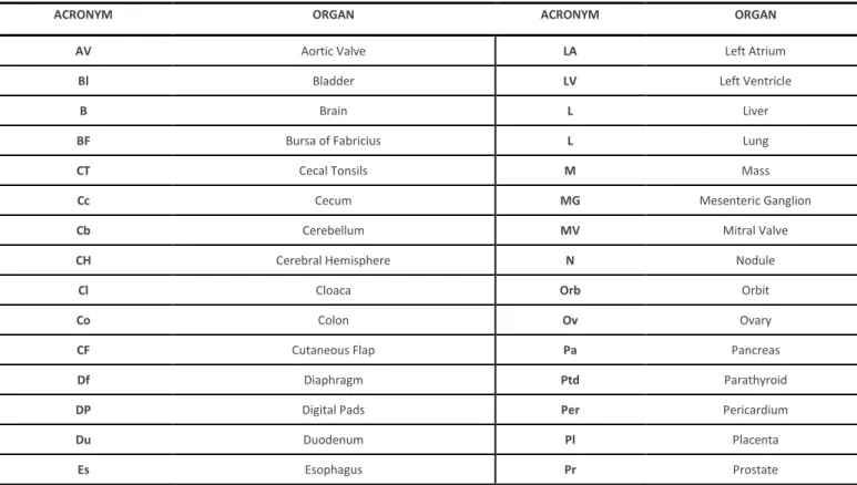
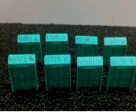
Thus, to minimize analytical and post-analytical errors in the identification of the samples inside the laboratory, and allow rapid progression for further histopathological processing steps, as well as the correct macroscopic description, each cut sample should generate a histological preparation in appropriate anatomical context and it is essential never to lose its precise identification. To achieve this, each histological cassete is identified with the respective serial number of analysis, obtained when the sample first entered the laboratory. In this present work, it is proposed to add an acronym corresponding to each organ (Figure 13) for the maintenance of its correct identification in the following steps of the histological processing and to make easier the interpretation and reading in the microscopic examination. It is also proposed that in the same organ/lesion, the sections needed to represent the sample/lesion, are ordered alphabetically, following the proposal of the Table nº. 2.
Table 2. Acronyms for quick identification of organs in blocks and slides
ACRONYM ORGAN ACRONYM ORGAN
AV Aortic Valve LA Left Atrium
Bl Bladder LV Left Ventricle
B Brain L Liver
BF Bursa of Fabricius L Lung
CT Cecal Tonsils M Mass
Cc Cecum MG Mesenteric Ganglion
Cb Cerebellum MV Mitral Valve
CH Cerebral Hemisphere N Nodule
Cl Cloaca Orb Orbit
Co Colon Ov Ovary
CF Cutaneous Flap Pa Pancreas
Df Diaphragm Ptd Parathyroid
DP Digital Pads Per Pericardium
Du Duodenum Pl Placenta
Vala, H., Bernardo, S., Esteves, F & Garcia, C. (2016).
Application of strategies to minimize the error in pathological anatomy. Millenium, (ed espec nº2), 95-106.
103
e
2
ACRONYM ORGAN ACRONYM ORGAN
F Fat Pv Proventriculus
G Ganglion RA Right Atrium
G Gizzard SN Sciatic Nerve
Gl Gland SI Small Intestine
H Heart S Spleen
IV Ileocecal Valve S Stomach
Il Ileum T Tendon
I Intestine Tes Testicle
Je Jejunum Ti Thymus
K Kidney Td Thyroid
LI Large Intestine Tr Trachea
The number of mammary chains, pieces of regional or individual mastectomies sent is increasing, due to the increase in mammary oncological diseases in companion animals. Since the mammary glands are not sent properly identified, nor the beginning or end of the mammary chain is indicated, in order to allow their correct identification and orientation, added to the fact that a multi-node chain has very often different diagnoses or similar but with different graduation, the authors propose the adoption of terminology of Table 3 to be adopted in the LPAV for mammary chains.
Table 3. Acronyms for quick identification of mammary chains in blocks and slides
ACRONYM MAMMARY GLAND CORRECTLY IDENTIFIED crTM cranial Thoracic Mammary gland
cdTM caudal Thoracic Mammary gland
crAM cranial Abdominal Mammary gland
cdAM caudal Abdominal Mammary gland
IGM Inguinal Mammary gland
ACRONYM MAMMARY GLAND CORRECTLY UNIDENTIFIEDa
M1 Mammary gland 1
M2 Mammary gland 2
M3 Mammary gland 3
M4 Mammary gland 4
M5 Mammary gland 5
aNote: in case of mammary gland is not identified, the authors suggest
Vala, H., Bernardo, S., Esteves, F & Garcia, C. (2016). Application of strategies to minimize the error in pathological anatomy. Millenium, 2(ed espec nº2), 95-106.
e
2
104
Figure 13. Identification of the histological cassetes with respective serial number of analysis and the acronym corresponding to the organ.
4. DISCUSSION
The errors most frequently identified in this study included the incomplete filling of the analysis request form, as well as its illegibility, which is in agreement with the consulted bibliography (Dilworth et al., 2014; Zarbo et al., 2005). These categories of error are equivalent to hiding of information, essential for the histopathological diagnosis and result in an irremediable delay in the issuance of the final diagnostic report (Cree et al., 2014).
The second most incident error, also described in the bibliography consulted (Zarbo et al., 2005; Cree et al., 2014; Dilworth et
al., 2014; Stratman et al., 2016) was improper conditioning of the sample, either at the level of inadequate container storage, or
in the hermeticity of the container used in the sample conditioning, events that often culminate with the leakage of the fixative liquid. This will damage the analysis request forms which followed together, making them illegible and leading to the consequences already described above, for which simple rules of waterproofing of the analysis request and reinforcing the hermeticity of the sample container forms, were pointed out.
Still in this category of pre-analytical errors, the third most frequently verified error, was associated with the inadequate amount of formaldehyde for fixation and transport of the sample, whether for reasons of savings or ignorance of good conditioning rules, which causes delays in sample fixation, preventing immediate processing. Taking into account that the processing of tissues takes about three days after the complete fixation, this problem adds a time interval of one or two days to the elaboration of the final diagnosis, which represents a serious risk to the animal's health. This error could be easily minimized by simply following the rules outlined (Foucar, 2001; Dilworth et al., 2014; Vala & Pires, 2016).
Last but not least, it was verified that the marking and orientation of the surgical specimen send to LPAV, still lacks the implementation of strategic measures, which can be easily implemented at low cost, either in VMC or in the laboratory itself, making it a useful tool for describing the final diagnosis by the pathologist, fact also corroborated by Galosi and co-workers (2011).
In clinical context, the correct orientation of the sample can be solved through basic measures that consist on the application of paints, coloured pins, needles or sutures, which take only two minutes to perform, being a simple and low cost process. This is even facilitated by the recent creation of the multidisciplinary Veterinary Medical Team, where all the tasks are no longer only in charge of the Veterinarian Surgeon, as it was before the integration of new professionals, as is the case of the Veterinarian Nurse (Vala et al., 2016), which could prove to be a crucial opportunity in the implementation of good practices, in this field of packaging and shipment of samples to the laboratory.
In laboratorial context, the introduction of a system of acronyms that identify the organs sent, can minimize problems with sample identification, thus constituting a strategy of easy adoption in the LPAV and that can minimize the category of analytical and post-analytical errors.
It also reinforces the idea of Valenstein and co-workers (2004) that any error in the packaging and shipment of samples to the LPAV may compromise the health, and therefore, the life of the animal.
CONCLUSIONS
The potential errors that could influence the quality of diagnosis in the LPAV-ASV were mainly related to the incorrect filling of analysis request form, as well as incorrect conditioning of the sample. Such errors belong to the pre-analytical category, for which it is suggested the adoption of simple strategies that include the correct filling of the analysis request form, the assurance
Vala, H., Bernardo, S., Esteves, F & Garcia, C. (2016).
Application of strategies to minimize the error in pathological anatomy. Millenium, (ed espec nº2), 95-106.
105
e
2
of the waterproofing and hermeticity conditions of the sample container, which should be adequate, resistant and wide-mouthed, as well as able to contain volume of fixative liquid ten to fifteen times greater than the sample volume.
The correct identification of the sample and its orientation also deserves attention from the VMC, which can adopt the described strategies. However, in the laboratory itself, the adoption of a system of acronyms for the identification of the samples could guarantee the analytical and post-analytical quality in the histopathological diagnosis and minimize errors, since in this field, an exchange of samples can seriously compromise the emission of the report with the final diagnosis.
ACKNOWLEDGMENTS
This work is financed by national funds through FCT - Fundação para a Ciência e Tecnologia, I.P., under the project UID/Multi/04016/2016. Furthermore we would like to thank the Instituto Politécnico de Viseu and CI&DETS for their support. This work is supported by European Investment Funds by FEDER/COMPETE/POCI– Operacional Competitiveness and Internacionalization Programme, under Project POCI-01-0145-FEDER-006958 and National Funds by FCT - Portuguese Foundation for Science and Technology, under the project UID/AGR/04033/2013. Furthermore we would like to thank to UTAD and CITABS for their support.
REFERENCES
Cree, I. A., Zandra, D., Marjolijn, J. L., Ligtenberg, M. J. L., Normanno, N., Edsjö, A., Rouleau, E., Solé, F., Thunnissen, E., Timens, W., Schuuring, E., Dequeker, E., Murray, S., Dietel, M., Groenen, P., & Krieken, J. H. (2014). Guidance for laboratories performing molecular pathology for cancer patients. Journal of Clinical Pathology, 67(11), 923–931. doi:10.1136/jclinpath-2014-202404.
Dilworth, L. L., McGrowder, D. A., & Thompson, R. K. (2014). Identification of Pre-examination Errors in the Chemical Pathology Laboratory at the University Hospital of the West Indies. Indian Journal of Clinical Biochemistry, 29(2), 227–231. doi:10.1007/s12291-013-0348-6.
Elston, D. M., Stratman, E. J., & Miller, S. J. (2016). Skin biopsy: Biopsy issues in specific diseases. Journal of the American
Academy of Dermatology, 74(1), 1-16. doi:10.1016/j.jaad.2015.06.033.
Foucar, E. (2001). Error in anatomic pathology. American Journal of Clinical Pathology, 116 (suppl 1), S34-S46. Retrieved from https://www.ncbi.nlm.nih.gov/pubmed/11993701.
Galosi, A. B., Muzzonigro, G., Lacetera, V., & Mazzucchelli, R. (2011). Specimen orientation by marking the peripheral end:
(potential) clinical advantages in prostate biopsy. Prostate Cancer, ID 270403, 1-7. doi:10.1155/2011/270403.
Marques, A. J., Areia, F. R., Marques, V. M., Casimiro, P., & Mendonça, P. (2007). Actuação dos Técnicos de Anatomia Patológica no Exame Macroscópico: Caraterização em 2006. Revista Técnica de Anatomia Patológica, 10(14), 16-19. Retrieved from http://hdl.handle.net/10400.21/359.
Padley, L. (2012). Error Reduction in Anatomic Pathology. MedicalLab Management, 1 (2), 6-10. Retrieved from https://www.medlabmag.com/digitalmag/Main.php?MagNo=2&PageNo=8#page/10.
Raab, S, S., & Grzybicki, D. M. (2008). Multi-institutional database of anatomic pathology errors. Mini-symposium Quality
management in pathology. Diagnostic histopathology 14(7), 316-324. doi: 10.1016/j.mpdhp.2008.06.005.
Stratman, E. J., Elston, D. M., & Miller, S. J. (2016). Skin biopsy: Identifying and overcoming errors in the skin biopsy pathway.
Journal of the American Academy of Dermatology, 74(1), 19-25. doi:10.1016/j.jaad.2015.06.034.
Tomé, P., & Vala, H. (2012). How Experience can be Useful in Veterinary Pathological Anatomy. In Perez-Marin, C. (Ed.) A
bird's-eye view of Veterinary Medicine. Rijeka: InTech: pp. 51-70. Retrieved from
http://www.intechopen.com/books/a-bird-s-eye-view-of-veterinary-medicine.
Valenstein, P. N., & Sirota, L. (2004). Identification errors in pathology and laboratory medicine. Clinics in laboratory medicine, 24, 979-996. doi:10.1016/j.cll.2004.05.013.
Vala, H., & Pires, M. A. (2016). Recolha e Envio de Material para o Laboratório de Anatomia Patológica. In Carreira, R. P., & Pires, M. A. (Eds.). Descrição Anatomopatológica em Medicina Veterinária. Retrieved from
Vala, H., Bernardo, S., Esteves, F & Garcia, C. (2016). Application of strategies to minimize the error in pathological anatomy. Millenium, 2(ed espec nº2), 95-106.
e
2
106
Vala, H., Nóbrega, C., Mega, A. C., Santos C., Cruz, R., Esteves, F., & Mesquita, J. R. (2016). Interação Médico Veterinário – Enfermeiro Veterinário no exercício da profissão. In Livro de conferências do 7º Encontro de Formação da Ordem dos
Médicos Veterinários (pp. 365-366). Lisboa: Centro de Congressos. Retrieved from
http://hdl.handle.net/10400.19/3507.
Westra, W. H, Hruban, R. H., Phelps,T. H, & Isacson, C. (2003). Surgical Pathology Dissection: An illustrated guide. 2nd ed. New
York: Springer.
Zarbo, R. J., Meier, F. A., Raab, S. (2005). Error Detection in Anatomic Pathology Pathology. Archives of Pathology & Laboratory
Medicine, 129, 1237-1245. Retrieved from http://www.archivesofpathology.org/doi/full/10.1043/1543-2165(2005)129[1237:EDIAP]2.0.CO;2.