GPCRs Direct Germline Development and
Somatic Gonad Function in Planarians
Amir Saberi1, Ayana Jamal1, Isabel Beets2, Liliane Schoofs2, Phillip A. Newmark1*
1Howard Hughes Medical Institute and Department of Cell and Developmental Biology, University of Illinois at Urbana-Champaign, Urbana, Illinois, United States of America,2Department of Biology, Functional Genomics and Proteomics Unit, KU Leuven, Leuven, Belgium
Abstract
Planarians display remarkable plasticity in maintenance of their germline, with the ability to develop or dismantle reproductive tissues in response to systemic and environmental cues. Here, we investigated the role of G protein-coupled receptors (GPCRs) in this dynamic germline regulation. By genome-enabled receptor mining, we identified 566 putative planar-ian GPCRs and classified them into conserved and phylum-specific subfamilies. We per-formed a functional screen to identify NPYR-1 as the cognate receptor for NPY-8, a neuropeptide required for sexual maturation and germ cell differentiation. Similar to NPY-8, knockdown of this receptor results in loss of differentiated germ cells and sexual maturity. NPYR-1 is expressed in neuroendocrine cells of the central nervous system and can be activated specifically by NPY-8 in cell-based assays. Additionally, we screened the comple-ment of GPCRs with expression enriched in sexually reproducing planarians, and identified an orphan chemoreceptor family member,ophis, that controls differentiation of germline stem cells (GSCs).ophisis expressed in somatic cells of male and female gonads, as well as in accessory reproductive tissues. We have previously shown that somatic gonadal cells are required for male GSC specification and maintenance in planarians. However,ophisis not essential for GSC specification or maintenance and, therefore, defines a secondary role for planarian gonadal niche cells in promoting GSC differentiation. Our studies uncover the complement of planarian GPCRs and reveal previously unappreciated roles for these receptors in systemic and local (i.e., niche) regulation of germ cell development.
Author Summary
G protein-coupled receptors (GPCRs) are the largest and most versatile family of cell-sur-face receptors. They play critical roles in various cellular and physiological systems and have emerged as a leading group of therapeutic targets. Due to their structural and func-tional conservation across animals, much has been learned about GPCRs from studies in laboratory models. Here, we performed genome-wide receptor mining to identify and cat-egorize the complement of GPCR-encoding genes in the planarianSchmidtea mediterra-nea, an emerging model organism for regeneration and germ cell biology. We then
a11111
OPEN ACCESS
Citation:Saberi A, Jamal A, Beets I, Schoofs L, Newmark PA (2016) GPCRs Direct Germline Development and Somatic Gonad Function in Planarians. PLoS Biol 14(5): e1002457. doi:10.1371/ journal.pbio.1002457
Academic Editor:Mariana Federica Wolfner, Cornell University, UNITED STATES
Received:December 22, 2015
Accepted:April 11, 2016
Published:May 10, 2016
Copyright:© 2016 Saberi et al. This is an open access article distributed under the terms of the
Creative Commons Attribution License, which permits unrestricted use, distribution, and reproduction in any medium, provided the original author and source are credited.
Data Availability Statement:All relevant data are within the paper and its Supporting Information files. GenBank accession numbers for planarian GPCR genes cloned in this work are KX018822—
KX018983.
conducted two studies implicating planarian GPCRs in the regulation of reproductive function. First, we found the receptor component of a central neuropeptide Y signaling pathway and demonstrated its involvement in the systemic control of reproductive devel-opment. Next, we showed that a novel chemoreceptor family member is expressed in somatic cells of the planarian gonads and directs germ cell maturation via the niche. We predict that future studies on the hundreds of other planarian GPCRs identified in this work will not only help us understand the conserved role of these receptors in various physiological pathways but also pave the way for identification of novel therapeutic targets in parasitic relatives of the planarian.
Introduction
G protein-coupled receptors (GPCRs) play critical roles in sexual reproduction, guiding germ cell migration, mediating hormonal regulation of gamete development, and facilitating the function of accessory reproductive tissues. For example, a complex network of peptidergic neu-rons in the mammalian hypothalamus controls the release of pituitary gonadotropins that sys-temically regulate gonadal function. GPCRs mediate various short- and long-range
communication events in this hormonal cascade, whether the target is another neuron or a gonadal cell. Mutations in several of these GPCRs and their ligands are associated with hypogo-nadotropic hypogonadism and other reproductive disorders [1]. GPCRs that act as receptors for follicle-stimulating hormone (FSH), luteinizing hormone (LH), gonadotropin-releasing hormone (GnRH), kisspeptin, prokineticin, and tachykinin play essential roles in systemic reg-ulation of gonadal function in mammals [2–6]. Despite extensive genetic information and molecular studies in mammalian models, much remains to be learned about the role of GPCR signaling in sensing physiological and environmental cues and the evolutionary conservation of these mechanisms in regulating reproduction across metazoans.
GPCRs also facilitate germ cell differentiation and maturation cell autonomously. Chemo-kine receptor CXCR4 in vertebrates and rhodopsin-like GPCR Tre1 inDrosophilaenable che-mokine-guided migration of primordial germ cells (PGCs) early in development [7–9]. Other olfactory, adhesion-like, and secretin-like GPCRs and their signaling partners are required for the maintenance and proliferation of germline progenitors and stem cells as well as gamete morphogenesis [10–15]. In many cases, however, GPCR ligands, signaling partners, and func-tional mechanisms are far from understood.
Platyhelminthes (flatworms) exhibit the remarkable ability to coordinate their reproductive development with systemic and environmental cues such as body size, nutritional status, and season. Upon starvation or severe injury, planarians are capable of reversibly disassembling their reproductive system, presumably to curb metabolic demand or to prepare for body-wide tissue remodeling [16]. Upon amputations that entirely remove the reproductive system, the remaining head fragments can re-specify germ cells and reproductive structures de novo [17,18]. Furthermore, a number of classic and recent studies suggest that planarian neuroendo-crine cells systemically influence reproductive development. For example, head amputation, which involves removal of the cephalic ganglia, results in regression of the male gonads to clus-ters of PGCs [19,20]. Owing to its reproductive plasticity and the availability of numerous functional genomic tools, we use the planarianSchmidtea mediterraneaas a model to study regulation of germ cell development and reproductive function, focusing here on the GPCR superfamily.
funders had no role in study design, data collection and analysis, decision to publish, or preparation of the manuscript.
Competing Interests:The authors have declared that no competing interests exist.
Flatworm GPCRs have been the focus of very few functional studies, which have been lim-ited mainly to neurotransmitter response, body patterning through the Wnt/frizzled pathway, or photoreception by opsins [21–25]. An earlier genome-wide study of flatworm GPCRs was based on an incomplete genome assembly and limited to in silico prediction of GPCR genes [26]. While planarian neuropeptides and other GPCR ligands have received some attention [27–31], receptor/ligand pairs and their specific physiological function in planarians have not been defined. Our recent genome-wide characterization of planarian neuropeptides identified NPY-8, a conserved neuropeptide Y homolog, that is required for proper development of the planarian germline [32]. Here, we characterized the planarian GPCR complement to identify the NPY-8 receptor and other GPCRs involved in regulating germ cell development. Our stud-ies suggest that GPCRs within the central nervous system (CNS) and the gonads are key com-ponents of the signal transduction mechanisms that regulate reproductive development in planarians.
Results
Genome-Wide Analysis Reveals Conserved and Phylum-Specific
GPCR Families
To explore the role of GPCRs in planarian reproductive biology, we generated a comprehensive database of planarian GPCR gene sequences, classes, and expression information. A previous annotation of flatworm GPCRs was based on an early draft of the planarian genome [26]. We found that many GPCR genes are absent from this list, while several genes encode proteins that more closely resemble other transmembrane protein families. To generate a complete database, we used extensive transcriptomic data to assemble a de novo transcriptome that we mined for putative seven-transmembrane receptor sequences (workflow shown inS1A Fig, de novo tran-scriptome can be found inS1 Data). Combining these transcriptomic data and sensitive pat-tern-discovery methods, we developed a comprehensive list of 566 GPCRs. We confirmed and improved the annotation of 343 previously identified GPCRs and discovered 223 new ones (S2 andS3Data). The availability of RNA-seq data from specific tissues or experimentally modi-fied planarians revealed a great deal of information about GPCR expression in planarians (S3 Data,S1D Fig). A partial in situ hybridization (ISH) screen revealed expression of planarian GPCRs in a variety of tissues, including nervous and reproductive systems, intestine, epithe-lium, and presumptive sensory organs (S2 Fig).
Other rhodopsin-like families includeRho-L,Rho-R, andRho-D(arbitrary designations) that have no known homologs outside of platyhelminthes. The largest and most expanded rho-dopsin-like family,Srf/w(for“Serpentine receptors of flatworms and srw”), contains 199 genes in four subclusters:Srfa,Srfb, andSrfcthat appear to be flatworm-specific, andSrw, members of which aggregate with thesrwsubfamily ofCaenorhabditis eleganschemoreceptors (S1B Fig). InC.elegans, members of thesrwsubfamily are the only chemoreceptors with a recognizable sequence similarity to the main rhodopsin family (specifically to FMRFamide receptors) [36]. While only oneC.eleganschemoreceptor has been experimentally de-orphanized [37], GPCRs of this family are called“chemoreceptors”because a large fraction of them are required for che-mosensation and/or expressed in chemosensory neurons that respond to environmental mole-cules [38]. For simplicity, we call theSrf/wfamily, which includesophis(GB: KX018822, see below), the planarian chemoreceptor family.
A subset of putative planarian GPCRs (48 rhodopsin-like and 50 others) that did not join any convex clusters include homologs of conserved adiponectin receptors, lung seven-trans-membrane receptors, leucine-rich repeat-containing GPCRs, and receptors with no known
Fig 1. Global view of planarian GPCRs and the NPY receptor family.(A) Similarity clustering of 566 planarian GPCRs revealing members of all five metazoan GPCR families. Only GPCRs of the convex clusters are counted and highlighted by filled circles (seeMaterials and Methods). (B) Co-clustering of conserved planarian and human rhodopsin-like GPCRs reveals large groups of amine and peptide receptors, as well as smaller groups of receptors with other functionalities. Planarians appear to lack homologs of humanγandδrhodopsins or lipid receptors. PlanarianRho-Cand human receptors are shown by filled circles and grey crossmarks, respectively. Darker edges indicate higher similarity (lowerp-value) between nodes.p-value scale shown at top-right corner. Area in dashed box is expanded in panel C. (C) Magnified view of the similarity network around NPY receptors (dashed box in B). Three groups of planarian receptors (16 total) are located adjacent to human NPY receptors. (D) Bayesian inference topology of candidate planarian NPY receptors (orange) and parasitic flatworm homologs (pink). Arrow indicates the root. Posterior probabilities are 1.00 at every node, except those with a value shown. Three monophyletic groups of NPY receptors are found that are parallel to groups identified by similarity clustering in panel C. Type 1 is conserved across flatworms, arthropods, and nematodes, type 2 receptors are found in flatworms and arthropods, and type 3 receptors are
lophotrochozoan-specific. The complete phylogenetic analysis is shown inS1E Fig. SeeS1andS2Figs for more information on the planarian GPCR complement.
homologs (S3 Data, not highlighted by a circle inFig 1A). Overall, our analyses reveal that major groups of planarian GPCRs fit within the conserved GRAFS classification; however, the rhodopsin-like family in planarians is highly diversified and includes multiple invertebrate-specific and potentially flatworm-invertebrate-specific subfamilies.
NPY Receptor
npyr-1
Is Required for Systemic Regulation of Germline
Development
We next wanted to know if planarian GPCRs are involved in signaling pathways that regulate development and maintenance of the germline. Our previous studies have shown that Smed-npy-8, an NPY homolog expressed in the planarian nervous system, is required for germ cell development and sexual maturity. RNA interference (RNAi) knockdown ofnpy-8during post-embryonic development (paradigm inFig 2A) blocks differentiation of both male and female germ cells, as well as formation of somatic accessory reproductive structures (S3A Fig) [32]. Our identification of the planarian GPCR repertoire allowed us to investigate the role of NPY receptors in planarian reproductive development. We hypothesized that if NPY-8 acts through a conserved NPY receptor to promote reproductive development, RNAi knockdown of at least one NPY receptor should phenocopynpy-8(RNAi), barring functional redundancy.
Since NPY receptors are conserved throughout metazoans, to identify planarian NPY recep-tor genes we focused onRho-Cand repeated the clustering analysis only using planarian and human rhodopsin-like GPCRs (Fig 1B). Our analysis identified 16 putative planarian NPY receptors that cluster with human NPY receptors (Fig 1C). Bayesian phylogenetic analyses sug-gest that planarian NPY receptors exist in three monophyletic groups: one that includes flat-worm, arthropod (includingDrosophilaNPFR-1 [39]), and nematode sequences, one that only includes flatworm and arthropod NPY receptors, and a third group that appears to be lopho-trochozoan-specific (including the snail NPY receptor GRL105 [40]) (Figs1DandS1E). Color-imetric ISH shows that most planarian NPY receptors are expressed in the CNS and the testes (S2H–S2P Fig).
To identify a candidate receptor for NPY-8, we individually knocked down each of the NPY receptor genes in planarian hatchlings (S3 Data,Fig 2A).Smed-germinal histone H4(gH4) was used as a marker for spermatogonial cells, female germ cells, and neoblasts, andSmed-nanos
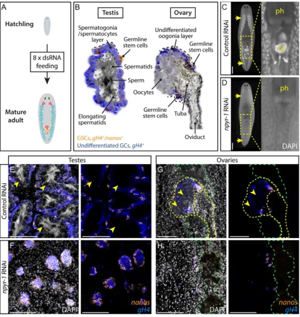
(nanos) was used to label male and female germline stem cells (GSCs) [18,41,42]. Knockdown ofSmed-npyr-1(GB: KX018969) prevented germ cell differentiation and formation of the reproductive system, regardless of the region of the gene targeted (Fig 2C and 2D,S4 Data). In control treatments, testes reach maturity and produce sperm with compact, elongated nuclei readily visualized by DAPI staining (Fig 2E). Innpyr-1(RNAi)planarians, testes only contain undifferentiated GSCs (nanos+/gH4+) and spermatogonia (nanos–/gH4+) (Fig 2F).npyr-1is also essential for female germ cell differentiation, as the ovaries innpyr-1(RNAi)worms only include GSCs and oogonia and lack mature oocytes seen in control worms (Fig 2G and 2H). Notably, thenanos+GSC pool was present in all RNAi animals (Fig 2E–2H) andnanosmRNA expression was unaffected as assayed by quantitative PCR (qPCR)(S3C Fig). We also found thatnpyr-1is not required for de novo germ cell specification (S3B Fig).
Next, we wanted to rule out the possibility that the phenotypes observed afternpy-8or
npyr-1RNAi are an indirect consequence of down-regulating the other gene. We knocked downnpy-8ornpyr-1and found that while the targeted genes are down-regulated at least 4-fold, expression of the other gene is not affected (S3C Fig). Collectively, our experiments indicate thatnpyr-1knockdown phenotypes closely resemble those ofnpy-8, suggesting that
Fig 2. NPY receptornpyr-1is required for germ cell maturation.(A) Post-embryonic development RNAi paradigm used to determine the function of genes during normal planarian growth. Hatchlings (2 wk old) were fed dsRNA corresponding to each gene eight times to ensure that control worms achieve sexual maturity. (B) Schematic showing planarian testis and ovary structures. In both testes and ovaries,nanos+/gH4+GSCs (orange) andnanos-/gH4+spermatogonia/oogonia (blue) are located on the periphery, while more differentiated spermatids,
sperm, or oocytes (grey) are in the middle of the gonads. (C, D) DAPI staining showing testes and stored sperm in whole-mount samples. Insets show the copulatory apparatus region. Pharynx and copulatory apparatus are marked by“ph”and“ca”, respectively. RNAi treatment followed the paradigm in A. Control worms (C) develop a complete reproductive system, whilenpyr-1(RNAi)worms (D) lack developed reproductive tissues and mature gametes.n= 5/ 5 for each of the threenpyr-1clones and control. RNAi quantification data can be found inS4 Data. (E–H) Double-FISH labeling GSCs (nanos+/gH4+, orange) and spermatogonia (nanos-/gH4+, blue) in whole-mount control and
npyr-1(RNAi)samples. GSCs and spermatogonial cells are present in both conditions. Testis germ cells differentiate into sperm and spermatids (arrowheads) in control worms (E), but not innpyr-1(RNAi)worms (F). In ovaries, mature oocytes with large cytoplasm (arrowheads) are surrounded bygH4+oogonia in control planarians (G), but are
absent innpyr-1(RNAi)animals (H). DAPI (grey) labels nuclei. Yellow dashed lines indicate ovaries and oviducts in G and H. Green dashed lines indicate cephalic ganglia near the ovaries. Scale bars are 1 mm in C and D and 100μm in E–H. See alsoS3 Fig.
NPY-8 Specifically Activates NPYR-1 In Vitro
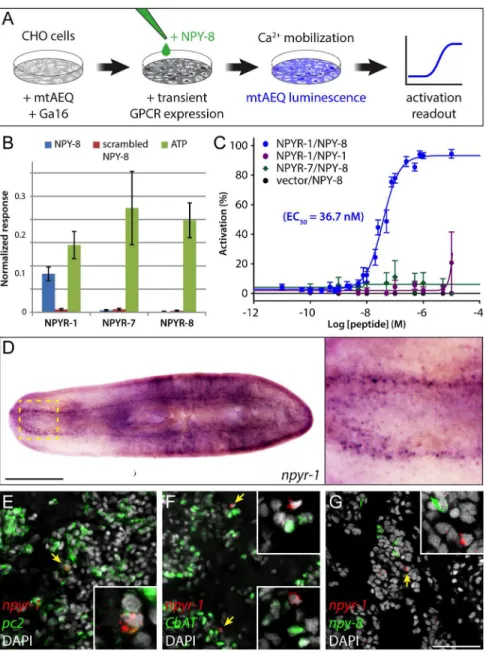
To test whether NPY-8 is able to functionally activate NPYR-1, we performed a cell-based receptor-activation assay. We individually expressed three NPY receptors encoded by the npyr-1,npyr-7, andnpyr-8genes in CHO cells co-expressing the promiscuous Gα16subunit and mitochondrially targeted apoaequorin (CHO/mtAEQ/G16) [43], which enable sensitive moni-toring of intracellular calcium responses to exogenous ligands. We then assayed receptor acti-vation after addition of various concentrations of synthetic NPY-8, as well as a closely related family member, NPY-1, and scrambled NPY-8 as controls (Fig 3A). NPYR-1 was activated by NPY-8, but not by the control ligands; by contrast, cells expressing NPYR-7 or NPYR-8, or transfected with an empty vector were not activated (Fig 3B and 3C). Moreover, concentra-tion-response assays showed that NPY-8 activates NPYR-1 at nanomolar concentrations, with an EC50value of 36.7 nM (Fig 3C). Taken together, our results suggest that NPYR-1 is the
cog-nate receptor for NPY-8.
npyr-1
Is Expressed in Neuroendocrine Cells in the CNS
To identify tissues potentially targeted by NPY-8 signaling, we characterized the expression pattern ofnpyr-1. Colorimetric ISH revealed thatnpyr-1is expressed specifically in a subset of cells in the brain and ventral nerve cords (Fig 3D). We did not detectnpyr-1expression in the gonads or accessory reproductive tissues, suggesting that NPY-8 signaling does not directly tar-get reproductive tissues.S.mediterraneaexists in two distinct biotypes: hermaphroditic sexuals that reproduce by cross-fertilization, and asexuals that reproduce by fission. Asexual planarians specify PGCs but lack differentiated germ cells and accessory reproductive tissues [44]. Inter-estingly, asexual planarians expressnpyr-1at levels slightly higher than sexuals (S3D Fig). Asexuals, however, expressnpy-8at ~50-fold lower levels, likely not enough to activate the
npyr-1receptor (S3D Fig).
To identify thenpyr-1+cells in the CNS, we examined coexpression ofnpyr-1and two other nervous system markers:Smed-prohormone convertase 2(pc2, peptidergic neural cells) [45] andSmed-choline acetyltransferase(ChAT, cholinergic neurons) [46]. Fluorescent in situ hybridization (FISH) indicated that mostnpyr-1+cells arepc2+/ChAT-, suggesting that these cells are not cholinergic neurons, but rather neuroendocrine cells that express and release other neuropeptides or hormones (Fig 3E and 3F). Moreover,npyr-1+cells do not express
npy-8, inconsistent with an autocrine NPY-8 signaling loop (Fig 3G). Together, our results suggest that NPY-8 targets CNS peptidergic cells through thenpyr-1receptor, resulting in downstream signaling that eventually regulates germ cell maturation. Although the identity of the signal(s) mediating communication between the CNS and reproductive system remains unknown, the latter must have the capacity to receive and interpret such cues to regulate reproductive output.
A Subset of Planarian GPCRs Is Enriched in Reproductive Tissues
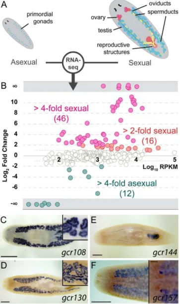
Because GPCRs are the largest group of cell-surface receptors, we expected to identify addi-tional GPCRs expressed in the reproductive tissues, enabling responses to local or systemic cues. To select such candidate genes, we compared transcriptomes of theS.mediterraneasexual and asexual biotypes (Fig 4A). Genes enriched in the germline and reproductive tissues account for the majority of the differences between transcriptomes of the two biotypes [47].
Of 566 GPCRs, 46 (~8%) are up-regulated in sexual planarians (4-fold andp-value<0.05,
Fig 3. NPY-8 targetsnpyr-1+neuroendocrine cells in the CNS.(A) Schematic of receptor-activation assay performed in CHO/mtAEQ/G16 cells. Candidate GPCRs were expressed transiently in a cell line that enables visualization of calcium mobilization upon receptor activation. (B) Normalized activation response of CHO cells expressing NPYR-1 or control receptors, challenged with NPY-8 or control peptides. NPY-8 specifically activates NPYR-1, while two other NPY receptors (NPYR-7 and NPYR-8) are not activated upon NPY-8 treatment. Scrambled NPY-8 was used as a negative control. Calcium responses were normalized to the total calcium response after addition of 0.1% Triton X-100. ATP, which activates an endogenous CHO receptor, was used to test the functionality of the assay. Peptides and ATP were tested at 10 and 1μM, respectively. (C) Concentration-response curves for the activation of NPYR-1 by NPY-8 and control peptides. Data are shown as a percentage of the highest normalized response of the concentration series. NPY-8 activates NPYR-1 at EC50= 36.7 nM (blue). Closely related NPY-1 fails to activate NPYR-1 (purple). Empty
pcDNA3.1 vector and NPYR-7 were used as negative controls. Error bars in B and C represent standard error of the mean (SEM) (n4). The underlying data for receptor assays can be found inS4 Data. (D) Colorimetric ISH showing expression ofnpyr-1in a subset of cells in the brain and along the ventral nerve cords. Inset shows the brain region at higher magnification. (E–G) Double-FISH labelingnpyr-1(red) and other neural markers (green).npyr-1+cells express neuroendocrine cell markerpc2(E, 28/30 expresspc2) but not the cholinergic neuronal markerChAT(F, 0/30 expressChAT).npy-8andnpyr-1are expressed in distinct populations of cells (G, 0/30npyr-1cells expressnpy-8and 0/30npy-8cells expressnpyr-1). DAPI (grey) labels nuclei. Scale bars are 1 mm in D and 50μm in E–G.
exception ofgcr108, which is expressed in spermatocytes and spermatids (Fig 4C), all the other examined receptors are enriched in spermatogonial cells (e.g.,gcr130,Fig 4D). Other GPCRs are expressed in accessory reproductive organs, such as oviducts and copulatory apparatus (e.g.,
gcr144,Fig 4E). Only one of the tested GPCRs,gcr157, is enriched in both female and male germ cells (Fig 4F). These results implicate GPCRs in reception of signals by germ cells and their associated somatic tissues.
Fig 4. A subset of planarian GPCRs is enriched in reproductive tissues.(A) Schematic of the reproductive system in the two biotypes ofS.mediterranea. Sexual planarians (right) develop a complete reproductive system, including mature gonads and accessory reproductive organs. The asexuals (left) contain only presumptive gonads with PGCs. (B) Normalized RNA-seq RPKM ratios between sexual and asexual planarians plotted against relative abundance of each GPCR gene. Only data points withp -value<0.05 are shown. (C–F) Representative colorimetric ISH experiments used to validate RNA-seq results (n= 24/27 genes tested expressed in sexual organs). Sexually enriched genes are expressed in various reproductive tissues, including spermatids (C), spermatogonia (D and F), oviducts and female copulatory apparatus (E), and ovaries (F). Scale bars are 1 mm. Insets in C and D show the area inside the dashed box. Inset in F shows the ventral side and the scale bar is 200μm. See alsoS4 FigandS3 Data.
The Orphan Receptor
ophis
Is Required for Germ Cell Differentiation
and Reproductive Maturity
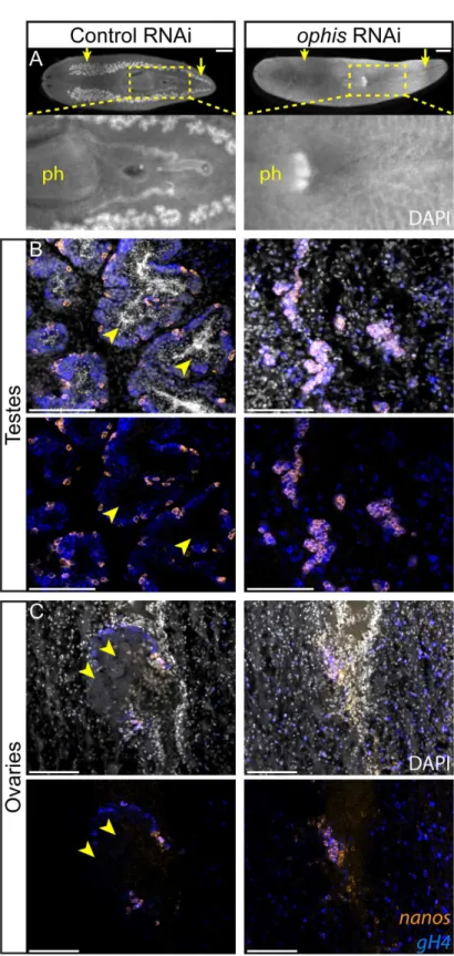
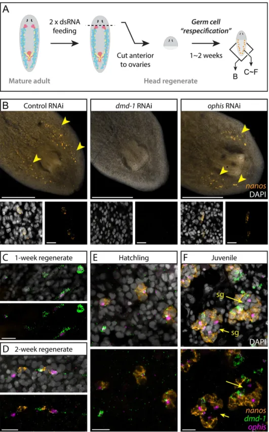
To determine whether any of the sexually enriched GPCRs are required for reproductive devel-opment, we performed an RNAi screen starting with planarian hatchlings (Fig 2A). We found that knockdown of a serpentine receptor family (Srf/w) member we named“ophis”(after the mythological serpent wrapping around the Orphic Egg), resulted in animals with immature testes that lack differentiatinggH4+spermatogonial cells, spermatocytes, spermatids, and sperm (Fig 5A and 5B,S4 Data). Ovaries were also affected, revealed by the absence of mature oocytes (Fig 5C,S4 Data). Seminal vesicles with stored sperm were not observed inophis (RNAi)worms (Fig 5A). Despite the loss of all differentiating germ cells, allophis(RNAi) ani-mals retained their pool ofnanos+GSCs (Fig 5B and 5C). Therefore,ophisis required for dif-ferentiation, but not maintenance, ofnanos+GSCs.
ophis
Is Expressed in Somatic Reproductive Tissues, Including Gonadal
Niche Cells
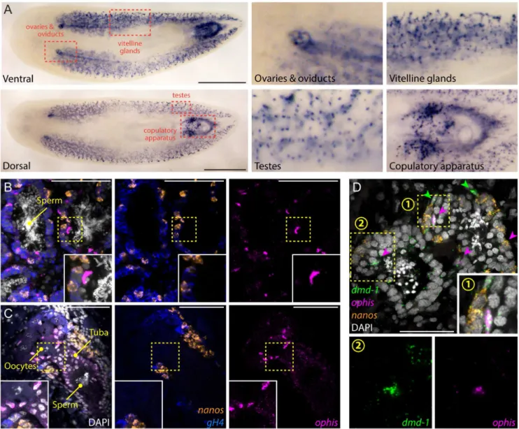
To determine whereophisis expressed in sexual planarians, we performed whole-mount ISH. We detectedophisexpression in several accessory reproductive tissues, including oviducts, tuba, vitellaria, and parts of the copulatory apparatus, as well as in discrete cells in testes (Fig 6A). To characterize the cell types in whichophisis expressed in the gonads, we performed FISH. In both testes and ovaries,ophisis detected in somatic cells (nanos-/gH4-) closely associ-ated with germ cells (Fig 6B and 6C). We previously showed that a few somatic cells within each testis lobe express a conserved sex-specific transcription factor (Smed-dmd-1) and lack known germline markers [48]. Thesedmd-1+cells are required for specification and mainte-nance ofnanos+GSCs and are thought to contribute to a presumptive germline niche. FISH experiments revealed coexpression ofophisanddmd-1transcripts in the somatic cells of the testes (Fig 6D). The nuclei of these cells have an elongated and angular shape, distinct from the round nuclear morphology of spermatogonia and spermatids. Furthermore, somatic cells of the testes seem to have an expanded cytoplasm, delineated bydmd-1+puncta that stretch between germ cells (Fig 6D).
A population ofdmd-1+cells also exists in the dorsal mesenchyme (between testis lobes) and are potential progenitors of somatic gonadal cells [48]. By FISH, these cells seem to express higher levels ofdmd-1compared to the somatic gonadal cells and do not expressophis(Fig 6D). On the ventral side,ophisis expressed abundantly in vitellaria, copulatory organs, ovi-ducts, and ovaries (Fig 6A and 6C). In oviducts and ovaries, expression ofophisresembles that ofSmed-nhr-1, a nuclear hormone receptor required for planarian germ cell development [49].
ophis+cells in the ovary are alsonanos-/gH4-, suggesting that they represent somatic cells of planarian ovaries (Fig 6C).
ophis
Knockdown Does Not Affect Male GSC Specification
SinceophisRNAi did not affect the maintenance ofnanos+GSCs, we tested whether initial specification of GSCs required somaticophisexpression. We analyzedophis(RNAi)worms through the de novo GSC re-specification paradigm shown inFig 7Ausingdmd-1(RNAi)
worms as positive controls. Whiledmd-1(RNAi)worms failed to re-specifynanos+PGCs, re-specification appeared normal in control andophisknockdown worms (Fig 7B). Our results indicate that althoughdmd-1andophisare both expressed in the somatic testis cells, their func-tions differ in thatophisknockdown does not affect specification of GSCs or their
A Subset of
dmd-1
+Cells Expresses
ophis
and Establishes New Testes
The appearance ofnanos+GSCs has been the earliest known event marking the establishment of new testes in planarians, and previous work ondmd-1did not directly address the role of the undifferentiated germ cells (nanos-/gH4+, blue) in control andophis(RNAi)worms. In testes (B),ophis(RNAi)
worms contain onlynanos+GSCs and are devoid ofnanos-/gH4+spermatogonial cells and DAPI-rich
spermatids and sperm. Control worms have fully developed testes with spermatids and sperm in the middle of lobes (arrowheads). In ovaries (C), mature oocytes are observed in control animals (arrowheads) but not in
ophis(RNAi)worms. SeeFig 2Bfor a schematic representation of the spatial organization of the gonads. DAPI (grey) labels nuclei. Scale bars are 1 mm in A and 100μm in B and C.
doi:10.1371/journal.pbio.1002457.g005
Fig 6.ophisis expressed in the somatic gonadal niche.(A) Colorimetric ISH shows expression ofophisin somatic reproductive structures. Insets show magnified view of specific tissues indicated by red dashed boxes. (B and C) Triple-FISH labelingophis(magenta),gH4(blue), and
nanos(orange). Within gonads,ophisexpression is exclusive to somatic cells in the periphery of testis lobes (B) and in presumptive follicular cells of the ovaries (C). (D) Triple-FISH labelingophis(magenta),nanos(orange), anddmd-1(male somatic gonad cells, green) in the testes.
ophisanddmd-1are co-expressed inside testes (magenta arrowheads).dmd-1+/ophis-cells can be seen outside the testes (green
arrowheads). Insets 1 and 2 show magnification of regions indicated by numbered yellow dashed boxes. DAPI (grey) labels nuclei in B–D. Scale bars are 1 mm in A, 100μm in B and C, and 50μm in D.
Fig 7. Somaticdmd-1+/ophis+cells facilitate testis regeneration and development in planarians.(A)
Germ cell re-specification paradigm used to challenge worms to specify a germline de novo. Indicated genes were knocked down in worms prior to amputation. After 1 to 2 wk of posterior regeneration, regenerates were fixed and labeled to detectnanosexpression. Schematic shows areas imaged in panel B and panels C–F. (B) FISH labelsnanos+GSCs in 2-wk head regenerates of control,dmd-1(RNAi), andophis(RNAi)planarians.
somatic gonadal cells in early gonadogenesis. With identification ofophisas a second marker for the somatic gonadal cells, we re-examined the developmental events leading to formation of a new testis. With more sensitive ISH techniques [50] we find that the majority of sexual pla-narians possessnanos+cells at hatching. To recapitulate the earliest stages of gonadogenesis (before expression ofnanosin PGCs) we forced the worms to specify new gonads de novo. To this end, we allowed head fragments from wild-type planarians to regenerate new tails and simultaneously monitored expression ofdmd-1,ophis, andnanosby FISH in the regenerated tissues. We observed thatnanos+cells are rarely present one week after amputation, however somaticdmd-1+/ophis-anddmd-1+/ophis+cells appear dorsally (Fig 7C). After 2 wk of regener-ation,nanos+GSCs are present adjacent to thedmd-1+/ophis+cells on the dorsal side (Fig 7D). Notably, nonanos+cells can be found isolated from presumptive somatic testis cells, suggesting that direct contact with the somatic niche is required for GSC specification and maintenance.
We also followed the progression of gonadogenesis by performing FISH on planarian hatch-lings. Early hatchlings (<2 wk) resemble 2-wk head regenerates in that they possessdmd-1+
anddmd-1+/ophis+cells as well asnanos+GSCs (Fig 7E). In juvenile planarians (>2 wk),
prim-itive gonads containing differentiatingnanos-germ cells can be observed (Fig 7F). In regenerat-ing head fragments, early hatchlregenerat-ings, and juveniles,dmd-1+/ophis+cells can be found outside of presumptive testes (in the mesenchyme,Fig 7C–7F), which is not the case with adult planar-ians (i.e.,ophisexpression can only be detected in somatic cells within the gonads of adult pla-narians) (Fig 6D, sheet“Fig 7C–7F”inS4 Data). This suggests that during homeostasis, dmd-1+cells either join pre-existing testis lobes before expressingophis, or that de novo testis forma-tion around admd-1+/ophis+cell occurs very rapidly. Asexual planarians, which only specify GSCs but are unable to produce gametes, possessdmd-1+gonadal niche cells associated with clusters ofnanos+GSCs (S5A Fig). Consistent with the RNA-seq data,ophisexpression is lower in asexual worms (~5-fold) and cannot be detected by FISH (S5A and S5B Fig). These results support the hypothesis that GSCs can give rise to differentiated germ cells only in asso-ciation with somatic gonadal cells that expressophisat detectable levels.
Discussion
We investigated the function of planarian GPCRs in different aspects of germline function. We performed a comprehensive bioinformatics analysis to identify and classify GPCRs of the planarianS.mediterranea, followed by expression and functional studies to characterize roles for these genes in reproductive development. We identified homologs of the NPY receptor family and showed that the CNS-expressednpyr-1is required for differentiation of germ cells into mature gametes in a manner similar to that of the previously identified NPY-like peptide,
npy-8[32]. By in vitro receptor assays, we demonstrated that synthetic NPY-8 can specifically activate NPYR-1. Next, to identify receptors that act to regulate the reproductive tissues, we focused on GPCRs enriched in the sexual strain ofS.mediterranea. We found that genes in this category are mainly associated with the germline and somatic reproductive tissues. One fragments. Insets show earlynanos+GSCs. (C-F) FISH showing expression ofdmd-1,ophis, andnanos
during de novo gonad regeneration. At one week post-amputation (C),dmd-1+/ophis-anddmd1+/ophis+
cells are detected at the posterior half of head regenerates. Most worms are devoid ofnanos+germ cells (n= 8/10). At 2 wk (D),nanos+cell clusters appear adjacent todmd-1+/ophis+cells (n= 10/10). Early hatchlings (<2 wk old, E) express clusters ofnanos+cells neardmd-1+/ophis+somatic cells. No differentiated
germ cells (nanos-) are observable within the clusters. In juveniles (F), in addition to all of the previous
combinations, testis lobes with more differentiated spermatogonial cells (“sg”and arrows,nanos-) appear in the middle of the clusters. Quantification of the observations in C–F can be found inS4 Data. DAPI (grey) labels nuclei. Scale bars are 500μm in B and 20μm in insets and C–F. See alsoS5 Fig.
sexually enriched gene,ophis, is expressed in somatic gonadal niche cells (among other repro-ductive tissues) and is required for differentiation of both male and female GSCs. We also found that, in testes,ophisis co-expressed withdmd-1. However, unlikedmd-1,ophisis not essential for GSC specification or maintenance. Therefore, theophisphenotype uncovers a sec-ondary role for somatic gonadal cells in supporting GSC differentiation.
Novel Subfamilies of Rhodopsin-Like GPCRs Have Evolved in
Flatworms
Due to the near-completeness of the genome and abundance of high-quality transcriptomic data, our bioinformatics analysis has likely identified the full complement of planarian GPCRs. This collection has increased the number of known planarian GPCRs from 343 to 566, and sig-nificantly improved the average number of discovered transmembrane (TM) domains to over 6.8. Using a combination of similarity-based clustering and phylogenetic methods, we were able to classify 516 GPCR genes (91%) into five conserved GRAFS families; contrary to a previ-ous report [26], no significant clusters of non-GRAFS GPCRs were identified in the planarian genome.
The rhodopsin family of GPCRs is remarkably expanded in planarians. Only 143 out of 461 of planarian rhodopsin-like GPCRs (Rho-C) cluster with vertebrate counterparts and the rest form divergent subfamiliesSrf/w,D,L, andR.Srf/wincludes representatives of thesrwfamily of chemoreceptors. Our data support the hypothesis that the invertebrate chemoreceptor fam-ily split from the peptide subfamfam-ily of receptors sometime around the divergence of the proto-stome ancestor [51]. Other chemoreceptor-like genes identified in this study (Srfa,Srfb, and
Srfc) as well asRho-L,D, andNhave no previously reported homologous families, rendering them as potential flatworm-specific groups.S.mediterraneais among the most experimentally tractable members of the lophotrochozoan superphylum, the biology of which is relatively unexplored compared to vertebrates or ecdysozoans. Study of a potentially vast number of functionalities (e.g., neurotransmission, pheromone signaling, structural roles) facilitated by the GPCR subfamilies discovered in this work will enrich our understanding of the diversity of strategies utilized in metazoans development and physiology.
Central NPY Signaling May Have a Conserved Role in Reproductive
Development
Previous studies in mammals andDrosophilahave failed to depict a clear and consistent pic-ture of how NPY and its receptors are involved in reproductive function. NPF expression in
Drosophilabrain is sexually dimorphic and is believed to be centrally involved in mating behavior [52]. Also, NPF-deficient flies show a decrease in egg laying capacity, but the same effect is not observed in NPFR-1-deficient flies [39]. In mammals, injection of NPY into sex steroid-primed ovariectomized rats induces secretion of LH and GnRH [53,54]. Conversely, in intact rats, NPY has an inhibitory effect on reproduction by suppressing the pituitary-gonadal axis [55]. This effect is exacerbated under conditions of negative energy balance, when the endogenous hypothalamic NPY levels are high, suggesting that NPY is responsible for coordi-nating reproductive function with energy availability [56].
positively modulates GnRH neuronal output in mammals and teleosts [61,62], implying a sys-temic pro-germline regulatory function for NPY. Further complicating the picture, deletion of NPY or its receptors in many other studies has led to no obvious changes in reproductive func-tion, presumably due to functional redundancy (among NPY and its paralogs or the NPY receptors) or compensatory mechanisms [63,64].
Our studies have shown that neurally expressed NPY-8 and its receptor within the CNS, NPYR-1, are required for proper germline development in planarians (Fig 8). This is consistent with, and may help explain the regression of the reproductive system observed upon head amputation in planarians [19,20]. Our findings suggest that NPY signaling plays a conserved role in regulation of reproductive development and expression of thenpyr-1receptor in the planarian CNS makes for an even more compelling case of evolutionary conservation of NPY signaling function. Furthermore, the NPY receptor identified in this work,npyr-1, provides an entry point for cellular and molecular studies of NPY receptor signaling and its downstream pathways and binding partners.
Diversity of Planarian Rhodopsin Family Reveals Novel Insights into
Invertebrate Chemoreception
We show that the large metazoan rhodopsin family has split into multiple diverse subfamilies in planarians, of which, only one (Srw) has been reported in other animals. Much like in nema-todes, planarian GPCRs can potentially act as a versatile toolbox enabling them to respond to a wide range of molecules and ligands associated with food, predators, or mates [65]. Chemosen-sation through sensory neurons can coordinate germ cell development with population size and food abundance inC.elegans[66]. However,ophisrepresents an interesting case in which a chemoreceptor family member expressed within the somatic niche, rather than specialized sensory neurons, regulates germ cell development. No direct homologs ofophishave been identified in other organisms so far and characterization of its ligand, mechanism of action, and downstream pathways will require further studies. Although a majority of invertebrate chemoreceptors are as yet orphan, some have been directly linked to odorant or pheromone sensation with developmental consequences [37,67]. Whetherophisresponds to a pheromone-like molecule, a systemic hormone, or a feedback signal from the developing germ cells remains to be discovered. The receptor-activation assay used in this work can be used in conjunction with biochemical fractionation methods to de-orphanizeophisand other planarian
chemoreceptors.
A Dual Role for Planarian Somatic Gonadal Niche Cells
GSCs are dependent on interactions with specialized somatic cells to maintain their identity and proper function [68]. In the mammalian testis, the environment created in the vicinity of blood vessels by myoid cells, the basement membrane, and specialized domains within Sertoli cells provides the niche for male GSCs [69]. The expansive cytoplasm of Sertoli cells also sur-rounds germ cells and supports their differentiation throughout spermatogenesis [70]. How-ever, inDrosophilaandC.elegans, there is a division of labor among somatic cells of the gonad, with cap/hub cells and distal tip cells providing the niche for GSCs and escort/cyst/follicle cells and sheath cells supporting differentiation of germ cells [71–73]. In the planarian testes, the
Planarian GPCR Research Can Guide Similar Studies in Parasitic
Flatworms
Parasitic members of the phylum Platyhelminthes are equally impressive with regards to their reproductive development, managing to maintain reproductive potential throughout the many stages of their life cycle and reaching sexual maturity at the appropriate time and place within the suitable host [74,75]. Furthermore, parasitic flatworms infect most vertebrate species, caus-ing major health concerns in developcaus-ing countries [76]. Currently, praziquantel is the only therapeutic agent available for schistosomiasis treatment [77], and the threat of developing resistance in patients warrants a systematic search for novel therapeutic targets. Characteriza-tion of the planarian GPCR complement will guide similar studies in related parasitic flat-worms such asSchistosoma mansoniin which flatworm-specific receptor families could constitute potential drug targets [26].
Conclusions
Our analyses characterized the complement of planarian GPCRs and pave the way for studying how this major group of cell-surface receptors is involved in developmental and physiological functions, such as reproduction, regeneration, organogenesis, chemosensation, and adaptation. In this work, we examined two cases in which GPCRs are involved in regulation of the germ-line: central NPY signaling that is required for sexual maturation, and a niche-level mechanism in which a novel chemoreceptor family member promotes germ cell differentiation. In a
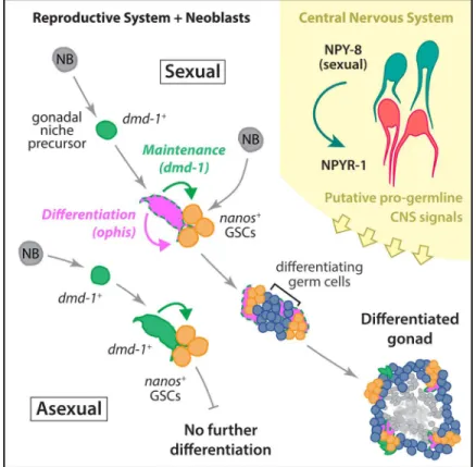
Fig 8. Schematic of the developmental mechanisms involved in planarian testis formation.dmd-1+
cells in both sexual and asexual worms are required for specification ofnanos+GSCs. In sexual planarians, thesedmd-1+cells expressophis, which is required for further differentiation of GSCs into mature gametes.
NPY-8 signaling, which occurs in the CNS, systemically promotes later stages of germ cell maturation.
broader context, this work lays the groundwork for future characterization of parasitic flat-worm GPCRs for basic biology and drug discovery purposes, and provides insights into the diversity and evolutionary history of metazoan GPCRs.
Materials and Methods
Planarian Husbandry
Sexual and asexualS.mediterraneawere maintained at 18°C in 0.75X Montjuïc salts [78] or 0.5 g/L Instant Ocean Sea Salts (Spectrum Brands), respectively. Animals were starved at least one week prior to use. For all experiments with sexualS.mediterranea, worms8 mm length were used, unless otherwise specified.
Discovery of Planarian GPCRs
To maximize coverage of the sexual and asexual strains of the planarian, we developed two strain-specific de novo transcriptomes which we then combined and used for receptor mining (FASTA file inS1 Data). For some gene curations, we also used a publicly available assembly that was generated through a different methodology (PlanMine v2.0beta, http://planmine.mpi-cbg.de/planmine/). Transcriptomes were mined using tBLASTx (BLOSUM62) and HMMer [79] to identify potential receptor sequences. Conserved GPCRs from other organisms and pre-viously published planarian GPCR sequences [26] were used as a seed dataset to discover pla-narian GPCRs. Where possible, we expanded and curated the transcripts to reach the ends of the open reading frames (ORFs). ORFs were marked“N-term OK”if we found a stop codon closely followed by an AUG start codon at the 50
-end of the transcript. ORFs were marked“ C-term OK”if we found a stop codon at the 30-end. If both of these conditions were met, the entry was marked“ORF confident.”A pool of potential receptor sequences was then filtered to exclude non-GPCR sequences, namely, those that showed significant similarity to other types of transmembrane proteins, such as ion channels or solute carrier proteins (E-value<1E-10
for at least half of the top-fifty tBLASTx hits against NCBI nucleotide collection). We also removed genes that appeared to have a complete ORF but encoded a protein that did not show the correct topology: seven TM domains flanked by an extracellular N-terminus and an intra-cellular C-terminus (determined by TMHMM2.0) [80]. To complement the topologies calcu-lated by TMHMM2.0, we inspected the posterior probability graphs generated by the program looking for potential TM domains that did not pass the 50% threshold to be reported by the program. Such curations are noted in the table inS3 Data. After a second round of receptor mining and filtering, a set of 566 GPCR genes was finalized for downstream analyses. SeeS1A Figfor the workflow we followed. Additional recursive searches did not expand the number of putative receptors, suggesting that our database contains nearly all of the expressed GPCR genes and the possibility of a significant number of unidentified GPCRs is slim.
Classification of Planarian GPCRs
subsequently analyzed to extract convex clusters of four or more genes with the attraction value limit set at 0.5 standard deviation.
Next, we pooled the rhodopsin-like genes from the previous analysis and aligned them using ClustalW [81] within CLC Genomics Workbench 7. We then generated a phylogenetic tree by the neighbor-joining method (Jukes-Cantor distance measure), with 1000 replicas to deduce bootstrap values. We used this tree to validate the clustering analysis results. Two small clusters, containing 13 and five genes, were merged with clustersSrfaandSrw, respectively, because they were bound within the representatives of their parent clusters on the tree. On the other hand, clusterR1was extracted from clusterRho-C, because its members were more closely related to those of clusterR2than the original cluster. Finally,gcr081,gcr089,gcr442, andgcr500were moved fromL1toL2based on their positions on the phylogenetic tree.
NPY Receptor Phylogeny
The 16 putative NPY receptor genes were used to perform an alignment search against Refer-ence Proteomes with the EMBL-EBI HMMER tool (http://www.ebi.ac.uk/Tools/hmmer/). Representative hits with E-value<1E-30 were aligned and phylogenetic trees were
con-structed. In a separate analysis, only the planarian NPY receptors and their parasitic flatworm homologs were used. ClustalW (within CLC Genomics Workbench 7.0) was used to align amino acid sequences with default parameters. The resulting alignments were subjected to phy-logenetic analysis by Bayesian inference in MrBayes v.3.2 [82], using the Whelan and Goldman (WAG) evolutionary model [83] to construct a 50% majority rule tree and assign posterior probabilities to tree nodes. Markov Chain Monte Carlo (MCMC) analyses ran on two indepen-dent chains (default) for 200,000 iterations and finished with an average standard deviation of split frequencies of 0.01 or less. The first 25% of sampled trees were discarded as the burnin period. Phylogenetic trees were visualized by FigTree v.1.4 (http://tree.bio.ed.ac.uk/software/ figtree/). All trees were rooted with a group of two human and two planarian amine receptors.
Receptor Cloning
For the purpose of ISH and RNAi experiments, 500–1,000 bp segments of the GPCR genes were amplified (primers inS5 Data) using Platinum Taq DNA Polymerase (Invitrogen). Prod-ucts were TA-cloned into pJC53.2 and sequenced to determine the directionality of cloning. pJC53.2 allows for probe synthesis using SP6 or T3 RNA polymerases, as well as dsRNA syn-thesis using T7 RNA polymerase [32].
A number of full-length NPY receptor coding sequences were cloned into pcDNA3.1 for the purpose of in vitro receptor assays. Primers corresponding tonpyr-1(forward: ACAGGGATC CACCATGGATTTGTGTAAGGATAATC, reverse: TATAGAATTCAGGACGACGATAC TTCACTTTTG),npyr-7(forward: ACAGGGATCCACCATGAATTCTATGAAAAATC, reverse: TATAGAATTCATAAAGATGATATTTTGAATCTTC),npyr-8(forward: ACAGG GATCCACCATGATTTTATCGAATGGC, reverse: TATAGAATTCAATTTACTAATCC AATATGAGAATC),ophis(forward: ACAGGGATCCACCATGGTTTTCTGTAGACTAAT, reverse: TATAGAATTCAATTTATCGTTGAAGATTG) were used to amplify from sexual planarian cDNA. Forward primers had an extension containing aBamHIsite and a Kozak sequence before the start codon, while reverse primers contained a stop codon followed by an
RNAi Knockdown
Knockdowns were generated by feeding in vitro transcribed dsRNA, as previously described [32], and using dsRNA matching theccdBandcamR-containing insert of pJC53.2 as a control. Worms were fed with 5μg of dsRNA combined with 45μL of a 3:1 calf liver:water mixture. For
the post-embryonic development paradigm, cut and regenerated worms smaller than 8 mm were fed dsRNA eight times, every 6 d (Fig 2A). For the re-specification paradigm, mature worms were fed two doses of dsRNA, cut anterior to the ovaries, and the head fragments were allowed to regenerate for 2 wk, unless otherwise specified (Fig 7A).
Peptide Synthesis
NPY-1 (LNEYFAIVGRPRF-amide), NPY-8 (PMFDSADAFRNYLRKLNNEYMIAGRPRF-amide), and scrambled NPY-8 (LFRMRFDAMKDELRANNNRYFIPSYPGA-amide) were syn-thesized by New England Peptide (Gardner, MA). Peptides were designed and C-terminally amidated based on in silico prediction of their bioactive form after enzymatic processing [32]. For each peptide, purity of>95% was confirmed through HPLC by the vendor. Peptides were
soluble in pure water (aided by brief sonication if needed).
In Vitro Receptor-Activation Assay
In vitro receptor-activation assays were done as previously described [84]. Briefly, calcium responses were measured in CHO-K1 cells transiently transfected with a receptor::pcDNA3.1 construct of interest. Cells also stably expressed the promiscuous Gα16protein and
mitochond-rially targeted apoaequorin. After loading with the cofactor coelenterazine, transfected cells were challenged with synthetic peptides (>95% purity) and calcium responses were
simulta-neously monitored on a Mithras LB940 luminometer. In each case, calcium responses evoked by peptides were normalized to the total calcium response (i.e., response evoked by the peptide plus response evoked by a second addition of 0.1% Triton X-100). For concentration-response curves, the normalized calcium responses are plotted as a percentage of the highest normalized response of the concentration series. Data were averaged from at least two independent experi-ments. Dose-response curves were constructed with a nonlinear regression analysis using a sig-moidal dose-response equation in Sigmaplot 12.0.
In Situ Hybridization
Whole-mount ISH was performed with a formaldehyde-based fixation procedure as previously described [50]. The protocol was optimized for larger sexual planarians: formaldehyde fixation was increased to 30 min, proteinase K treatment was increased to 30 min, and post-fixation was increased to 20 min. Colorimetric and FISH samples were imaged on an Axio Zoom.V16 and a Zeiss LSM 710 confocal microscope (Carl Zeiss, Germany), respectively. ISH probes were synthesized according to the methods previously described [50]. Single-stranded RNA probes forSmed-gH4(GB: DN306099) andSmed-npy-8(GB: BK007010) were labeled with fluorescein isothiocyanate (FITC), forSmed-nanos(GB: EF035555) andSmed-npyr-1with dinitrophenol (DNP), and forSmed-pc2(GB: BK007043),Smed-ChAT(PlanMine,
AP-conjugated antibodies and developed with nitro-blue tetrazolium (NBT) and 5-bromo-4-chloro-3'-indolyphosphate (BCIP).
RNA-seq Expression Analyses
Approximately 400,000 reads from 12 independent control samples (six sexual and six asexual worms;S6 Data) were mapped to the GPCR database using 0.9 as minimum similarity and cov-erage fractions. Base 2 logarithm of the RPKM values [85] were used as a relative measure of expression comparing the two strains. False Discovery Rate-correctedp-values [86] smaller than 0.05 were considered significant. Read mapping and statistical analyses were performed using CLC Genomics Workbench 7. Because sexual planarians express sexually enriched GPCRs at very high levels compared to asexuals (increasing the denominator in the RPKM fraction), other GPCRs falsely appear to be enriched in asexuals. To correct this bias, we graphed log2(fold change) against log10(RPKM) for the 376 GPCRs with significantp-values.
We then calculated a linear regression trend line for the middle 282 (75%) data points and used it to transform all fold change values so that the majority of them are around zero. The following transformation was applied: normalized log2(fold change) = original log2(fold
change)−(0.411log10(RPKM)) + 2.29.
Quantitative PCR
Total RNA was extracted from whole individual worms using TRIzol Reagent (Invitrogen) according to the manufacturer’s instructions, using the high-salt step for RNA precipitation. RNA was DNAse-treated, purified, and concentrated using the DNA-free RNA kit (Zymo Research). About 1μg total RNA was used to prepare cDNA using iScript cDNA Synthesis Kit
(Biorad) according to the kit protocol. GoTaq qPCR Master Mix (Promega) was used for qPCR reactions in a StepOnePlus Real-Time PCR machine (Applied Biosystems). Smed-beta-tubulinwas used as an internal control. Primers corresponding tonpy-8(forward: TGACT CAGCTGATGCCTTTC, reverse: GCCAAATCTTGGTCTTCC),npyr-1(forward: ACGA CATTCAACGACAGAGG, reverse: GTAACGACATCGGACCAACA),nanos(CAAGGA CAAATGTTGCCTGTA, reverse: CAACCCATCGATCCAACTCT),beta-tubulin(forward: TGGCTGCTTGTGATCCAAGA, reverse: AAATTGCCGCAACAGTCAAATA), andophis
(forward: ATCGTCTATTGGCCCGTAAG, reverse: AAACGACTGAGCGGAACAAC) were used. Three technical replicates were assayed for each sample. At least four individual animals were used as biological replicates for each condition tested, unless mentioned otherwise. For each gene,ΔCtwas calculated as the difference between Ctvalues of the gene of interest and
beta-tubulin. Error bars indicate the range of relative quantities calculated fromΔΔCt± SEM,
where SEM is the standard error of the mean ofΔCtvalues of the test (and not the reference)
biological replicates.
Supporting Information
S1 Data.S.mediterraneatranscriptome assembled de novo from sexual and asexual reads.
(ZIP)
S2 Data. Planarian genes encoding putative GPCRs in FASTA format.
(FA)
Number of exons detected based on mapping of GPCR genes toSchmidtea mediterranea
genome version 2.0 [87]. The actual number of exons may be higher as some gene structures are incomplete.Cluster:Results of clustering analysis using CLANS 2.0.Human:Homologous human GPCR class.BLAST description:Top BLAST (protein vs. protein) hit against human proteins.Lowest E-value:E-value corresponding to the top BLAST hit against human proteins.
ORF confident:Whether either end or both ends of the open reading frame are confidently discovered.#TM domain:Number of transmembrane domains detected by TMHMM 2.0.
Max. control RPKM:Highest RPKM value resulting from mapping sexual or asexual RNA-seq reads to the GPCR RNA-sequence list.Log2(FC(sex/asex)) [normalized]:Log base 2 RPKM fold-change between sexual and asexual reads, normalized as described in methods. Values shown only if the associatedp-value is smaller than 0.05.Asex/Sex (Graveley):RPKM fold-change between asexual and sexual RNA-seq reads from a previous study [88]. Values shown only if the associatedp-value is smaller than 0.05.Sexual irradiation (Graveley):RPKM fold-change between irradiated and control sexual planarian RNA-seq reads [88]. Values shown only if the associatedp-value is smaller than 0.05.Asexual irradiation (Graveley):RPKM fold-change between irradiated and control asexual planarian RNA-seq reads [88]. Values shown only if the associatedp-value is smaller than 0.05.X1 (SCs) (Pearson):RPKM fold-change between X1 and differentiated sorted cell populations RNA-seq reads [89]. Values shown only if the associatedp-value is smaller than 0.05.X2 (Progeny) (Pearson):RPKM fold-change between X2 and differentiated sorted cell populations RNA-seq reads [89]. Values shown only if the associatedp-value is smaller than 0.05.in situ pattern:Expression patterns that were observed in at least two independent colorimetric ISH experiments. In all RPKM ratio values, “10,000”represents division by zero—i.e., no reads mapped to reference in the control sample. InLog2(FCsex/asex) [normalized],“13.29”, which is log2(10000), represents division by zero. In
in situ pattern,the following abbreviations were used: BR, brain; INT, intestine; TE, testis; OV, ovary; OVD, oviduct; VIT, vitellaria; SPD, sperm duct; CO, copulatory apparatus; NE, no expression detected.
(XLSX)
S4 Data. Quantification of FISH, RNAi, and receptor assay experiments.
(XLSX)
S5 Data. Primers used to clone the GPCRs analyzed.
(XLSX)
S6 Data. RNA-seq reads corresponding to the de novo assembly of GPCR genes.
(ZIP)
rhodopsin family, suggesting that its members may retain affinity to small molecule ligands. Some members ofSrfbhave been previously identified as the PROF1 family of GPCRs [26]. (C) Neighbor-joining phylogenetic tree showing the hypothetical evolutionary relationship between planarian rhodopsin-like GPCRs. Conserved (D/E)R(Y/F) motifs are depicted in sequence logos. (D) Relative abundance of planarian GPCRs grouped according to their fami-lies or, in case of the rhodopsin family, separated by subfamifami-lies.Y-axis shows RPKM values based on a mapping where only the GPCR database (and not a transcriptome) was used as the reference. For GPCRs that are differentially expressed between sexual and asexual strains, the higher values were used. Bars indicate the median and quartiles. GPCRs of theRho-Lsubfamily are noticeably less abundant compared to the other groups.Rho-R2GPCRs are the most het-erogeneous in terms of relative abundance. Frizzled and secretin GPCRs are on average the most abundant groups. (E) Bayesian inference topology of planarian NPY receptors with their closest counterparts throughout metazoans. Non-planarian GPCRs were selected only accord-ing to highest similarity in HMMER search (irrespective of the species of origin). Three types of planarian NPY receptors are identified: Type 1 including NPYR-1 to 6 and their arthropod and nematode homologs.C.elegansNPR-11 andDrosophilaNPFR-1 are in this group. Type 2 includes planarian NPYR-8 to 10, in addition to many arthropod homologs. Type 3 includes planarian NPYR-11 to 16 and appears to be lophotrochozoan-specific. The snail NPY receptor GRL105 [40] is a member of this group. Vertebrate NPY receptors form a fourth monophyletic group that appears to be outside of the invertebrate clade (although with a lower 0.62 posterior probability). Posterior probabilities are 1.00 at every node, except those with a value shown. Common names or sequence identification numbers (GI) are shown for proteins on the tree. Tree is rooted with human and planarian amine receptors.
(TIF)
S2 Fig. Planarian GPCRs are enriched in an assortment of tissues and organ systems; related toFig 1.Representative colorimetric ISH experiments show GPCRs of different classes enriched in the nervous system, reproductive structures, and the intestine. (A)gcr102 (unclus-tered) is expressed in a subset of cells in the ventral brain region (left) and putative sensory organs around the edge of the head on the dorsal side (right). (B)gcr158(Rho-R2) is expressed in cells associated with the cephalic ganglia. (C)gcr121(adhesion) is expressed in a handful of anterolateral cells. (D)gcr106(metabotropic glutamate receptor) is expressed both in the brain (left) and in the secretory glands around the copulatory apparatus (right). (E)gcr084(related to human transmembrane protein 181) is highly enriched in and around the penis papilla. (F)
gcr160(Rho-L1) is expressed in a variety of epithelial tissues, including pharynx, seminal vesi-cles (left), around the head (middle), and the vitellaria (right). (G)gcr153(unclustered) in highly enriched in the intestine. (H–P) Expression patterns of representative NPY receptor genes.npyr-1,3, and7are expressed in subsets of cells in the brain.npyr-2,5,6,8,9, and15are enriched in the testes.npyr-4and10did not produce a specific ISH pattern.npyr-11to14and
16were not tested or did not show specific expression. SeeS3 Datafor a summary of expression patterns. Scale bars are 1 mm where whole animals are shown. Scale bars are 200μm for insets.
(TIF)
S3 Fig. Characterization of thenpyr-1knockdown phenotype; related toFig 2.(A) Double-FISH detectsnanos(orange) andgH4(blue) expression in ovaries of control andnpy-8(RNAi)
worms. While control worms develop a complete ovary with mature oocytes (arrowheads),
npy-8(RNAi)worms only displaynanos+/gH4+GSCs andgH4+oogonia. Scale bars are 100μm.
cells expressing low levels ofdmd-1can be detected indmd-1(RNAi)regenerating worms, they were not able to re-specifynanos+GSCs. Scale bars are 500μm and 20μm (insets). (C) qPCR
experiments showingnpy-8andnpyr-1mRNA levels after four feedings ofnpy-8ornpyr-1
dsRNA in homeostatic mature sexuals. RNAi knockdown ofnpy-8ornpyr-1only reduces the expression of the targeted gene. Neither knockdown significantly affectsnanosexpression. (D) qPCR experiments showingnpy-8andnpyr-1expression levels in sexual and asexual planari-ans. Whilenpy-8is enriched ~50-fold in sexuals compared to asexuals,npyr-1is expressed at comparable levels across asexuals and hatchling and mature sexuals. Error bars in C and D are SEM for four individual worms in each treatment.
(TIF)
S4 Fig. Validation of sexually enriched GPCRs by colorimetric ISH; related toFig 4. Colori-metric ISH of representative sexually enriched GPCRs.gcr108(unclustered) is expressed in the inner layer of the testes, suggesting thatgcr108expression is enriched in spermatids. Expression of 16 other GPCRs (gcr124-141; members ofRho-L,Rho-C, orSrf/w, or unclustered;gcr140was ruled out as a GPCR) are shown in the outer layer of the testes where spermatogonial cells are located.gcr143(unclustered) is expressed in the brain (top) as well as the vitellaria (bottom).
gcr144(secretin) is expressed in the oviducts and copulatory apparatus.gcr157(Srfb) is enriched in the ovaries.
(TIF)
S5 Fig. Low levels ofophisexpression in asexual planarians; related toFig 6.(A) FISH label-ingnanos(orange),dmd-1(green), andophis(magenta) in whole-mount asexual planarians. Clusters ofnanos+cells are present adjacent todmd-1+somatic cells on the dorsal side.ophis
mRNA is not detectable in somatic cells. Imaging settings used were identical to otherophis
FISH experiments. DAPI labels nuclei (grey). Scale bars are 100μm. (B) qPCR analysis ofophis
expression in asexual and hatchling and mature sexual planarians. Expression is comparable between asexuals and hatchling sexuals, but about 4-fold enriched in mature sexuals. Expres-sion levels were averaged between four individual animals in each treatment and normalized to the expression level ofophisin asexual worms. Error bars represent SEM among biological rep-licates.
(TIF)
Acknowledgments
We thank Rachel Roberts-Galbraith, Melanie Issigonis, Tania Rozario, Marla Tharp, and Umair Khan for comments on the manuscript, and Tracy Chong, Ryan S. King, Harini Iyer, and Labib Rouhana for sharing RNA-seq data.
Author Contributions
Conceived and designed the experiments: AS IB LS PAN. Performed the experiments: AS AJ IB. Analyzed the data: AS IB LS PAN. Wrote the paper: AS PAN.
References
1. Noel SD, Kaiser UB. G protein-coupled receptors involved in GnRH regulation: molecular insights from human disease. Mol Cell Endocrinol. 2011; 346: 91–101. doi:10.1016/j.mce.2011.06.022PMID: 21736917
3. Millar RP, Lu ZL, Pawson AJ, Flanagan CA, Morgan K, Maudsley SR. Gonadotropin-releasing hormone receptors. Endocr Rev. 2004; 25: 235–275. PMID:15082521
4. Cole LW, Sidis Y, Zhang C, Quinton R, Plummer L, Pignatelli D, et al. Mutations inprokineticin 2and
prokineticin receptor 2genes in human gonadotrophin-releasing hormone deficiency: molecular genet-ics and clinical spectrum. J Clin Endocrinol Metab. 2008; 93: 3551–3559. doi:10.1210/jc.2007-2654 PMID:18559922
5. Topaloglu AK, Reimann F, Guclu M, Yalin AS, Kotan LD, Porter KM, et al.TAC3andTACR3mutations in familial hypogonadotropic hypogonadism reveal a key role for Neurokinin B in the central control of reproduction. Nat Genet. 2008; 41: 354–358. doi:10.1038/ng.306PMID:19079066
6. Messager S, Chatzidaki EE, Ma D, Hendrick AG, Zahn D, Dixon J, et al. Kisspeptin directly stimulates gonadotropin-releasing hormone release via G protein-coupled receptor 54. Proc Natl Acad Sci U S A. 2005; 102: 1761–1766. PMID:15665093
7. Molyneaux KA, Zinszner H, Kunwar PS, Schaible K, Stebler J, Sunshine MJ, et al. The chemokine SDF1/CXCL12 and its receptor CXCR4 regulate mouse germ cell migration and survival. Development. 2003; 130: 4279–4286. PMID:12900445
8. Knaut H, Werz C, Geisler R, The Tübingen 2000 Screen Consortium, Nüsslein-Volhard C. A zebrafish homologue of the chemokine receptorCxcr4is a germ-cell guidance receptor. Nature. 2003; 421: 279– 282. PMID:12508118
9. Kunwar PS, Starz-Gaiano M, Bainton RJ, Heberlein U, Lehmann R. Tre1, a G protein-coupled receptor, directs transepithelial migration ofDrosophilagerm cells. PLoS Biol. 2003; 1: e80. PMID:14691551 10. Pesce M, Canipari R, Ferri GL, Siracusa G, De Felici M. Pituitary adenylate cyclase-activating
polypep-tide (PACAP) stimulates adenylate cyclase and promotes proliferation of mouse primordial germ cells. Development. 1996; 122: 215–221. PMID:8565832
11. Vaudry D, Gonzalez BJ, Basille M, Yon L, Fournier A, Vaudry H. Pituitary adenylate cyclase-activating polypeptide and its receptors: from structure to functions. Pharmacol Rev. 2000; 52: 269–324. PMID: 10835102
12. Agnese M, Valiante S, Angelini F, Laforgia V, Andreuccetti P, Prisco M. Pituitary adenylate cyclase-activating polypeptide and its receptor PAC1 in the testis ofTriturus carnifexandPodarcis sicula. Gen Comp Endocrinol. 2010; 168: 256–261. doi:10.1016/j.ygcen.2010.03.016PMID:20338177
13. Goto T, Salpekar A, Monk M. Expression of a testis-specific member of the olfactory receptor gene fam-ily in human primordial germ cells. Mol Hum Reprod. 2001; 7: 553–558. PMID:11385110
14. Schneider LE, Spradling AC. TheDrosophilaG protein-coupled receptor kinase homologueGprk2is required for egg morphogenesis. Development. 1997; 124: 2591–2602. PMID:9217001
15. Davies B, Baumann C, Kirchhoff C, Ivell R, Nubbemeyer R, Habenicht UF, et al. Targeted deletion of the epididymal receptor HE6 results in fluid dysregulation and male infertility. Mol Cell Biol. 2004; 24: 8642–8648. PMID:15367682
16. Newmark PA, Sánchez Alvarado A. Not your father’s planarian: a classic model enters the era of func-tional genomics. Nat Rev Genet. 2002; 3: 210–219. PMID:11972158
17. Morgan TH. Growth and regeneration inPlanaria lugubris. Archiv für Entwickelungsmechanik der Orga-nismen. 1901; 13: 179–212.
18. Wang Y, Zayas RM, Guo T, Newmark PA.nanosfunction is essential for development and regenera-tion of planarian germ cells. Proc Natl Acad Sci U S A. 2007; 104: 5901–5906. PMID:17376870 19. Ghirardelli E. Differentiation of the germ cells and generation of the gonads in planarians. In: Kiortsis V,
Trampusch H, editors. Regeneration in Animals and Related Problems. Amsterdam: North–Holland; 1965. pp. 177–184.
20. Fedecka-Bruner B. [Differentiation of the male gonads in the planarian,Dugesia lugubris, during regen-eration]. C R Seances Soc Biol Fil. 1967; 161: 21–23. PMID:4234324
21. Taman A, Ribeiro P. Characterization of a truncated metabotropic glutamate receptor in a primitive metazoan, the parasitic flatwormSchistosoma mansoni. PLoS ONE. 2011; 6: e27119. doi:10.1371/ journal.pone.0027119PMID:22069494
22. El-Shehabi F, Taman A, Moali LS, El-Sakkary N, Ribeiro P. A novel G protein-coupled receptor of
Schistosoma mansoni(SmGPR-3) is activated by dopamine and is widely expressed in the nervous system. PLoS Negl Trop Dis. 2012; 6: e1523. doi:10.1371/journal.pntd.0001523PMID:22389736 23. Lapan SW, Reddien PW. Transcriptome analysis of the planarian eye identifiesovoas a specific
regu-lator of eye regeneration. Cell Rep. 2012; 2: 294–307. doi:10.1016/j.celrep.2012.06.018PMID: 22884275