cop
yr
ight
© ABE&M todos os dir
eitos r
eser
v
ados
apresentação de caso
FELIPE HENNING GAIA DUARTE
MARCIO CARLOS MACHADO
JOILMA RODRIGUES DE LIMA
LUIZ ROBERTO SALGADO
Endocrine Service, Hospital do Câncer A.C. Camargo,
Fundação Antonio Prudente, (FHGD, MCM, JRL);
Neuroendocrine Unit, Division of Endocrinology and Metabolism, University of São Paulo School of Medicine (FHGD, MCM, LRS); São Paulo, SP, Brazil.
Recebido em 31/3/2008 Aceito em 22/7/2008 ABSTRACT
Sellar and parasellar masses blocking inhibitory hypothalamic dopaminergic to-nus can produce hyperprolactinemia. One of these conditions, seldom reported, is internal carotid artery aneurysm causing pituitary stalk compression and hy-perprolactinemia, the majority of which is related to small increases in serum prolactin levels. The aim of this study is to report the case of a patient with an internal carotid aneurysm and severe hiperprolactinemia. A 72 years old female patient, on oncology follow-up for clinically controlled cervical carcinoma, was evaluated due to worsening chronic headaches. During the investigation, com-puted tomography and magnetic resonance imaging (MRI) showed a sellar mass associated with high prolactin level (1.403 µg/L) that initially was considered a macroprolactinoma, and treated with bromocriptine. However, subsequent pitu-itary MRI suggested an internal carotid aneurysm, which was confirmed by an angioresonance imaging of cerebral vessels. On low bromocriptine dose (1.25 mg/day), there was a prompt normalization of prolactin levels with a great in-crease (> 600 µg/L) after withdrawal, which was confirmed several times, sug-gesting HPD. We report a patient with internal carotid artery aneurysm with severe hyperprolactinemia never reported before in patients with HPD, and the need for a differential diagnosis with macroprolactinomas even considering high prolactin levels. (Arq Bras Endocrinol Metab 2008; 52/7:1189-1193)
Keywords: Hyperprolactinemia; Internal carotid artery aneurysm; Hypothalamic-pituitary disconnection; Prolactinoma; Differential diagnosis of Hypothalamic-pituitary tumor
RESUMO
Hiperprolactinemia Severa Associada a Aneurisma Interno da Artéria Caróti-da: Diag nóstico Diferencial entre Prolactinoma e Desconexão Hipotálamo-hipofisária.
Massas selares e parasselares podem produzir hiperprolactinemia por bloquear o tônus inibitório hipotalâmico de dopamina. Uma destas condições, raramente reportada, é o aneurisma de artéria carótida interna causando compressão da haste hipofisária e hiperprolactinemia, a maioria com pequenas elevações da prolactina. O objetivo deste estudo é descrever o caso de uma paciente com an-eurisma de carótida interna e grave hiperprolactinemia. Paciente feminina, 72 anos, em acompanhamento oncológico por carcinoma de colo de útero clinica-mente controlado, avaliada por causa da piora de cefaléia crônica. Durante inves-tigação, tomografia computadorizada e ressonância magnética (RM) de hipófise mostraram massa selar associada com altos níveis de prolactina (1.403 µg/L), sendo avaliado como macroprolactinoma e tratado com bromocriptina. Entretan-to, RM subseqüente sugeriu aneurisma de carótida interna que foi confirmado por angiorressonância de vasos cerebrais. Em uso de baixas doses de bromocrip-tina (1,25 mg/dia), houve pronta normalização da prolacbromocrip-tina com grande elevação (> 600 µg/L) após a retirada do medicamento, sendo confirmado por várias vezes sugerindo DHH. Reporta-se uma paciente com aneurisma de artéria carótida in-terna com grave hiperprolactinemia, nunca descrita anteriormente em pacientes com DHH, e a necessidade do diagnóstico diferencial com macroprolactinoma, mesmo considerando altos níveis de prolactina. (Arq Bras Endocrinol Metab 2008; 52/7:1189-1193)
© ABE&M todos os dir
eitos r
eser
v
ados
P
rolactin differs from other pituitary hormones as its secretion is regulated mainly through an inhibitory dopamine tonus, produced by neurons located in the hypothalamus (1). In addition to dopamine, other hy-pothalamic factors (TRH, oxytocin, VIP, angiotensin II, NPY and galanin) or even steroid hormones (estro-gen) modulate the secretion of prolactin (2). The hy-pothalamic dopamine reaches the pituitary through the pituitary stalk and interacts with the dopamine receptor (D2), located in the cell membrane of lactotropes via protein Gi. After its binding, dopamine exerts an inhi-bitory effect on prolactin gene causing a reduction in secretion and hormone production (1).Sellar and parasellar masses may hinder transporta-tion of hypothalamic dopamine to the pituitary, leading to secondary hyperprolactinemia due to an absence of its inhibitory action on normal lactotropes (3). This process has been called hypothalamic-pituitary disconnection (HPD) and is classically associated to small or moderate serum prolactin increases, usually less than 100 µg/L.
We report the case of a patient with sellar and supra-sellar mass associated with a very high serum prolactin levels (1.403 µg/L) initially considered as a macropro-lactinoma. This is an atypical case of severe hyperprolac-tinemia due to HPD, and it should be stressed that prolactin levels as high as in this case has never been re-ported before. The aim of this paper is to discuss the importance of the differential diagnosis between internal carotid aneurysm and macroprolactinoma.
CASE REPORT
A female patient, age 72 years, was evaluated in 1987 for cervical carcinoma and submitted to radiotherapy treat-ment with clinical remission. After three years, she presen-ted worse chronic headaches, nausea and arterial hypertention. A cranial computed tomography (CT) re-vealed the presence of a sellar and suprasellar mass with a 20 mm diameter, which was confirmed by pituitary MRI. The patient had no complaints of decreased libido, abnor-mal visual fields or galactorrhea. Initial laboratory analysis excluded hematological, renal and hepatic abnormalities. Patient denied having taking medications that could lead to hyperprolactinemia. Hormonal assessment showed a prolactin level of 1403 µg/L (NV 2-15 µg/L) (Table 1) and panhypopituitarism (T4L 0.69 ng/dL, NV: 1.0-1.8
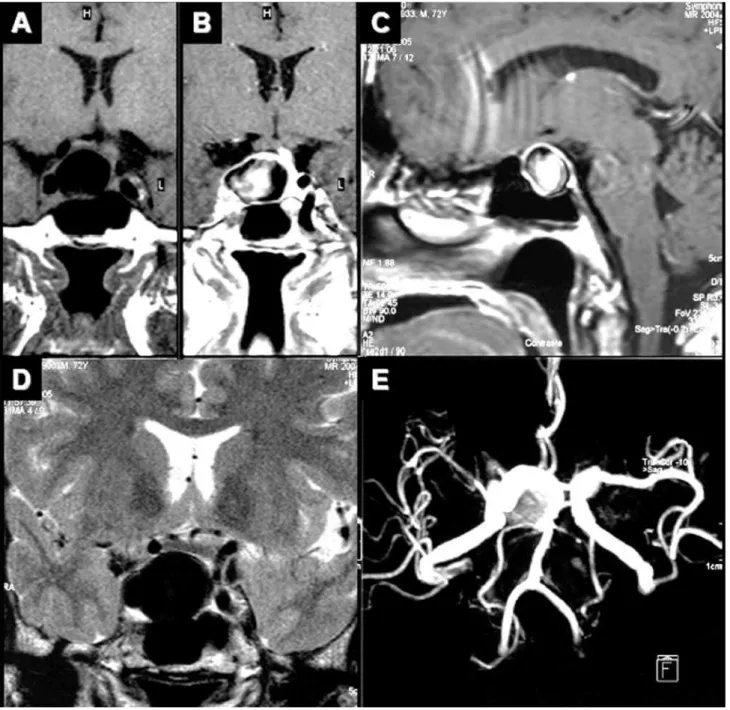
µg/L, NV: 96-502 µg/L). Screening for macroprolacti-nemia was negative as well. Thepatient was treated with replacement therapy of prednisone 5 mg/day and thyro-xine 100 µg/day. As the high prolactin levels suggested a macroprolactinoma, she also received bromocriptine (BRC) starting with 1.25 mg/day. A new pituitary MRI confirmed a sellar and suprasellar mass of similar dimen-sions of the previous one, but with heterogeneous con-tent, which raised suspicion of a giant aneurysm of the internal carotid artery with a thrombus inside (Figure 1). This was confirmed by an angioresonance imaging of ce-rebral vessels (Figure 1). On BRC (1.25 mg/day), a dra-matic reduction of prolactin levels was observed and the dosage was increased to 2.5 mg/day when serum prolac-tin levels became almost undetectable (0.6 µg/L) (Table 1). After multi-disciplinary discussion, a conservative ap-proach was chosen instead of surgery.
The patient continued BRC use for approximately one year when it was discontinued. Shortly after hyper-prolactinemia as high as 657 and 437 µg/L was obser-ved, bromocriptin was restarted with a dose of 2.5 mg/day with prolactin returning to normal values (Table 1). The drug was then gradually reduced to 0.625 mg/day, keeping serum prolactin in normal range levels (8.5 µg/L). During imaging assessment, the aneurysm remained with the same diameter as in the initial exam, and the patient was kept in full repla-cement for hypopituitarism.
Table 1. Outcome of prolactin levels with bromocriptine use.
Date Bromocriptine (mg/day) Prolactin (µg/L)
September 2000 Before 1403.0
November 2000 1.25 38.0
January 2001 2.5 2.5
May 2001 2.5 0.6
September 2001 Stop 657.0
September 2001 Stop 437.0
October 2001 2.5 0.6
February 2003 1.25 6.0
July 2003 0.625 3.0
September 2004 0.625 1.0
November 2004 Stop 449.0
August 2005 0.625 2.3
January 2006 0.625 5.1
March 2006 Stop 405.0
cop
yr
ight
© ABE&M todos os dir
eitos r
eser
v
ados
© ABE&M todos os dir
eitos r
eser
v
ados
The Research Projects and Graduate Studies Commission and the Ethics Commission of our insti-tutions approved the study, and the patient provided informed consent.
Hormonal assay
Serum prolactin was measured by commercial immu-noassay (Bayer Advia Centaur, Indiana, USA).
DISCUSSION
The differential diagnosis of hyperprolactinemia inclu-des a large number of clinical physiological situations (pregnancy, lactation, stress, sleep, exercise, maxillary stimulation) or pathological conditions (1). Among the pathological causes, the two most common etiologies are prolactinomas and use of anti-dopamine drugs. Other pathologic conditions such as polycystic ovary syndrome, hypothyroidism, adrenal failure, hepatic cir-rhosis, chronic kidney failure, brain trauma injuries and clinically non-functioning pituitary adenomas are kno-wn causes of hyperprolactinemia (3). Furthermore, other lesions of the hypothalamic-pituitary region also can cause increased secretion of prolactin due to HPD. In addition to HPD, the rise in intrasellar pressure cau-sed by pituitary masses can also lead to increacau-sed levels of serum prolactin (4). However, both increases of in-trasellar pressure and suprasellar masses causing HPD are usually associated with moderated increases of se-rum prolactin levels (4-6).
Classically, serum levels of prolactin higher than 200 µg/L are related to macroprolactinomas, but tho-se levels should not exclude other etiologies or even use of anti-dopamine drugs as been reported in litera-ture (7). Therefore, any sellar masses among cranio-pharyngiomas, optical gliomas, secondary metastatic implants, chordomas and internal carotid aneurysms can be associated with moderated increases of serum prolactin levels and should be differentiated from pro-lactinomas (8,9).
It should be mentioned that co-existence of aneu-rysm of internal carotid and hyperprolactinemia is usu-ally iatrogenic (10), and descriptions of its primary association is rare (11). Another considered possibility was the presence of a co-existing prolactinoma. Al-though rare, few reports described this association
ruled out in our case. The first published case of inter-nal carotid aneurysm and primary hyperprolactinemia was reported by Verbalis et al.(5), in 1982, in a 59 years old patient with hypopituitarism and prolactin serum levels of 177 µg/L related to internal carotid aneurysm. After the aneurism surgical decompression serum prolactin levels returned to normal. Thereafter, a number of new cases were reported over the subse-quent years (6,8,9,15-21).
An important clinical aspect of these reported cases is the female prevalence and age ranged between 42 and 76 years, exceptions being a case reported by Kaya-th et al. (18-year-old male patient) and two oKaya-ther pa-tients reported by Fonseca et al.(17), both males, age 57 and 68 years. Serum prolactin levels ranged between 4 and 182 µg/L (5,6,8,9,16,17,19-21) associated with hypopituitarism. Clinically, the most common com-plaints were headaches and blurred vision attributed to the mass effect.
In only two reported cases, serum prolactin levels were higher than 200 µg/L. The first was reported by Fernandez-Real et al.(18), who described a case of a 52-year-old women with a giant aneurysm associated with subarachnoid hemorrhage and serum prolactin le-vels of 1.093 µg/L who presented sudden headaches, a decline in consciousness, hemiparesis and signs, and symptoms relate to hypopituitarism. In the second case, published by Kahn et al.(15), a 42 years old patient presented galactorrhea, headaches, abnormal visual fields and serum prolactin levels of 365 µg/L.
In the case reported, the patient presented higher serum prolactin levels, associated with internal carotid aneurysm (1.403 µg/L), than reported in the literature to date. Our case was noticed to be similar to that case reported by Kahn et al. (15) where there were no other factors besides the internal carotid aneurysm that could cause compression leading to HPD and hyperprolacti-nemia. It should be stressed that there is no histological proof excluding a macroprolactinoma extremely sensi-tive to dopamine agonist. However, the absence of imaging suggestive of pituitary adenoma, an angioreso-nance confirming a giant aneurysm of internal carotid artery and the extreme sensibility to the BRC presented by the patient, strongly suggest hyperprolactinemia se-condary to the HPD.
inter-cop
yr
ight
© ABE&M todos os dir
eitos r
eser
v
ados
sidering the presence of sellar masses. A test with low dose of dopamine agonist, independent of serum prolac-tin levels, could be a clue to the diagnosis of HPD.
REFERENCES
1. Ben-Jonathan N, Hnasko R. Dopamine as a prolactin (PRL) inhibitor. Endocr Rev. 2001;22:724-63.
2. Freeman ME, Kanyicska B, Lerant A, Nagy G. Prolactin: struc-ture, function, and regulation of secretion. Physiol Rev. 2000;80:1523-31.
3. Melmed S, Kleinberg D. Anterior pituitary. In: Larsen PR, Kro-nenberg HM, Melmed S, Polonsky KS, editores. Willians text-book of endocrinology. 10ª ed. Philadelphia: Saunders; 2002. p. 205-7.
4. Arafah BM, Prunty D, Ybarra J, Hlavin ML, Selman WR. The dominant role of increased intrasellar pressure in the patho-genesis of hypopituitarism, hyperprolactinemia, and heada-ches in patients with pituitary adenomas. J Clin Endocrinol Metab. 2000;85:1789-93.
5. Verbalis JG, Nelson PB, Robinson AG. Reversible panhypopi-tuitarism caused by a suprasellar aneurysm: the contribution of mass effect to pituitary dysfunction. Neurosurgery. 1982;10:604-11.
6. Borges FZ, Ferreira BP, Resende EA, Neto EN, Borges WA, Oli-veira RS, Borges MF. Aneurisma gigante de carótida interna simulando macroadenoma de hipófise. Arq Bras Endocrinol Metab. 2006;50:558-63.
7. Kleinberg DL, Gordon LN, Frantz AG. Galactorrhea: a study of 235 case, including 48 pituitary tumors. N Engl J Med. 1977;296:589-600.
8. Kayath MJ, Lengyel AM, Nogueira R, Tella Junior O, Czepie-lewski MA. Giant aneurysms of the sellar region simulating pituitary adenomas: a diagnosis to be considered. J Endocri-nol Invest. 1991;14:975-9.
9. Arlot S, Lalau JD, Galibert P, Quichaud J. Intrasellar carotid aneurysm simulating prolactin adenoma. Rev Med Interne. 1985;6:505-9.
10. Reddy K, Lesiuk H, West M, Fewer D. False aneurysm of the cavernous carotid artery: a complication of transsphenoidal surgery. Surg Neurol. 1990;33:142-5.
11. Wakai S, Fukushima T, Furihata T, Sano K. Association of cere-bral aneurysm with pituitary adenoma. Surg Neurol. 1979;12: 503-7.
12. Yang MY, Chen C, Shen CC. Cavernous aneurysm and pituitary adenoma: management of dual intrasellar lesions. J Clin Neu-rosci. 2005;12:477-81.
13. Chuang CC, Chen YL, Pai PC. A giant intracavernous carotid ar-tery aneurysm embedded in pituitary macroadenoma presen-ting with pituitary apoplexy. Cerebrovasc Dis. 2006;21:142-4. 14. Sony A, de Silva SR, Allen K, Byrne JV, Cudlip S, Wass JA. A
case of macroprolactinoma encasing an internal carotid aneu-rysm, presenting as pituitary apoplexy. Pituitary. 2007 [Epub ahead of print].
15. Kahn SR, Leblanc R, Sadikot AF, Fantus IG. Marked hyperpro-lactinemia caused by carotid aneurysm. Can J Neurol Sci. 1997;24:64-6.
16. Romano A, Chibbaro S, Marsella M, Ippolito S, Benericetti E. Carotid cavernous aneurysm presenting as pituitary apoplexy. J Clin Neurosci. 2006;13:476-9.
17. Fonseca AL, Souto AA, Domingues F, Vaisman M, Gadelha MR, Chimelli L, et al. Hormonal dysfunction of nonpituitary lesions from midline and perisellar área. Arq Neuropsiquiatr. 2001;59:905-12.
18. Fernandez-Real JM, Fernandez-Castaner M, Villabona C, Sa-garra E, Gomez-Saez JM, Soler J. Giant intrasellar aneurysm presenting with panhypopituitarism and subarachnoid he-morrhage: case report and literature review. Clin Investig. 1994;72:302-6.
19. Mindel JS, Sachdev VP, Kline LB, Sivak MA, Bergman DA, Yang WC, et al. Bilateral intracavernous carotid aneurysms mi-micking a prolactin-secreting pituitary tumor. Surg Neurol. 1983;19:163-7.
20. Ooi TC, Russell NA. Hypopituitarism resulting from an intra-sellar carotid aneurysm. Can J Neurol Sci. 1986;13:70-1. 21. Michils A, Baleriaux D, Mockel J. Bilateral carotid aneurysm
unmasked by severe hypopituitarism. Postgrad Med J. 1991;67:285-8.
Correspondence to:
Luiz Roberto Salgado
Av. Dr. Eneas de Carvalho Aguiar #155, 8th floor, Cerqueira Cesar
University of São Paulo School of Medicine 05403-060 São Paulo, SP – Brazil