cop
yr
ight
© ABE&M todos os dir
eitos r
eser
v
ados
original
VANESSA PRADODOS SANTOS ROBERTO AUGUSTO CAFFARO GEANETE POZZAN
MAURO AJAJ SAIEG VALTER CASTELLI JÚNIOR
Vascular Surgery, School of Medical Sciences, Santa Casa de São Paulo, São Paulo, SP, Brazil (VPS, RAC, VCJ); Pathology, School of Medical Sciences, Santa Casa de São Paulo, São Paulo, SP, Brazil (GP, MAS)
Recebido em 29/10/2007 Aceito em 19/8/2008 ABSTRACT
Objetive: To perform a comparative analysis of atherosclerotic lesions and capillaries changes in diabetic and nondiabetic patients. Methods: Leg ar-teries and skin of 57 amputated lower limbs of diabetic (47.3%) and nondia-betic patients were histologically examined. The percentage of arterial stenosis of infrapopliteal arteries and the histological classification of ath-erosclerotic lesions were determined. Capillary thickening was classified into four categories. Results: Diabetic group showed more than 75% steno-sis in 57% (vs. 56% in nondiabetic) of the anterior tibial; 78% (vs. 68%) of the posterior tibial; 58% (vs. 50%) of the peroneal leg arteries. Diabetic and non-diabetic patients have predominance of type VI atherosclerotic lesions. The comparison of both groups showed no significant differences in atheroscle-rotic lesions. Diabetic patients had significantly more PAS positive capillary thickening (63% vs. 23%). Conclusions: There were no differences in histo-logical characteristics of atherosclerosis between the two groups. Capillary thickening has been more observed in diabetics. (Arq Bras Endocrinol Me-tab 2008; 52/7:1115-1123)
Keywords: Diabetes mellitus; Atherosclerosis; Lower extremity; Diabetic an-giopathies; Diabetes complications
RESUMO
Estudo Histológico Comparativo das Lesões Ateroscleróticas e Alterações Microvasculares em Membros Inferiores Amputados de Pacientes Diabéticos e Não-diabéticos.
Objetivo: Comparar as lesões ateroscleróticas das extremidades de diabé-ticos e não-diabédiabé-ticos, estudando a ocorrência de espessamento capilar.
Métodos: Examinou-se segmentos arteriais e da derme de 57 membros
in-feriores amputados de diabéticos (47,3%) e não-diabéticos. Analisou-se a porcentagem de estenose das artérias infra-poplíteas e a classificação his-tológica da placa. A presença de espessamento capilar foi classificada em quatro categorias. Resultados: Entre os diabéticos 57% (versus 56% dos
não-diabéticos) apresentavam estenose maior que 75% da artéria tibial an-terior; 78% (versus 68%) da tibial posterior; 58% (versus 50%) da fibular.
Houve predominância em ambos de lesões ateroscleróticas do tipo VI. Com-parando os grupos, não houve diferença significante na porcentagem de obstrução arterial ou na classificação da placa aterosclerótica. Os diabéticos apresentaram significativamente mais espessamento capilar (63% versus
23%). Conclusões: Não houve diferença nas características das lesões ate-ros cle róticas em diabéticos e não-diabéticos. O espessamento capilar foi mais prevalente entre os diabéticos. (Arq Bras Endocrinol Metab 2008; 52/7:1115-1123)
cop
yr
ight
© ABE&M todos os dir
eitos r
eser
v
ados
INTRODUCTION
D
iabetes mellitus (DM) is the main cause of non-traumatic amputations worldwide (1). DM pa-tients have 15 times greater risk of lower extremity amputation than nondiabetic patients (2). Atheroscle-rotic macrovascular disease in patients with DM usually affects the infragenicular arteries (3). An arteriographic study has shown that diabetic patients have a greater prevalence of diffuse atherosclerosis simultaneously affecting the femoral, popliteal and tibial sections than nondiabetic patients (4).Histologically, however, atherosclerotic lesions of the lower extremities seem to have the same morpholo-gy and distribution in both diabetic and nondiabetic patients. Differences are often found only in the occur-rence of calcification in the media or Monckeberg’s medial calcific sclerosis, which is more common in dia-betic patients (3,5).
Diabetes mellitus accelerates the development of macrovascular atherosclerotic complications and leads to microvascular complications such as diabetic nephropa-thy and retinopanephropa-thy (6). However, the existence of a mi-crovascular disease affecting the lower extremities of diabetic patients has been questioned in literature (7).
Diabetic microangiopathy is characterized by de-position of PAS positive hyaline material in arterioles and capillaries of the lower extremities. Goldenberg e cols. (8) analyzed 152 fragments of lower extremities of diabetic and nondiabetic amputated patients, for di-fferent diseases. No differences were found in the in-tensity of atherosclerosis between diabetic and nondiabetic individuals; however, the authors descri-bed the presence of “diabetic lesions” in arterioles and capillaries of diabetic patients, which were characteri-zed by deposition of a periodic acid Schiff (PAS) posi-tive material. Other studies in the literature, however, do not confirm these findings. Strandness e cols. (9), in a histological study of vessels and tissues of diabetic and nondiabetic patients found no differences in the histo-logical presentation of the smaller arteries when the two groups were compared. Most of the studies publi-shed, however, included small samples and used tissue fragments of patients with a diagnosis of trauma, vascu-litis or tumors. Therefore, it remains unclear whether the microvascular changes found in the skin fragments studied by Goldenberg e cols. (8) were secondary to severe peripheral arterial occlusive disease (PAOD) or to diabetic microangiopathy.
The objective of this study is to investigate histologi-cal differences between the atherosclerotic occlusive dise-ase of amputated lower limbs from diabetic and nondiabetic patients, and to observe the morphological structure of arterioles and capillaries in these groups (pre-sence of diabetic microangiopathy). We compared the degree of obstruction and the classification of the athe-rosclerotic lesions of below-the-knee arteries (popliteal, anterior tibial, posterior tibial, and peroneal) of amputa-ted lower limbs to PAOD of diabetic and nondiabetic patients, and described the histopathological changes of skin arterioles and capillaries of these patients.
METHODS
Fragments of leg arteries and skin of 57 lower extre-mities amputated from 55 patients were examined at Santa Casa de São Paulo, Brazil, from June 2004 to June 2006.
This study included patients that had undergone abo-ve-knee amputations due to diabetic and nondiabetic se-vere PAOD ischemic lesions. Patients were excluded if their amputations were motivated by acute ischemia or any other disease than PAOD, like trauma and vasculitis.
Detailed patient information was recorded follo-wing a protocol. Patients were divided into two groups, diabetic and nondiabetic, for the analysis of histopatho-logical differences in leg arteries, arterioles and capilla-ries. Diabetic patients were defined as those with a prior diagnosis of DM and under treatment for it.
After femoral superficial clamping, during above-knee amputation, the arterial trifurcation was dissected, the popliteal artery were cut above the bifurcation and the proximal portion of anterior tibial, tibial-fibular trunk, posterior tibial and peroneal arteries were collec-ted. Skin and subcutaneous tissue from the first or se-cond toe, without gangrene, were also collected to analyze the arterioles and capillaries of the sample.
The specimens were placed in 10% formaldehyde solution. Four cross-sections of about 0.2 cm each were obtained from each of the arteries: popliteal artery abo-ve its bifurcation, and anterior tibial, tibial-fibular trunk, posterior tibial, and peroneal arteries below their origins. Four skin fragments were obtained from each toe and sent to paraffin blocks.
cop
yr
ight
© ABE&M todos os dir
eitos r
eser
v
ados
The samples were analyzed by blinded observers to DM diagnoses. The material was examined using an Axioskop 40 Zeiss optical microscope connected to a DSC-S85 Sony camera (4 megapixels).
The atherosclerotic lesions of arteries in diabetic and nondiabetic patients were compared according to two parameters: degree of lumen stenosis (1=0 to 50% stenosis; 2=50 to 75% stenosis; and 3=more than 75% stenosis); and histological classification of the atheros-clerotic lesions according to Stary e cols. (10). This clas-sification describes six types of lesions: Type I: isolated macrophage foam cells; Type II: mainly intracellular li-pid accumulation; Type III: pools of foam cells and small pools of extracellular lipids; Type IV: (atheroma) well-formed lipid nucleus with foam cells and extra-cellular deposits of lipids in the form of cholesterol crys-tals; Type V: (fibroatheroma) well-formed lipid nucleus covered by fibrous capsule; Type VI: plaque complica-tions with ulceration, fissure, thrombosis, inter-plaque hemorrhage or intense calcification. For analytical pur-poses, the absence of atherosclerotic lesions was classi-fied as Type 0, usually characterized only by intimal fibrous thickening.
The arteries of the two groups were also examined to identify Monckeberg’s medial calcific sclerosis and PAS-positive thickening of the arterial vasa vasorum.
Skin fragments were examined to identify ca-pillary PAS positive thickening and fragments were classified into four categories: 0 or absent: no ca-pillary wall thickening in the dermis (Figure 1); 1 or mild: rare capillaries with discrete PAS-positive wall thickening, scarcely distributed in the dermis (Figu-re 2); 2 or moderate: f(Figu-requent capillaries with mo-derate PAS-positive wall thickening, irregularly distributed in the dermis (Figure 3); 3 or intense: capillaries with intense and diffuse PAS-positive wall thickening, homogeneously distributed in the der-mis (Figure 4). The arteriolar thickening was consi-dered present or absent.
The sample size for the comparison of capillary thi-ckening between diabetic and nondiabetic patients was calculated using the chi-square test, considering a po-wer of 80% and a 5% of significance level.
The chi-square and the Fisher exact tests were used to investigate the association between the histo-logical variables and the disease (DM). Analysis of va-riance (ANOVA) was used to compare quantitative variables. Backward stepwise logistic regression (Wald)
Figure 1. Skin fragment with normal capillaries, classified as 0 or no thickening (AO:400x).
Figure 2. Skin fragment with capillaries showing thickening classified as mild or category 1 (AO:400x)
was used to calculate which of the study variables was associated with capillary thickening. A 5% significance level (p ≤ 0.05) was considered in order to reject the null hypothesis.
This study was approved by the Research Ethics Committee of Santa Casa de São Paulo, São Paulo, Brazil. All patients included in the study provided in-formed consent.
cop
yr
ight
© ABE&M todos os dir
eitos r
eser
v
ados
bilateral amputation was performed in two patients (3.6%) in different times. Mean age (67.38±15.12 vs. 68.34±13.73 years; p=0.80) and initial white blood cells count (13.362±4.801 vs. 13.155±5.188 mL/u; p=0.87) did not show statistically significant differen-ces between diabetic and nondiabetic patients. Wo-men were statistically more prevalent in the diabetic group (p=0.02) and arterial hypertension too (p=0.006). Smoking was significantly more prevalent among nondiabetic patients (p=0.03). Only patients with diabetes had kidney disease requiring dialysis (p=0.04). DM patients had a significantly smaller inci-dence of aortoiliac PAOD and a greater inciinci-dence of below-knee PAOD (with pedal pulses absent and po-pliteal pulses present) at physical examination (p=0.002). The comparison results between the two groups are shown in Table 1.
Table 1. Comparative analysis of the demographic profiles of patients with and without DM.
Non-DM DM
Variables N % N % p
Sex 0.02
Male 21 72.4 11 42.3
Female 8 27.6 15 57.7
Smoker 0.03
No 11 37.9 17 65.4
Yes 18 62.1 9 34.6
PAOD segment 0.002
Aortoiliac 11 37.9 1 3.8
Femoral-popliteal 18 62.1 21 80.8
Below-knee 0 0.0 4 15.4
Hypertension 0.006
No 13 44.8 3 11.5
Yes 16 55.2 23 88.5
Kidney disease requiring dialysis
0.04
No 29 100 22 84.6
Yes 0 0 4 15.4
DM=diabetes mellitus; PAOD=peripheral atherosclerotic occlusive disease.
Figure 3. Skin fragment with capillaries showing PAS-positive thickening classified as moderate or category 2 (AO:400x)
Figure 4. Skin fragment with capillaries showing PAS-positive thickening classified as intense or category 3 (AO:400x)
RESULTS
cop
yr
ight
© ABE&M todos os dir
eitos r
eser
v
ados
We collected and studied 54 popliteal artery seg-ments (94.7% of 57 limbs), 41 anterior tibial arteries (71.9%), 36 tibial-fibular trunks (63.2%), 42 posterior tibial arteries (75%), and 35 of inter-bone arteries (62.5%). There was no significant difference among the number of arteries analyzed between the two groups (diabetic and nondiabetic patients).
The comparative analysis of the atherosclerotic im-pairment of the below-knee arteries between the two groups is shown in Tables 2 and 3.
Table 2. Comparative analysis of percentage of lumen steno-sis in five types of below-knee arteries between patients with and without DM.
Non-DM DM
% stenosis N % N % p
Popliteal artery 0.53
1 8 28.6 10 40.0
2 9 32.1 5 20.0
3 11 39.3 10 40.0
Anterior tibial artery 0.90
1 5 27.8 7 30.4
2 3 16.7 3 13.0
3 10 55.5 13 56.6
Tibial-fibular trunk 0.16
1 1 5.6 4 22.2
2 4 22.2 1 5.6
3 13 72.2 13 72.2
Posterior tibial artery 0.75
1 2 10.5 2 8.7
2 4 21.1 3 13.0
3 13 68.4 18 78.3
Peroneal artery 0.78
1 5 31.2 4 21.1
2 3 18.8 4 21.1
3 8 50.0 11 57.8
DM=diabetes mellitus; 1=0 to 50% stenosis; 2=50 to 75% stenosis; 3=more than 75% stenosis.
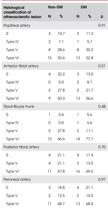
Table 3. Comparative analysis of histological classification of atherosclerotic lesions in below-knee arteries in patients with and without DM.
Histological classification of atherosclerotic lesion
Non-DM DM
N % N % p
Popliteal artery 0.91
0 3 10.7 3 11.3
Type IV 2 7.1 1 5.7
Type V 8 28.6 8 30.2
Type VI 15 53.6 13 52.8
Anterior tibial artery 0.51
0 4 22.2 3 13.0
Type IV 0 0.0 2 8.7
Type V 5 27.8 5 21.7
Type VI 9 50.0 13 56.6
Tibial-fibular trunk 0.48
0 1 5.6 1 5.6
Type IV 0 0.0 1 5.6
Type V 5 27.8 2 11.1
Type VI 12 66.6 14 77.7
Posterior tibial artery 0.70
0 4 21.1 4 17.4
Type V 4 21.1 3 13.0
Type VI 11 57.8 16 69.6
Peroneal artery 0.97
0 3 18.8 4 21.1
Type V 2 12.5 2 10.5
Type VI 11 68.7 13 68.4
DM=diabetes mellitus.
cop
yr
ight
© ABE&M todos os dir
eitos r
eser
v
ados
The distribution of variables studied for the ante-rior tibial artery, the tibial-fibular trunk and the pero-neal artery did not show significant differences between the two groups either.
The percentage of obstruction, classification of the atherosclerotic lesion and thickening of the vasa-vaso-rum of the posterior tibial artery was similar in both groups, and the only difference found was the greater prevalence of Monckeberg’s medial calcific sclerosis among diabetic patients (47.8 vs. 15.8%, p=0.02).
No significant differences were found in the per-centage of arterial obstruction or classification of athe-rosclerotic lesions in the analysis of the 208 below-knee arteries of diabetic and nondiabetic patients with severe lower extremity PAOD (Tables 2 and 3). Most diabetic and nondiabetic patients had advanced atherosclerotic lesions, types V and VI, in the below-knee arteries.
Fifty-three skin fragments were analyzed (93% of the 57 fragments of amputated toes). Histological analysis of the other 4 fragments was not possible due to extensive tissue necrosis. The analysis of the arterio-les did not show any significant differences related to the presence of thickening between the two groups (63% vs. 50%; p=0,25).
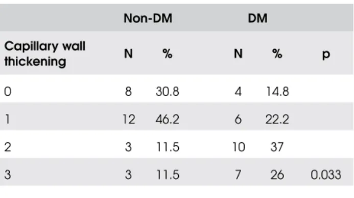
Diabetic patients showed a significantly greater occurrence (p=0.03) of moderate and intense PAS-positive capillary thickening than nondiabetic pa-tients (Table 4). Other variables did not show statistic correlation with capillary thickening: 28% of hyper-tensive patients had moderate and 18% had intense capillary wall thickening, contrasting with 14% and 21% of patients with normal blood pressure (p=0,4). 21% of smokers had moderate and 21% had intense capillary wall thickening, contrasting with 28% and 17% of non-smokers (p=0,9). 25% of women and 24% of men had moderate, and 29% of women and 10% of men had intense capillary wall thickening (p=0,2). All women were post-menopausal and none used hormonal replacement therapy. Logistic regres-sion showed that only DM was significantly associa-ted with moderate or severe capillary wall thickening (p=0.005), whereas no association was found on the other variables (sex, smoking, segment affected by PAOD, kidney disease requiring dialysis, and hyper-tension). Patients with diabetes had a 5.7 (odds ra-tio) greater chance (95% CI: 1.7-18.8) of having moderate or severe PAS-positive capillary thickening than patients without diabetes.
Table 4. Comparative analysis of PAS-positive thickening of capillary walls in patients with and without DM.
Non-DM DM
Capillary wall
thickening N % N % p
0 8 30.8 4 14.8
1 12 46.2 6 22.2
2 3 11.5 10 37
3 3 11.5 7 26 0.033
DM=diabetes mellitus.
DISCUSSION
The mean age of our patients was over 60 years and did not differ between groups. This is in agreement with the literature and with previous findings by our group (11-17). We believe that, even though PAOD occurs sooner in diabetic patients than in patients without dia-betes (6,7,18), the disease in advanced form, which re-sults in critical ischemia and greater rates of amputation, occurs at a later age after a long period of disease, as in nondiabetic patients.
Women were statistically more prevalent in the group of diabetic patients. Other authors also found a larger number of women among their diabetic patients with critical ischemia who underwent amputations (19-21). This finding may be associated with the reported in coronary atherosclerotic disease studies, which have shown that the protective effect of the female sex is eliminated by diabetes, and that diabetes doubles the risk of cardiovascular disease in men and triples it in women (22,23).
The arterial segment affected by PAOD diagnosed by pulses physical examination showed that most pa-tients in both groups had femoral-popliteal disease. DM patients had significantly less aortoiliac disease and a greater incidence of impairment of the below-knee segment, which is in agreement with findings reported in the literature (3,7,24,25).
ar-cop
yr
ight
© ABE&M todos os dir
eitos r
eser
v
ados
terioles and capillaries of diabetic patients, called “small vessel disease”. However they used a heterogeneous nondiabetic control group with patients amputated for different diseases including trauma, which brought up the question if those capillary thickening they found could be secondary only to severe atherosclerotic macro-vascular disease in leg arteries of diabetic patients.
Nevertheless, in our sample, no histological diffe-rences were found between diabetic and nondiabetic patients in percentage of obstruction of the below-knee arteries (popliteal, anterior tibial, tibial-fibular trunk, posterior tibial, or peroneal), which is in agreement with findings by Conrad (26), and Ferrier (27), but differs from those reported by Strandness e cols. (9), who found a greater percentage of trifurcation disease in the legs of diabetic patients (9). Arteriographic study also showed greater prevalence of infrapopliteal disease in diabetic patients (4).
In our study the most prevalent degree of obstruc-tion was greater than 75% in infra-popliteal arteries stu-died in diabetic and nondiabetic patients, which showed that advanced PAOD and ischemia were present in all patients of our sample. However, the arteries were not examined along the entire leg but only close to their origin, which is the segment where atherosclerotic le-sions tend to be more severe (close to the bifurcations). The arteriogram - not analized in this work - provided a guide to the sites of obstruction but do not show the characteristics of the atherosclerotic plaque surface accu-rately, and may also fail to detect plaques, because of the technique used for examination or due to the presence of lesions that do not cause significant stenosis (10).
The histological classification of the atherosclerotic lesions also demonstrated the severity of peripheral atherosclerotic disease in our patients. We used the his-tological classification of advanced atherosclerotic le-sions proposed by Stary e cols. (10) in 1995 to classify the severity of the disease. Types IV, V, and VI, the predominant types in our two groups, are advanced le-sions that occlude the arterial lumen and cause symp-toms, but may fail to be detected by angiography. However, type VI atherosclerotic plaques or complica-ted lesions are associacomplica-ted with symptoms of arterial obstruction, fissures, ulceration, hemorrhage and grea-ter thrombosis. Our diabetic and nondiabetic patients had a predominance of type VI atherosclerotic lesions in all five types of arteries studied (popliteal and trifur-cation of the leg). We did not find this comparison in other studies.
The calcification of the medial layer or Monckeberg’s sclerosis, a characteristic of diabetes, was more preva-lent in patients with DM as previously reported, but was not exclusive of this group (3,27). This difference was statistically significant in the comparisons of the popliteal and posterior tibial arteries, for which we had a larger sample. However, the examination of the ante-rior tibial and the tibial-fibular trunk also showed that the calcification of the medial layer was more prevalent among diabetic patients, although this difference was not statistically significant.
A statistically greater prevalence of thickening of the vasa-vasorum in the arterial adventitia in the DM groups was found only in the popliteal artery, in agreement with findings by Goldenberg e cols. (8). This may be due to the fact that the popliteal artery was the largest sample in our study. The percentage of patients without vasa-vaso-rum thickening of the posterior tibial, anterior tibial and peroneal arteries was greater than the percentage of pa-tients with thickening, even in the DM group. Therefo-re, we believe that there is no specific diabetic pattern of vasa-vasorum thickening in adventicia.
cop
yr
ight
© ABE&M todos os dir
eitos r
eser
v
ados
Results of logistic regression showed that only DM was significantly associated with moderate or severe ca-pillary thickening and ruled out hypertension, sex, seg-ment affected by PAOD and chronic kidney disease as confounding variables. In agreement with other stu-dies, we found no correlation between hypertension and capillary wall thickening (8,26,36). Hypertension, however, is frequently associated with microvascular complications of diabetes, such as retinopathy and ne-phropathy (37), which raises the hypothesis that it may be also associated with DM in the progression of peri-pheral microangiopathy. Analyzing only the nondiabe-tic group, 13% of the hypertensive patients had intense capillary thickening, while 9% of the normal blood pressure patients had intense capillary wall thickening, a difference without statistic significance. Three diabe-tic patients with normal levels of blood pressure had capillary wall thickening: two in intense category and one in moderate, confirming the role of diabetes in PAS positive capillary wall thickening.
Our study, however, differs from previous investi-gations because all patients have severe ischemia and histological qualitative study of severity of PAOD, it included a larger number of patients and obtained all samples of skin capillaries from amputations performed at the same level (above-knee). Our findings confirmed the presence of peripheral diabetic microangiopathy, but we may not forget that capillary thickening has been observed in some nondiabetic patients too. Aage-naes and Moe (36) and Banson and Lacy (28) showed that capillary thickening was not uniform in distribu-tion, and found capillaries with and without wall thi-ckening in the same samples. We included all capillaries of the sample in our capillary thickening classification (absent, mild, moderate or intense).
Although we found that there is a pattern of ca-pillary changes more frequent in diabetic than in non-diabetic patients, it does not justify maintaining an ischemic limb when there is the possibility of revascula-rization, but this may be the explanation for the unfa-vorable evolution of some patients. Other studies in literature have already demonstrated that, although the opportunity of undergoing arterial reconstruction is lo-wer in diabetic patients, the results of infrainguinal re-vascularization are similar for diabetic individuals and other patients with PAOD (3,38). Further studies are needed before diabetic peripheral microangiopathy can be totally understood.
Based on the results taken, we conclude that there are no differences in the degree of stenosis or
histologi-cal classification of atherosclerotic lesions in below-knee arteries between diabetic and nondiabetic patients with advanced PAOD and critical ischemia. Diabetic patients show a PAS-positive capillary thickening that is more intense and diffuse than in nondiabetic patients.
Acknowledgment. We are grateful to the Support Center for Scientific Publications of Santa Casa de São Paulo – Faculty of Medical Sciences for the editorial assistance. No potencial con-flict of interest relevant to this article was reported.
REFERENCES
Slovenkai MP. Foot problems in diabetes. Med Clin North 1.
Am. 1998;82:9948-70.
Most RS, Sinnock F. The epidemiology of lower extremity 2.
amputations in diabetic individuals. Diabetes Care. 1983; 6:87-91.
Mueller MP, Wright J, Klein SR. Diabetes and peripheral vas-3.
cular disease. In: Veith FJ, Hobson RW, Williams RA, Wilson S, editors. Vascular surgery: principles and practice. 2. ed. New York: McGraw Hill; 1994. p. 514-22.
Gensler SW, Haimovici H, Hoffert P, Steinman C, Beneventa-4.
no TC. Study of vascular lesions in diabetic, nondiabetic pa-tients. Arch Surg. 1965;91:617-22.
Colwell JA, Lopes-Virella M, Halushka PV. Pathogenesis of 5.
atherosclerosis in diabetes mellitus. Diabetes Care. 1981; 4:121-33.
Drouet L. Atherothrombosis in diabetes – its evolution and 6.
management. Diabetes Obes Metab. 1999;1:37-47.
Lo Gerfo FW, Coffman JD. Vascular and microvascular disea-7.
se of the foot in diabetes: implications for foot care. N Engl J Med. 1984;311:1615-9.
Goldenberg S, Alex M, Joshi RA, Blumenthal HT. Nonathero-8.
matous peripheral vascular disease of the lower extremity in diabetes mellitus. Diabetes. 1959;8:261-73.
Strandness Jr. DE, Priest RE, Gibbons GE. Combined clinical 9.
and pathologic study of diabetic and nondiabetic peripheral arterial disease. Diabetes. 1964;13:366-72.
Stary HC, Chandler AB, Dinsmore RE, Fuster V, Glagov S, In-10.
sull Jr W, et al. A definition of advanced types of atheroscle-rotic lesions and a histological classification of atherosclerosis. A report from the Committee on Vascular Lesions of the Council on Arteriosclerosis, American Heart Association. Cir-culation. 1995;92:1355-74.
Loue TJ, Bartlett JG, Tally FP, Gorbach SL. Aerobic and anae-11.
robic bacteria in diabetic foot ulcers. Ann Intern Med. 1976;85:461-3.
Sharp CS, Bessman AN, Wagner Jr FW, Garland D, Reece E. 12.
Microbiology of superficial and deep tissues in infected dia-betic gangrene. Surg Gynecol Obstet. 1979;149:217-9. Nicholas GG, Myers JL, Demuth WE. The role of vascular la-13.
boratory criteria in the selection of patients for lower extre-mity amputation. Ann Surg. 1982;195:469-73.
Pittet D, Wyssa B, Herter-Clavel C, Kursteiner K, Vaucher J, 14.
Lew PD. Outcome of diabetic foot infections treated conser-vatively. Arch Intern Med. 1999;159:851-6.
Gamba MA, Oliveira O, Fadlo Filho F, Martinez C, Kajita MY. A 15.
vascula-cop
yr
ight
© ABE&M todos os dir
eitos r
eser
v
ados
res de extremidades inferiores de pessoas com diagnóstico de diabetes mellitus – campanha da detecção e educação da ANAD. Diabetes Clinica. 2001;6:414-8.
Adler AI, Stevens RJ, Neil A, Stratton IM, Boulton AJM, Hol-16.
man RR. UKPDS 59: hyperglycemia and other potentially mo-difiable risk factors for peripheral vascular disease in type 2 diabetes. Diabetes Care. 2002;25:894-9.
Santos VP, Silveira DR, Caffaro RA. Risk factors for primary 17.
major amputation in diabetic patients. Sao Paulo Med J. 2006;124:66-70.
Pratt TC. Gangrene and infection in the diabetics. Med Clin 18.
North Am. 1965;49:987-1004.
Cameron HC, Leonard J, Robinson MP. Amputations in the 19.
diabetic: outcome and survival. Lancet. 1964;2:605-7. Lange E. Current surgery: drug combination treatment of dia-20.
betic gangrene of the foot. Infection. 1991;19:S351-4. Lira JRS, Castro AA, Pitta GBB, Figueiredo LFP, Lage VMM, 21.
Miranda Jr F. Prevalence of sensorimotor polyneuropathy in the feet at the moment of diabetes mellitus diagnosis. J Vasc Bras. 2005;4:22-6.
Nathan DM. Long-term complications of diabetes mellitus. N 22.
Engl J Med. 1993;328:1676-84.
Kannel WB, Daniel LM. Diabetes and cardiovascular disease: 23.
the Framinghan Study. JAMA. 1979;241:2035-8.
Caputo GM, Cavanagh PR, Ulbrecht JS, Gibbons GW, Karch-24.
mer AW. Assessment and management of foot disease in patients with diabetes. N Engl J Med 1994;331:854-60. Faglia E, Favales F, Quarantiello A, Calia P, Clelia P, Brambilla 25.
G, et al. Angiographic evaluation of peripheral arterial occlu-sive disease and its role as a prognostic determinant for ma-jor amputation in diabetic subjects with foot ulcers. Diabetes Care. 1998;21:625-30.
Conrad MC. Large and small artery occlusion in diabetics and 26.
nondiabetics with severe vascular disease. Circulation. 1967;36:83-91.
Ferrier TM. Comparative study of arterial disease in amputa-27.
ted lower limbs from diabetics and non-diabetics (with spe-cial reference to feet arteries). Med J Aust. 1967;1:5-11.
Banson BB, Lacy PE. Diabetic microangiopathy in human 28.
toes – with emphasis on the ultrastructural change in dermal capillaries. Am J Pathol. 1964;45:41-58.
McMillan DE. Deterioration of the microcirculation in diabe-29.
tes. Diabetes. 1975;24:944-57.
Raskin P, Pietri AO, Unger R, Shannon A. The effect of diabe-30.
tic control on the width of skeletal-muscle capillary basement membrane in patients with type I diabetes mellitus. N Engl J Med. 1983;25:1546-50.
Camerini-Davalos RA, Velasco C, Glasser M, Bloodworth Jr 31.
JMB. Drug-induced reversal of early diabetic microangiopa-thy. N Engl J Med. 1983;25:1551-6.
Tilton RG, Faller AM, Burkhardt JK, Hoffmann PL, Kilo C, 32.
Williamson JR. Pericyte degeneration and acellular capilla-ries are increased in the feet of human diabetic patients. Dia-betologia. 1985;28:895-900.
Zatz R, Brenner BM. Pathogenesis of diabetic microangiopa-33.
thy – the haemodynamic view. Am J Med. 1986;80:443-53. Dinh TL, Veves A. Microcirculation in the diabetic foot: an 34.
update. Int J Low Extrem Wounds. 2004;3:60-1.
Friederici HHR, Tucker R, Schwartz TB. Observations on small 35.
blood vessels of skin in the normal and diabetic patients. Dia-betes. 1960;15:233-50.
Aagenaes O, Moe H. Light and electron microscopic study of 36.
skin capillaries of diabetics. Diabetes 1961;10:253-9.
Neely KA, Quillen DA, Schachat AP, Gardner TW, Blankenship 37.
GW. Retinopatia diabética. Clin Med Am Norte. 1998;4:195-226. Fratezi AC, Albers M, De Luccia ND, Pereira CA. Outcome and 38.
quality of life of patients with severe chronic limb ischaemia: a cohort study on the influence of diabetes. Eur J Vasc Endo-vasc Surg. 1995;10:459-65.
Correspondence to: