cop
yr
ight
© ABE&M todos os dir
eitos r
eser
v
ados
apresentação de caso
JOSÉ A. M. MARCONDES DANIELA D. G. CURI CEZAR N. MATSUZAKI CRISTIANO R. G. BARCELLOS MICHELLE P. ROCHA SYLVIA A. Y. HAYASHIDA EDMUND C. BARACAT
Endocrine Unit (JAMM, CRGB, MPR); Gynecological Unit of the Hospital das Clínicas de São Paulo (DDGC, SAYH, ECB); Gynecological Unit of Hospital Nipo-Brasileiro de São Paulo (CNM), São Paulo, SP, Brasil.
Recebido em 1/2/2008 Aceito em 19/8/2008
ABSTRACT
Adrenal incidentaloma is not infrequent and can be found in hirsute women. We report a case of a 54-year-old woman with amenorrhea and hirsutism of abrupt onset and mild signs of virilization that had an adrenal incidentaloma coexisting with ovarian hyperthecosis. Basal total and free testosterone were 191 ng/dL and 179 pmol/L. Pelvic ultrasonography disclosed a right ovary with 10.3 cc and a left ovary with 9.8 cc without nodules or cysts, and computerized tomography of the abdomen disclosed a normal right adrenal gland. On the left adrenal gland a solid nodule with 0.8 cm was seen. After GnRHa adminis-tration, total testosterone was 23 ng/dL and free testosterone was 17 pmol/L. In view of a suppression of testosterone by GnRHa, the patient was submitted to a hystero-oophorectomy by laparoscopy. Symmetrically enlarged ovaries were seen. No tumor was apparent. Histology showed hyperthecosis, with foci of luteinized stromal cells. Only atretic follicles were detected. No hilar cell hyperplasia was seen. In conclusion, the presence of an adrenal mass in a hirsute woman can lead to a wrong diagnosis. In this case the suppression GnRHa test was fundamental to determine the origin of hyperandrogenemia.
(Arq Bras Endocrinol Metab 2008; 52/7:1184-1188)
Keywords: Hirsutism; Ovarian hyperthecosis; Adrenal incidentaloma; Menopause
RESUMO
Coexistência de Incidentaloma Adrenal e Hipertecose de Ovário em Mulher Menopausada.
Os incidentalomas adrenais não são infreqüentes e podem ser encontrados em pacientes com hirsutismo. Nesse relato, apresentamos o caso de coexis-tência de um incidentaloma adrenal com hipertecose de ovário, em uma mu- lher com 54 anos de idade com amenorréia e hirsutismo de início abrupto e sinais leves de virilização. As testosteronas total e livre basal foram de 191 ng/dL e 179 pmol/L, respectivamente. O ultra-som pélvico demonstrou o ovário direito com 10,3 cc e ovário esquerdo com 9,8 cc, sem nódulos ou cistos e a tomografia computadorizada de abdome demonstrou adrenal di- reita adrenal e nódulo sólido de 0,8 cm na adrenal esquerda. Após a adminis-tração de análogo de GnRH, as testosteronas total e livre foram de 23 ng/dL e 17 pmol/L, respectivamente. Considerando a supressão da concentração de testosterona pelo análogo de GnRH, a paciente foi submetida a histero- ooforectomia por via laparoscópica. O diagnóstico histológico foi de hiperte-cose, com focos de células estromais luteinizadas. Somente folículos atréticos foram visualizados. Não se detectou hiperplasia de células hilares. Em con-clusão, a presença de massa adrenal em uma paciente com hirsutismo pode levar ao diagnóstico errado. Neste caso, o teste de supressão com análogo de GnRH foi fundamental para se determinar a origem da hiperandrogenemia.
(Arq Bras Endocrinol Metab 2008; 52/7:1184-1188)
cop
yr
ight
© ABE&M todos os dir
eitos r
eser
v
ados
H
irsutism can be defined as excessive growth of androgen-dependent sexual hair. It is one of the manifestations of hyperandrogenic syndrome, a state of increase androgen production (1). It can be isolated or associated with other signs of increase in androgen le-vel, as menstrual disturbances, acne, deepening of the voice, and increase in muscle mass, clitoromegaly, bre-ast hypotrophy and frontoparietal balding.The mainly causes of hirsutism in young women, polycystic ovary syndrome and idiopathic hirsutism, are not prevalent after the menopause. Apart from iatroge-nia, hirsutism in menopause women can be arising out of nonclassical congenital adrenal hyperplasia, ovarian hyperthecosis or ovarian or adrenal tumors. This diffe-rential diagnosis is important, since adrenal and ovarian tumors can be life-threatening conditions and deserve surgical treatment. Hyperthecosis is characterized by hyperplasia and luteinization of the cortical stroma, and can occur in both premenopausal women, as a part of the HAIR-AN syndrome, and after menopause (2,3). While in the first, basic physiological mechanism is severe insulin resistance, in the second it is due to the continuous stimuli of high gonadotropins.
The following is a case report of a hyperandrogenic woman in menopause, where the finding of an adrenal adenoma could lead to a misleading treatment.
Case report
A 54-year-old woman had a 4-year history of excessi-ve hair growth, hair loss and acne. She had had nor-mal menstrual cycle since menarche and became amenorrheic with hot flushes and increase in libido 4 years ago. She denied any changes in her voice and muscle mass. She had two normal pregnancies pre-viously. A thyroidectomy was performed for papillary thyroid carcinoma at the ‘Hospital Nipo-Brasileiro’, in 2002. Her family history was unremarkable. At presentation she was under treatment with l -thyroxi-ne sodium.
Physical examination revealed a grade I obese wo-man (body mass index of 30.4 kg/m2), with male
pat-tern baldness, acne in her face, parietal temporal baldness and marked hirsutism with terminal hair on face, chest, abdomen and extremities (grade 15 of the Ferriman & Gallwey score, normal < 9) (4). Her voice was feminine and her muscle mass was slightly enlar-ged. Blood pressure was 150 × 98 mmHg and heart rate was 80/min. There were no striae or bruises. No acanthosis nigricans was noted. Breasts were atrophic,
without masses or galactorrhea. Abdomen was normal. Pelvic examination revealed clitoromegaly. No anexial mass was palpable.
The initial laboratory investigation revealed normal blood count and liver and renal function were within normal range.
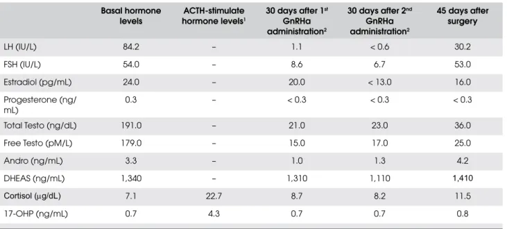
Measurements of circulating hormone levels un-der basal and dynamic condition are shown in Table 1. All studies were conducted between 7:30 and 8:30 a.m. after 8 hours of fasting. For the ACTH stimula-tion test, blood was collected for cortisol and 17-hydroxyprogesterone (17-OHP) determination 60 minutes after the administration of 0.25 mg of ACTH-(1-24) (Cortrosyn™, Organon, USA) by IV route over 60 seconds. The GnRHa suppression test was performed though the administration of 3.75 mg of leuprolide acetate (lupron® depot) by IM route
every 30 days, for 2 months, and blood collect after 30 days of each administration for determination of gonadotropins (LH and FSH), estradiol, cortisol, DHEAS and total and free testosterone.
Pelvic ultrasonography disclosed a right ovary with 10.3 cc and a left ovary with 9.8 cc, without nodules or cysts. Computerized tomography of the abdomen dis-closed a normal right adrenal gland. On the left adrenal gland, a solid nodule with 0.8 cm was seen.
Cushing syndrome was ruled out by an overnight dexamethasone suppression test (1 mg – cortisol < 1.0 µg/dL), primary hyperaldosteronism by the me-asurement of serum potassium (4.1 mEq/L), aldos-terone (3.1 ng/dL) and plasma renin activity (1.1 ng/mL/h) and pheochromocytoma by the measure-ments of urinary catecholamines (norepinephrine = 31 µg/24 h, epinephrine = 3 µg/24 h and dopamine = 185 µg/24 h) and metanephrine (0.22 metane-phrine µg/ mL of creatinine).
Patient was submitted to a hystero-oophorec-tomy by laparoscopy. Symmetrically enlarged ova-ries, with 3.0 cm and 10.0 g (right ovary) and 2.5 cm and 8.0 g (left ovary), with dense capsules were seen. No tumor was apparent. Histology showed hyperthecosis with foci of luteinized stromal cells. Only atretic follicles were detected. No hilar cell hyperplasia was seen.
cop
yr
ight
© ABE&M todos os dir
eitos r
eser
v
ados
On the other hand, male pattern baldness and hirsutism were not reduced.
DISCUSSION
The patient under discussion is a 54-year-old woman with a history of hyperandrogenic symptoms of a 4 year duration, coincident with the menopause. Relevant po-sitive physical findings included hirsutism, acne, fron-toparietal baldness and clitoromegaly. According to these signals, the patient was classified as a hyperandro-genic virilized woman. As expected, total and free tes-tosterone were markedly elevated at 191 ng/dL (normal value < 98 ng/dL) and 179 pmol/L (normal value < 45 pmol/L), respectively.
Considering the virilizing syndrome, the main hy-pothesis for this patient was adrenal or ovary virilizing tumor or hyperthecosis of the ovary. Although a small number of patients with polycystic ovary syndrome could have signals and symptoms of virilization, these, as well as idiopathic hirsutism, are diseases of preme-nopausal women. On the other hand, although the non-classical form of congenital adrenal hyperplasia due to 21-hydroxilase deficiency was rarely report in patients in menopause, this diagnosis was discarded through the appropriate test (ACTH stimulated 17-OHP < 17 ng/mL) (5).
Computerized tomography of the abdomen disclo-sed a solid nodule with 0.8 cm on the left adrenal gland, compatible with an adrenal tumor. These, mostly be-nign adenomas, are frequently in the general popula-tion and nowadays more often found incidentally (6), while adrenocortical cancer is rare, with an estimated prevalence between 0.6 to 2 cases per millionin the normal population (7). Adrenal tumors can be hormo-nally silent or hormone-secreting. Most of the adrenal tumors are benign and hormonally silent. Virilization secondary to hypersecretion of adrenal androgens oc-curs in less than 20% of adults with functional adrenal neoplasms in our institution (8). The others syndrome associated with adrenal tumors, as Cushing’s syndro-me, primary hyperaldosteronism and pheochromocyto-ma were discharged in this patient by the clinical pictures and the appropriate laboratory tests.
On the other hand, pelvic ultrasonography in this patient disclosed bilateral enlarged ovaries, wi-thout nodules or cysts. Although bilateral virilizing ovarian tumors have been described (9), the most common pathology associated with this finding is hyperthecosis of the ovary (10), a nonmalignant ova-rian disorder characterized by increased production of testosterone by luteinized theca cells in the stro-ma, due to the differentiation of the ovarian intersti-tial cells into steroidogenically active luteinized Table 1. Hormone levels under basal and dynamic condition and after surgery.
Basal hormone levels
ACTH-stimulate hormone levels1
30 days after 1st
GnRHa administration2
30 days after 2nd
GnRHa administration2
45 days after surgery
LH (IU/L) 84.2 – 1.1 < 0.6 30.2
FSH (IU/L) 54.0 – 8.6 6.7 53.0
Estradiol (pg/mL) 24.0 – 20.0 < 13.0 16.0
Progesterone (ng/ mL)
0.3 – < 0.3 < 0.3 < 0.3
Total Testo (ng/dL) 191.0 – 21.0 23.0 36.0
Free Testo (pM/L) 179.0 – 15.0 17.0 25.0
Andro (ng/mL) 3.3 – 1.0 1.3 4.2
DHEAS (ng/mL) 1,340 – 1,310 1,110 1,410
Cortisol (µg/dL) 7.1 22.7 8.7 8.2 11.5
17-OHP (ng/mL) 0.7 4.3 0.7 0.7 0.8
cop
yr
ight
© ABE&M todos os dir
eitos r
eser
v
ados
stromal cells (11), leading to increased serum testos-terone concentrations.
Hyperthecosis can occur during the reproductive years, usually associated with acanthosis nigricans and in the postmenopausal period, with different patho-physiological mechanism. In some patients with polycystic ovary syndrome, luteinized theca cells can be found confined to areas around cystic follicles and it is still unclear if hyperthecosis is a distinct disorder or is part of the spectrum of the polycystic ovary syn-drome. While in young women the precipitating ab-normality is thought to be insulin resistance (2), with a secondary increase in insulin levels and subsequent overproduction of androgens by the ovaries, in post-menopausal patients it is a gonadotropin dependent hyperandrogenic syndrome not related to insulin re-sistance, possibly due to continuous stimuli of luteini-zing hormone (11). The hallmark of this syndrome is an increase in bilateral ovarian volume, although uni-lateral increase in ovarian volume has been described (12). Although specificity of the ultrasound has not been determined, the ovaries appear more solid with very few or no cysts.
In view of the above finding, we considered that this patient had a nonfunctional adrenal adenoma coe-xisting with an ovary producing androgen pathology, most possibly ovarian hyperthecosis. We choose to submit this patient to a gonadotropin analogue (Gn-RHa) suppression test, with the purpose to separate a gonadotropin-dependent from a gonadotropin inde-pendent hyperandrogenemia. Usually, we administer GnRHa until complete LH suppression (< 0.6 IU/ mL), up to 3 doses. Although there are anecdotal re-ports of gonadotropin dependent adrenal tumors (13), the suppression of testosterone level by GnRHa could point to ovarian-secreting testosterone pathology, al-though we cannot separate ovarian hyperthecosis from virilizing ovarian tumor, as some are gonadotropin-dependent (14). Only a few women with hypertheco-sis have been treated with a GnRH agonist, but all had a substantial decrease in serum androgen concentra-tions (15-18). These results indicate that the ovarian hyperandrogenism in these women is at least partly gonadotropin-dependent.
In view of the finding of a suppressed testosterone after GnRHa administration we indicated bilateral oo-phorectomy, considering the menopausal status of the patient. Another approach could be a bilateral and
si-multaneous catheterism of the adrenal and ovary, but this is an invasive procedure with sometime misleading results (19). The histopathological diagnosis was com-patible with ovarian hyperthecosis.
This case illustrated the difficulty related to diffe-rential diagnosis in virilizing hyperandrogenic syndro-mes due to the high prevalence of nonfunctional adrenal adenoma in the population. The suppression GnRHa test was fundamental to determine the cause of hype-randrogenemia.
No potencial conflict of interest relevant to this article was reported.
REFERENCES
Rosenfield RL. Pilosebaceous physiology in relation to hirsu-1.
tism and acne. J Clin Endocrinol Metab. 1986;15:341-62. Khan CR, Flier JS, Bas RS, Archer JA, Gorden P, Martin MM, 2.
et al. The syndromes of insulin resistance and acanthosis ni-gricans. N Engl J Med. 1976;294:739-45.
Goldman JM, Kapadia JL. Virilization in a postmenopausal 3.
woman due to ovarian stromal hyperthecosis. Postgrad Med J. 1991;67:304-9.
Ferriman D, Gallwey JD. Clinical assessment of body hair 4.
growth in women. J Clin Endocrinol Metab. 1961;21:1440-7. Bachega TA, Billerbeck AE, Madureira G, Marcondes JA, Lon-5.
gui CA, Leite MV, et al. Molecular genotyping in Brazilian pa-tients with the classical and nonclassical forms of 21-hydroxylase deficiency. J Clin Endocrinol Metab. 1998;83:4416-9.
Mansmann G, Lau J, Balk E, Rothberg M, Miyachi Y, Borns-6.
tein SR. The clinically inapparent adrenal mass: update in diagnosis and management. Endocr Rev. 2004;25:309-40. Latronico AC, Chrousos GP. Extensive personal experien-7.
ce: adrenocortical tumors. J Clin Endocrinol Metab. 1997;82:1317-24.
Wajchenberg BL, Albergaria MAP, Medonca BB, Latronico 8.
AC, Campos Carneiro P, Alves VA, et al. Adrenocortical carci-noma: clinical and laboratory observations. Cancer. 2000;88:711-36.
Dicker D, Dekel A, Feldberg D, Goldman JA, Kessler E. Bilate-9.
ral sertoli-leydig cell tumor with heterologous elements: re-port of an unusual case and review of the literature. Eur J Obstet Gynecol Reprod Biol. 1986;22:175-81.
Henriksen HM, Fischer S, Guttorm E. Bilateral ovarian lipid 10.
cell hyperplasia in a young hirsute patient. Int J Gynaecol Obstet. 1981:19467-72.
Krug E, Berga SL. Postmenopausal hyperthecosis: functional 11.
dysregulation of androgenesis in climacteric ovary. Obstet Gynecol. 2002;99:893-7
Farber M, Madanes A, O’Briain DS, Millan VG, Turksoy RN, 12.
Rule AH. Asymmetric hyperthecosis ovari. Obstet Gynecol. 1981;57:521-5.
Werk Jr EE, Sholiton LE, Kalejs L. Testosterone-secreting 13.
cop
yr
ight
© ABE&M todos os dir
eitos r
eser
v
ados
Marcondes JA, Nery M, Mendonca BB, Hayashida SA, Halbe 14.
HW, Carvalho FM, et al. A virilizing Leydig cell tumor of the ovary associated with stromal hyperplasia under gonadotro-pin control. J Endocrinol Invest. 1997;20:685-9.
Barth JH, Jenkins M, Belchetz PE. Ovarian hyperthecosis, dia-15.
betes, and hirsutism in post-menopausal women. Clin Endo-crinol (Oxf). 1997;46:123-8.
Steingold KA, Judd HL, Nieberg RK, Lu JK, Chang RJ. Treat-16.
ment of severe androgen excess due to ovarian hypertheco-sis with a long-acting gonadotropin-releasing hormone agonist. Am J Obstet Gynecol. 1986;154:1241-8.
Parr JH, Abraham RR, Seed M, Short F, Wynn V. The treat-17.
ment of a hyperandrogenic and virilizing state in an elderly female with a synthetic LH RH agonist. J Endocrinol Invest. 1988;11:433-6.
Pascale MM, Pugeat M, Roberts M, Rousset H, Déchaud H, 18.
Dutrieux-Berger N, et al. Androgen suppressive effect of GnRH agonist in ovarian hyperthecosis and virilizing tumors. Clin Endocrinol (Oxf). 1994;41:571-6.
Kaltsas GA, Mukherjee JJ, Kola B, Isidori AM, Hanson JA, 19.
Dacie JE, et al. Is ovarian and adrenal venous catheterization and sampling helpful in the investigation of hyperandrogenic women? Clin Endocrinol (Oxf). 2003;59:34-43.
Correspondence to:
José Antonio Miguel Marcondes Rua Baronesa de Itu, 821, apto. 112 01231-001 São Paulo SP