©Revista Brasileira de Fisioterapia
STUDY OF HEART RATE AUTONOMIC MODULATION AT REST IN
ELDERLY PATIENTS WITH CHRONIC OBSTRUCTIVE PULMONARY
DISEASE
P
ANTONICBF
1, R
EISMS
1, M
ARTINSLEB
2, C
ATAIAM
1, C
OSTAD
1,3& B
ORGHI-S
ILVAA
11 Departament of Physical Therapy, Federal University of São Carlos - UFSCar, São Carlos, SP - Brazil
2 School of Physical Education, State University of Campinas, Campinas, SP - Brazil
3 School of Health Sciences, Piracicaba Methodist Univestity, Piracicaba, SP, Brazil
Correspondence to: Profa Dra Audrey Borghi Silva, Núcleo de Pesquisa em Exercício Físico, DeFisio, UFSCar, Rodovia
Washington Luis, km 235, CEP 13565-905, São Carlos, SP – Brazil, e-mail: [email protected]
Received: 14/02/2006 - Revised: 26/07/2006 - Accepted: 28/09/2006
ABSTRACT
Objective: To evaluate heart rate variability (HRV) among elderly patients with chronic obstructive pulmonary disease (COPD) and healthy elderly individuals, during postural change. Method: Nine individuals with COPD (70 years old) and eight healthy individuals (68 years old) were studied. Heart rate and electrocardiographic R-R intervals (iR-R) were recorded for 360 seconds in the supine and seated positions. HRV was analyzed in the time domain (TD) (RMSSD index, i.e. the root mean square of the squares of the differences between successive iR-R records, and the SDNN index, i.e. the mean standard deviation of normal iR-R in ms) and in the frequency domain (FD), from the low-frequency (LF) and high-frequency (HF) bands in absolute units (au) and normalized units (nu), and the LF/HF ratio. The Mann-Whitney and Wilcoxon Tests respectively were utilized for inter-group and intra-inter-group analysis, with a significant level of p< 0.05 (median values). Results: In TD, the control inter-group (CG) presented significantly higher values for the RMSSD index (14.6 versus 8.3 ms) and the SDNN index (23 versus 13.5 ms) in the seated position, in comparison with the COPD group (DG). In FD, the CG presented significantly higher values for HF components, in the supine position (39 versus 7.8 au), and for LF components (146.7 versus 24.4 au) and HF (67.6 versus
22.7 au), in the seated position, as well as for the total power spectrum (552.5 versus 182.9 ms2). Conclusion: Patients with COPD
presented reduced HRV with decreased sympathetic and vagal activity. Additionally, neither the COPD patients nor the healthy elderly participants presented autonomic alterations with postural change.
Key words: heart rate variability, chronic obstructive pulmonary disease, autonomic nervous system, resting condition.
INTRODUCTION
Chronic Obstructive Pulmonary Disease (COPD) is characterized by airflow limitation, not totally reversible. This limitation is usually progressive and associated to abnormal pulmonary inflammatory responses to particles or noxious gases1.
The COPD leads to important ventilatory limitations due to pulmonary dead space increase and gas exchange decrease2,3. Additionally, heart dysfunction may be present
due to right ventricle after-load increase imposed by the high pulmonary vascular resistance, with vascular damages and hypoxic constriction4. These changes cause the appearance
of the cor pulmonale, which may lead to right heart failure4.
Besides the changes of the cardiovascular system in COPD patients, autonomic alterations have also been described, evaluated by heart rate variability (HRV) of such patients5.
HRV represents the variations of R-R intervals (R-Ri) duration of the electrocardiogram (ECG), which depends on the sympathetic and parasympathetic nervous system6. This
method consists on a non-invasive autonomic assessment and its analysis can be performed either in time domain (TD) or in frequency domain (FD).
In the TD, statistical methods are used to quantify the variation of the standard deviation or the differences between successive R-Ri 7,8. The FD analysis decomposes the variability
in high frequency (HF), low frequency (LF) and very low frequencies bands (VLF)6,8.
The VLF components, with frequencies lower than 0.04 Hz, don´t have a defined physiological explanation and are related to renin-angiotensin-aldosterone system and thermoregulation8,9,10. The LF, between 0.04 and 0.15 Hz,
0.15 and 0.40 Hz, corresponds to the respiratory modulation and is mediated only by the parasympathetic nervous system8,9,11,12.
Some authors have ascribed that HRV is altered in
COPD13,14,15. The abnormal autonomic heart rate adjustments,
reflected by HRV alterations, may be related with the severity of disease. Heart autonomic function disorders may result in the appearance of arrhythmias in these patients16.
At rest condition, Volterrani et al.13 have observed that
COPD patients have autonomic nervous system function abnormalities, with HRV decrease on responses to vagal and sympathetic stimulus. Similarly, Paschoal, Petrelluzzi & Gonçalves17 observed HRV reduction in COPD patients. On
the other hand, Scalvini et al.18 observed that only COPD
patients with severe hypoxemia have abnormal autonomic nervous system (ANS) behavior, characterized by HRV decrease.
During the passive head-up tilt maneuver, it has been demonstrated that the sympathetic response becomes altered in COPD 13. However, regarding to active postural change,
it´s not known about the heart rate (HR) autonomic responses in COPD patients. Thus, the objectives of this study were to verify, by means of HRV, if elderly COPD patients present HR autonomic modulation damages in supine and seated position, as well as to compare these responses with healthy elderly subjects gender and age-matched.
METHODS
Subjects
A doctor referred to the study 53 patients who had previously been clinically diagnosed with COPD, with forced expiratory volume on the first second (FEV1) < 80% of the predicted and FEV1/ forced vital capacity (FVC) ratio < 70% of the predicted1, absence of reversibility after
post-bronchodilator spirometry test and presence of clinical stability. The subjects had clinical diagnosis for more than 5 years, were former smokers and had not been practicing regular physical activity at least for 6 months ago. In addition, healthy subjects also participated of this study and comprised the control group (CG).
All volunteers were submitted to a clinical evaluation, resting ECG, thorax X-ray and ergometric test. The volunteers signed a post-informed agreement term with previous explanation of the purposes of this study, in conformity to the 196/96 Resolution of the Health National Council. This study was approved by the Ethics Committee on Research with Human beings of the Federal University of São Carlos (025/2002).
Inclusion criteria
Patients with FEV1 < 50% of the predicted with an obstruction degree from moderate to severe1, who did not
present coronary arterial disease, arterial hypertension, diabetic
neuropathies, severe cardiopathies and cardiac arrhythmias that prevented R-Ri recording were included in this study. Moreover, patients who presented comprehension deficit, neurological sequelaes or associated respiratory diseases and used vasodilator drugs, angiotensin converter enzyme inhibitors, anti-hypertensive and corticoid systemic drugs were also excluded from this study.
The control group (healthy individuals) included subjects that did not present evidences of abnormalities in ECG, ergometric test and/or in laboratorial exams; without cardiovascular, respiratory, neuromuscular, musculoskeletal and metabolic diseases; non-medications users, non-smokers, alcoholic drinkers, regular physical activities non-practitioners, and who presented normal pulmonary function test.
The COPD patients and the healthy volunteers received orientations concerning the procedures of the proposed protocol and were familiarized with the equipments and the investigators. All subjects were oriented to refrain from caffeine and/or any other stimulating beverage and alcoholic drinks, to avoid moderate or excessive efforts on the day prior to the tests and to get a good night of sleep. The COPD patients were instructed to keep the medication prescribed by the doctor during the treatment. However, the use of inhalatory corticoids for 12 hours or bronchodilators of short duration 6 hours before the tests were interrupted. All procedures were carried out in acclimatized room, with temperatures from 22°C to 24°C and relative air humidity at 50 to 60%.
Experimental procedure
Spirometry: After measurement of the height and weight values on a biometrical scale (Soehnle) the spirometry was carried out using a Vitalograph model 2120 spirometer. The technical procedures, acceptability and reproducibility criteria were performed using the norms recommended by the Brazilian Spirometry Consensus19. The reference values used
were those from Knudson et al.20. Three forced expiratory
curves, technically acceptable for measures of slow vital capacity, FVC and FEV1 were obtained19. The volunteers
received orientations about the procedures before the maneuvers and remained seated with a nasal clip during the spirometry. The spirometric results were immediately expressed on volume-time graphs scaled in liters and seconds and represented on a BTPS scale (Body Temperature Pressure Standard).
Resting ECG: After skin abrasion and hair shaving, the electrodes were placed on the subjects to electrocardiography signal register. The ECG was performed under resting conditions, in supine position, in the 12 standard leads, using a monitor (Ecafix Model TC500, SP, Brazil) and an electrocardiographer (Ecafix).
transmitter, Polar Electro, Kempele, Finland) by an elastic belt placed on the lower third of the sternum. HR register, beat to beat, was performed under two conditions: a) Resting supine position (SU) and b) Resting seated position (SE), in which R-Ri was collected and recorded during 360 seconds, during spontaneous breathing in room air. The respiratory rate (RR) and the blood pressure (BP) were measured at the beginning and at the end of HRV recording and the peripheral oxygen saturation (SpO2) was continuously monitored by pulse oximetry (OX-P-10, Emai Transmai, SP, Brazil).
Data analysis
HRV was analyzed in TD and FD. In TD, HRV was analyzed based on R-Ri (ms) obtained on different conditions using RMSSD and the SDNN indexes. The RMSSD index corresponds to the root mean square of the squares of the differences between successive R-Ri (ms). The SDNN index corresponds to standard deviation of all R-Ri (ms)7.
In FD, data was analyzed from the total power spectrum (TP) in ms2, from the HF, LF bands, in absolute and
normalized units, and from the LF/HF ratio, representing the sympathetic-vagal balance12. This analysis consisted of Fast
Fourier Transform application to the time series data6,
implemented by specifically routine developed in “Matlab 6.1.1.451 Release 12.1.200” program.
Results were presented in tables containing median, maximum and minimum values. HR data and its variability were submitted to a frequency distribution analysis test (Kolmogorov – Smirnov) and, as it did not present normal distribution, non parametric tests were applied. For the intra-group analyses (SU versus SE) the Wilcoxon test was performed, while for the inter-group analyses (COPD versus control), Mann-Whitney test was applied, conducted on GraphPad InStat for Windows 3.0 version (1994-1999) program. The significance level was 5% (p< 0.05).
RESULTS
Only 9 out of the initial 53 patients referred to the experiments were included in the study. Considering the 44 subjects excluded, 21 presented FEV1 >50% of the predicted, 2 presented ischemic arterial disease, 5 presented arterial hypertension and/or used anti-hypertensive drugs, 1 with comprehension deficit, 1 aortic aneurism diagnosis, 1 with lung cancer diagnosis, 8 presented cardiac arrhythmias and 5 refused to participate in the study. Age, body mass, height, body mass index (BMI), RR and resting HR characteristics and spirometric variables of COPD patients (DG) and CG were presented in Table 1. No significant differences were found between groups concerning to age, body mass, height and BMI. However, the spirometric variables differed significantly between groups, with DG presenting moderate to severe obstruction. Despite the fact that RR was slightly higher in the DG than the CG, this difference was not
significant. Considering the SpO2, it was observed lower significantly values in DG when compared to the CG. In addition, the DG presented higher HR significantly values than the CG.
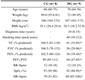
Table 1. Age, anthropometric and spirometric characteristics, respiratory rate, heart rate, and oxygen saturation of control group (CG) and COPD group (DG).
CG (n= 8) DG (n= 9)
Age (years) 68 (60-75) 70 (64-76)
Weight (kg) 69.8 (55.6-81) 72 (49-95)
Height (cm) 166 (164-174) 167 (161-173)
BMI (kg/m2) 25.1 (20.4-29.4) 26.3 (18.2-32.9)
Diagnosis time (years) - 10 (6-15)
Smoking time (pack/years) - 40 (10-212)
VC (% predicted) 104.5 (61-118) 61 (33-85)*
FVC (% predicted) 106.5 (78-132) 56 (29-86)*
FEV1 (% predicted) 102.5 (80-124) 36 (19-44)*
FEV1/FVC 99 (85-111) 64 (47-85)*
RR (bpm) 12 (10-18) 12 (10-23)
SpO2 (%) 97 (95-98) 92 (88-95)*
HR (bpm) 70 (51-82) 88 (85-106)*
Values in median, minimum e maximum. BMI= body mass index; VC= vital capacity; FVC= forced vital capacity; FEV1= forced expiratory volume in the first second; RR = respiratory rate; SpO2= peripheral oxygen saturation in seated rest; HR= heart rate; *= p< 0.05 vs. CG.
Table 2 shows TD and FD analyses data. In TD, in intra-group analysis, no significant differences were observed in the CG or in the DG, regarding to the position change from SU to SE. However, statistically significant differences in RMSSD and SDNN indexes values were observed in the inter-group comparison on the SE position, with the DG presenting lower values.
In FD, no significant differences were observed in intra-group analysis for both intra-groups. In inter-intra-group comparison, differences were found in HF bands, in absolute units, on the supine position, with the CG presenting higher values. In the seated position, the CG presented HF and LF bands higher values, as well as TP. However, no significant differences were observed in HF and LF bands (normalized units), as well as in the LF/HF ratio between groups.
CG (n= 8) DG (n= 9)
SU SE SU SE
TD RMSSD(ms) 9.9 14.6 8.2 8.3 *
(min-max) (5.2-44.1) (8.3-23.1) (4-13) (3.7-16.2)
SDNN (ms) 15.5 23 10 13.5 *
(min-max) (12.7-51.9) (14.7-53) (4.9-21) (7.1-27)
FD HF (ms2) 39 67.6 7.8 † 22.7*
(min-max) (10.6-532.7) (24.6-191.3) (5.1-69.6) (6.3-84.2)
LF (ms2) 72.4 146.7 39.6 24.4 *
(min-max) (22.6-989.5) (44.4-839.2) (4.1-94.9) (12.8-84.2)
TP (ms2) 241.3 552.5 100.8 182.9 *
(min-max) (160.4-2692.5) (215.6-2811.1) (23.8-441) (50.7-731.6)
HF (un) 0.37 0.29 0.29 0.4
(min-max) (0.14-0.6) (0.12-0.65) (0.11-0.64) (0.3-0.6)
LF (un) 0.63 0.71 0.71 0.6
(min-max) (0.4-0.9) (0.35-0.88) (0.36-0.89) (0.4-0.7)
LF/HF 1.7 2.5 2.4 1.5
(min-max) (0.7-6) (0.54-7.42) (0.6-8.5) (0.7-2.9)
Controle - supina DPOC - supina
DPOC - sentada Controle - sentada
(A) (B)
(C) (D)
Frequency (Hz) Frequency (Hz)
Frequency (Hz) Frequency (Hz)
To
ta
l P
o
w
e
r (
m
s²
)
Control - supine COPD - supine
Control - sitting COPD - sitting
To
ta
l P
o
w
er
(
m
s²
)
T
o
ta
l Po
we
r (
m
s²
)
T
o
ta
l
P
o
we
r (
m
s²
)
Table 2. HRV values in time domain (TD) and frequency domain (FD) of control group (CG) and COPD group (DG), in supine (SU) e seated (SE).
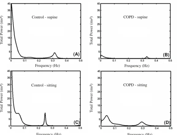
Figura 1. Graphic representation of power spectral density of a control group volunteer (A and C) and a chronic obstructive pulmonary disease group volunteer (B and D), in supine and seated position, respectively.
DISCUSSION
This study showed that stable COPD patients, from moderate to severe degree of airway limitation, present HRV indexes reduction, in TD and FD, when compared to healthy subjects. However, in this study, both elderly COPD patients and healthy elderly subjects did not present autonomic adjustments front postural changes.
Regarding to the studied sample, there were no statistically significant differences between the anthropometrical data and the age of CG and DG subjects, considering that the volunteers were paired according to the age. Considering the spirometric results, COPD patients from this study presented obstruction degree from moderate to severe19, and SpO2 values were reduced comparing to the
healthy volunteers.
These results are related to the clinical diagnosis time and smoking years of these patients. However, it lacks of a deeper scientific support about the relationship between the clinical diagnosis time and smoking year on previous studies, even though the degree of airway obstruction13 or
the hypoxemia18 have been frequently described as important
variables correlated to the cardiac autonomic system alterations.
In TD, assessed by RMSSD and SDNN index, the results showed that there were statistically significant differences between the groups in seated position, considering that the DG demonstrated lower values than the CG. The results from this study are in accordance to the results of Paschoal, Petrelluzzi & Goncalves17, which found significant
differences between the control group and the COPD patients, but only in the supine position. Additionally, Volterrani et al.13
verified significant decrease of the R-Ri standard deviation in these patients compared to the control group. On the same way, Pagani et al.14 demonstrated R-Ri variability reduction
in COPD patients in resting condition.
In FD, it was possible to observe that COPD patients presented lower values of HF components in absolute units, when compared to healthy subjects in supine position. Furthermore, taking into account the seated position, COPD patients presented lower values of HF and LF components (absolute units) when compared to the CG.
This is the first study that demonstrated that elderly COPD patients present reduction on both sympathetic and parasympathetic tonus. Volterrani et al.13 observed
parasympathetic tonus increase of COPD patients, while Stein et al.15 observed reductions in all HRV indexes, including the
LF and HF components in absolute units, but at a younger age.
Regarding to the postural change, differently from the present study, Volterrani et al.13 observed HF components
increase in COPD patients after the head-up tilt passive maneuver, indicating increase of parasympathetic tonus in these subjects. These authors inferred that the vagal tonus
increase could explain, in part, the FEV1 reduction and the bronchoconstriction increase.
Heindl et al.21, when studying chronic respiratory failure,
with COPD and pulmonary fibrosis, observed sympathetic activation increase in these patients. However, these authors evaluated a sample with ages ranging from 19 to 75 years old, not considering the changes brought by the aging process. Other authors have suggested that the aging process may alter the HRV responses22,23,24, leading to it´s attenuation with
time.
Some studies have shown that COPD patients have abnormal autonomic control of the cardiac function, represented by changes in HRV13,15,17, as well as of the pulmonary function
and bronchial tonus. Bartels et al.25 observed that COPD
patients presented HF components increase and LF/HF ratio decrease during exercise, suggesting an increase in the vagal activity in sinusal node, different from de control-subjects, who presented LF/HF ratio increase, as a response to LF components increase. For these authors25, these results
indicate an abnormal state of the parasympathetic tonus or a loss of the ability to activate the sympathetic response during the exercise, once the sympathetic tonus was already increased in the resting condition.
In the present study it was possible to observe that both the sympathetic and the parasympathetic activity (in absolute units) are reduced in COPD patients. The autonomic responses decrease during postural change reflects baroreflexive sensibility damages and reduction of the vagal activity upon the sinusal node. Some authors13 have suggested that the
autonomic control of such patients becomes “saturated” front stimulus. However, this fact cannot explain the loss of adjustments in response to the postural change of the control group in the present study.
Moreover, the literature has demonstrated that the reduction of variability indexes has a narrow relation with arrhythmias appearance16 and with increase of incidence of
sudden death26. Thus, HRV analysis in COPD patients may
have great importance on the initial evaluation of such patients in the beginning of a physical exercises program.
Regarding to the TP (Figure 1) it was observed that the COPD leads to a significant reduction of all frequency bands, when compared to the CG. Therefore, it is possible that other mechanisms, not yet well known, mediated by the VLF bands, may be altered in these patients.
complementary data. However, in the present study, only short duration registers were possible to be studied.
Moreover, the absence of autonomic adjustments during postural change in both groups indicates that the aging process may contribute to their attenuation. However, in this study, it was not possible to compare the autonomic responses with healthy young subjects, which would allow us to infer about the aging process and its importance on such adjustments. Finally, it may be concluded that elderly COPD patients present HRV reduction when compared to healthy elderly subjects gender and age-matched, with sympathetic and vagal activity reduction. Moreover, both COPD patients and healthy subjects did not present autonomic adjustments front postural change. Thus, it may be suggested that, in future studies, the HRV might become a useful tool to obtain parameters about the cardiovascular risk stratification of this population, as well as in the assessment of different physical therapeutic interventions designed to the treatment of these patients.
REFERENCES
1. Pauwels RA, Buist AS, Calverley PM, Jenkins CR, Hurd SS; GOLD Scientific Committee. Global strategy for the diagnosis, management, and prevention of chronic obstructive pulmonary disease. Am J Respir Crit Care Med. 2001;163(5):1256-76.
2. Celli BR, MacNee W. Standards for the diagnosis and treatment of patients with COPD: a summary of the ATS/ERS position paper. Eur Respir J. 2004;23(6):932-46.
3. O’Donnell DE. Ventilatory limitations in chronic obstructive pulmonary disease. Med Sci Sports Exerc. 2001;33 Suppl 7: S647-55.
4. Sietsema K. Cardiovascular limitations in chronic pulmonary disease. Med Sci Sports Exer. 2001;33 Suppl 7:656-61.
5. Silva AB, Marães VRFS, Pires Di Lorenzo VA, Costa D. Heart rate variability in chronic obstructive pulmonary disease during bilevel positive airway pressure. Critical Care. 2003; 7 Suppl 3:56.
6. Longo A, Ferreira D, Correia MJ. Variabilidade da freqüência cardíaca. Rev Port Cardiol. 1995;14(3):241-62.
7. Ramaekers D, Ector H, Aubert AE, Rubens A, Van de Werf F. Heart rate variability and heart rate in healthy volunteers. European Heart Journal. 1998;19:1334-41.
8. Task Force. Heart rate variability: standards of measurement, physiological interpretation and clinical use. Circulation. 1996;93:1043-65.
9. Akselrod S, Gordon D, Ubel FA, Shannon DC, Berger AC, Cohen RJ. Power spectrum analysis of heart rate fluctuation: a quantitative probe of beat-to-beat cardiovascular control. Science. 1981;213:220-2.
10. Akselrod S, Gordon D, Madwed JB, Snidman NC, Shannon DC, Cohen RJ. Hemodynamic regulation: Investigation by spectral analysis. Am J Physiol. 1985;249:H867-75.
11. Pomeranz B, Macaulay RJ, Caudill MA, Kutz I, Adam D, Gordon D, et al. Assessment of autonomic function in humans by heart rate spectral analysis. Am J Physiol. 1985;248:151-3.
12. Malliani A, Pagani M, Lombardi F, Cerutti S. Cardiovascular neural regulation explored in the frequency domain. Circulation. 1991;84:1482-92.
13. Volterrani M, Scalvini S, Mazzuero G, Lanfranchi P, Colombo R, Clark AL, et al. Decreased heart rate variability in patients with chronic obstructive pulmonary disease. Chest. 1994;106:1432-7.
14. Pagani M, Lucini D, Pizzinelli P, Sergi M, Bosisio E, Mela GS, et al. Effects of aging and of chronic obstructive pulmonary disease on RR interval variability. J Auton Nerv Syst. 1996; 59:125-32.
15. Stein PK, Nelson P, Rottman JN, Howard D, Ward SM, Kleiger RE, et al. Heart rate variability reflects severity of COPD in PiZ alpha1-antitrypsin deficiency. Chest. 1998;113:327-33.
16. Tukek T, Yildiz P, Atilgan D, Tuzcu V, Eren M, Erk O, et al. Effect of diurnal variability of heart rate on development of arrhythmia in patients with chronic obstructive pulmonary disease. Int J Cardiol. 2003;88:199-206.
17. Paschoal MA, Petrelluzzi KFS, Gonçalves NVO. Estudo da Variabilidade da Freqüência Cardíaca em Pacientes com Doença Pulmonar Obstrutiva Crônica. Rev Ciénc Méd. 2002;11:27-37.
18. Scalvini S, Porta R, Zanelli E, Volterrani M, Vitacca M, Pagani M, et al. Effects of oxygen on autonomic nervous system dysfunction in patients with chronic obstructive pulmonary disease. Eur Respir J. 1998;13:119-24.
19. Sociedade Brasileira de Pneumologia e Tisiologia. I Consenso Brasileiro sobre Espirometria. J Pneumol. 1996;22:105-64.
20. Knudson RJ, Leibowitz MD, Holberg CJ, Burrows B. Changes in the normal maximal expiratory flow-volume curve with growth and aging. Am Rev Respir Dis. 1983;127:725-34.
21. Heindl S, Lehnert M, Criée CP, Hasenfuss G, Andreas S. Marked sympathetic activation in patients with chronic respiratory failure. Am J Respir Crit Care Med. 2001;164:597-601.
22. Migliaro ER, Contreras P, Bech S, Etxagibel A, Castro M, Ricca R, et al. Relative influence of age, resting heart rate and sedentary life style in short-term analysis of heart rate variability. Braz J Med Biol Res. 2001;34:493-500.
23. Jensen-Urstad K, Storck N, Bouvier F, Ericson M, Lindblad LE, Jensen-Urstad M. Heart rate variability in healthy subjects is related to age and gender. Acta Physiol Scand. 1997;160:235-41.
24. Catai AM, Chacon-Mikahil MP, Martinelli FS, Forti VA, Silva E, Golfetti R, et al. Effects of aerobic exercise training on heart rate variability during wakefulness and sleep and cardiores-piratory responses of young and middle-aged healthy men. Braz J Med Biol Res. 2002;35:741-52.
25. Bartels MN, Jelic S, Ngai P, Basner RC, Demeersman RE. High--frequency modulation of heart rate variability during exercise in patients with COPD. Chest. 2003;124:863-9.