Cholesterol-dependent hemolytic activity
of
Passiflora quadrangularis
leaves
Departamento de Biofísica e Radiobiologia, Centro de Ciências Biológicas, Universidade Federal de Pernambuco, Recife, PE, Brasil
L.N. Yuldasheva, E.B. Carvalho, M.-T.J.A. Catanho and O.V. Krasilnikov
Abstract
Plants used in traditional medicine are rich sources of hemolysins and cytolysins, which are potential bactericidal and anticancer drugs. The present study demonstrates for the first time the presence of a hemo-lysin in the leaves of Passiflora quadrangularis L. This hemolysin is heat stable, resistant to trypsin treatment, has the capacity to froth, and acts very rapidly. The hemolysin activity isdose-dependent,with a slope greater than 1 in a double-logarithmic plot. Polyethylene glycols of high molecular weight were able to reduce the rate of hemolysis, while liposomes containing cholesterol completely inhibited it. In contrast, liposomes containing phosphatidylcholine were ineffective. The Passiflora hemolysin markedly increased the conductance of planar lipid bilayers containing cholesterol but was ineffective in cholesterol-free bilayers. Successive extraction of the crude hemoly-sin with n-hexane, chloroform, ethyl acetate, and n-butanol resulted in a 10-fold purification, with the hemolytic activity being recovered in the n-butanol fraction. The data suggest that membrane cholesterol is the primary target for this hemolysin and that several hemolysin molecules form a large transmembrane water pore. The properties of the Passiflora hemolysin, such as its frothing ability, positive color reaction with vanillin, selective extraction with n-butanol, HPLC profile, cholesterol-dependent membrane susceptibility, formation of a stable complex with cholesterol, and rapid erythrocyte lysis kinetics indicate that itis probably a saponin.
Correspondence O.V. Krasilnikov Departamento de Biofísica e Radiobiologia CCB, UFPE
Av. Prof. Moraes Rego, s/n 50670-901 Recife, PE Brasil
Fax: +55-81-2126-8560 E-mail: [email protected]
Research supported by CNPq.
Received June 3, 2004 Accepted April 26, 2005
Key words
•Passiflora quadrangularis •Leaf extract
•Liposomes •Bilayer permeation •Hemolysin
•Membrane cholesterol
Introduction
Pharmacological effects have been well established for many species of the genus
Passiflora, collectively known in Brazil as “maracujá”. Passiflora incarnata leaf ex-tracts have a potent anxiolytic and sedative effect. Moreover, this plant is used in ho-meopathic medicine for the treatment of in-somnia, epilepsy, tetanus, and muscle spasms (1). Hydroethanol extracts of P. alata and P.
are few publications about the cytotoxic ac-tivity of the family Passifloraceae (8-10).
Saponins are common constituents of plants that exhibit a broad spectrum of bio-logical activities (8,10-14), and frequently possess hemolytic, cytolytic and bactericidal activities (6,8,15). Furthermore, saponins have plasma cholesterol-lowering activity (5,12) and are widely utilized as a compo-nent of potent adjuvants to boost the im-mune response (16), principally when com-plexed with cholesterol (17). However, not all Passiflora species contain saponins in their leaves (18).
It is well known that saponins such as digitonin bind strongly to cholesterol to form a rigid equimolecular complex, and other saponins are also believed to interact with cholesterol (19,20). Saponins usually in-crease membrane permeability (15) and this property is widely used to control permeabi-lization of cell membranes (21-23). In the case of erythrocytes, complex formation re-sults in rapid cell lysis (24) and can lead to membrane disintegration in the presence of relatively large digitonin concentrations. The last effect suggests that digitonin has a deter-gent-like action. Digitonin forms so strong and stable a complex with cholesterol that it is used for cholesterol measurement in blood plasma, bile, and tissues. The purpose of the present study was to isolate a hemolytically active component from leaves of P. qua-drangularis and to characterize its physical and biological properties.
Material and Methods
Plant material
P. quadrangularis leaves were collected in September 2001, from a garden in Carpina, PE, Brazil. The identity of the plant was confirmed by Dr. Marlene Barbosa, Botany Department, Federal University of Pernam-buco. A voucher specimen of the plant was deposited in the IPA Herbarium (N56742)
and was identical to the voucher specimen in the UFP (Recife) Herbarium (RG: 13616).
Preparation of a crude extract containing saponins
Fresh leaves of P. quadrangularis were mixed 1:2 (w/v) with deionized water and homogenized in a blender for about 5 min. The homogenate was centrifuged at 3000 g
for 10 min at 4ºC. The supernatant was collected and 10 volumes were mixed with one volume of absolute ethanol (10:1, v/v) to stabilize the extract. This light yellow aqueous ethanol solution had relatively high osmolarity (~1,700 mOsm) and was acidic, pH 4.4. The concentration of solids in the supernatant was about 22.5 mg/ml, as deter-mined by drying to a constant weight at 60ºC.
Quantitative analysis of hemolytic activ-ity requires careful control of the osmolaractiv-ity and pH of all solutions to prevent swelling and nonspecific erythrocyte lysis. With this in mind, for the hemolytic assay, the primary aqueous ethanol solution was diluted 5.5 times with deionized water and the pH was adjusted to 7.4 with Tris base. The resulting slight sediment was removed by centrifuga-tion at 3000 g for 5 min. The final osmolarity of the diluted extract was 280 mOsm (Fiske Mark-3 Osmometer, Norwood, MA, USA). This adjusted extract possessed high hemo-lytic activity and will be referred to hereafter as crude hemolysin.
was dissolved in an appropriate amount of a solution containing 150 mM NaCl and 5 mM Tris-citrate, pH 7.4. All solutions were adjusted to 280 mOsm and to pH 7.4.
Chemicals
Egg yolk phosphatidylcholine (PC, type V-E) and cholesterol were purchased from Sigma (St. Louis, MO, USA). Trypsin was obtained from Boehringer (Ingelheim, Ger-many). Polyethylene glycols with average molecular masses of 6,000 and 35,000 kDa were purchased from Fluka (Buchs, Swit-zerland). Sucrose, glucose, Tris base, citric acid, EDTA, ethanol, NaCl, and Triton X-100 from various other suppliers were all of analytical grade. Milli-Q plus treated water (Millipore, Billerica, MA, USA) with resis-tivity of 18 MΩ-cm was used to prepare all buffer solutions.
Rabbit erythrocyte suspension
For the hemolysis assay, rabbit erythro-cytes were used as described previously (28). Briefly, fresh blood was mixed with 20 vol-umes of isotonic standard physiological so-lution (SPS, 150 mM NaCl, 5 mM Tris-citrate, 1% EDTA, pH 7.4). Rabbit erythro-cytes were obtained by centrifugation at 1000
g for 5 min. The supernatant was discarded and sedimented erythrocytes were then washed three times with the same buffer, centrifuged and resuspended in EDTA-free SPS. The hematocrit of the final suspension was measured with a capillary hematocrit centrifuge (model 210 I.E.C., FANEM, São Paulo, SP, Brazil) and adjusted to a 2% (w/ v) suspension, which was used throughout the study.
Liposome preparation
Lipids (20 mg PC, or a mixture of 10 mg PC with 10 mg cholesterol) were dissolved in Folch solution (chloroform:methanol, 2:1,
v/v), transferred to a round-bottom glass flask and dried with a nitrogen stream to form a thin film. To make the liposomes, 1 ml SPS was added to the flask and vigor-ously shaken to remove the lipid film from the flask wall. Finally the mixture was soni-cated (Mini-som, Thornton INPEC Eletró-nica, Vinhedo, São Paulo, SP, Brazil) to yield an aqueous vesicle suspension. The lipid concentration of this liposome suspen-sion was 20 mg/ml.
Hemolytic assay
Hemolysis was assayed at 25 ± 2ºC using a Bio-Rad plate reader (model 170-6638, with replacement lamp #3550: Hercules, CA, USA). The extent of lysis was quantified by direct measurement of cell suspension ab-sorbance at 655 nm. At this wavelength hemolysis causes a decrease in absorbance. All substances were dissolved in SPS. Ex-tracts were serially double diluted with SPS (which in other experiments was comple-mented with non-electrolytes or liposomes at the desired concentrations). The process was initiated by the addition of a 2% suspen-sion of rabbit erythrocytes. Absorbance was repeatedly measured in all wells at appropri-ate time intervals (usually 1-5 min). The final concentration of erythrocytes was 1% and the final volume in each microplate well was 100-200 µl. Wells containing only a 1% erythrocyte suspension in SPS were used for a negative control assay (0% hemolysis). Absorbance of wells with erythrocytes lysed with 2% Triton X-100 was taken as 100% hemolysis. The percentage of hemolysis in other wells was calculated relative to the Triton X-100 value. A multichannel pipette was used for simultaneous initiation of the assay and the first absorbance reading was taken immediately.
Hemolysis/K+-efflux assay
lysis and the estimation of K+-efflux from erythrocytes were measured in parallel in experiments carried out in glass tubes at 37 ± 1ºC in a final volume of 10 ml. At appropri-ate times a 1-ml aliquot of the erythrocyte suspension was centrifuged in Eppendorf tubes (30 s at 1000 g). The extent of lysis was estimated by determining the absorb-ance of the supernatant at 540 nm. The K+ concentration in the supernatant was meas-ured with a K+-selective electrode. All other conditions were as described for the hemo-lysis assay.
Transmembrane current assay
Planar bilayer lipid membranes (BLMs) were used to measure the change in mem-brane conductance in the presence of hemo-lysin. BLMs were formed at 25ºC by the technique of Mueller et al. (29) in an Ussing Teflon chamber, whose two compartments were separated by a 20-µm Teflon diaphragm with an orifice for bilayer formation ~300 µm in diameter. Bilayers were formed in the orifice of the chamber by applying a drop of 2% lipid solution in n-decane. Membrane formation was monitored using a binocular microscope and by observing a marked in-crease in capacitance. The electrical charac-teristics of BLMs were measured under volt-age clamp conditions as previously described (28). The amplifier signal was monitored with a Nicolet-2090-III storage oscilloscope (Nicolet Technologies, Madison, WI, USA) and recorded on an IBM-compatible 486/ 487 100 MHz computer with a DT01-EZ 12 bit A/D converter board (Data Translation, Marlboro, MA, USA). Whole Cell Electro-physiology software (WCP V1.7b) devel-oped by Dr. J. Dempster (University of Strathclyde, Glasgow, Scotland, UK) was employed for data analysis.
The trans-compartment of the experimen-tal chamber was connected to the virtual ground and voltage pulses (40 mV) were applied to the cis-compartment of the
cham-ber, to which the hemolysin was also added. The conductance of bilayer membranes (G) in symmetrical solutions was defined as
G = I/V,where I is the transmembrane cur-rent flowing through the membrane and
V, corresponds to the fixed potential. The basal conductance of BLMs was less than 5 pS.
Testing the properties of the hemolysin
To study the properties of the hemolysin and to clarify its target at the membrane level, hemolysin was pre-incubated with tryp-sin or with liposomes (prepared from PC or from a PC/cholesterol mixture) for 3 h in a water bath at 37ºC. The final concentra-tions of hemolysin, lipids and trypsin were 2, 2 and 0.02 mg/ml, respectively. The he-molytic activity of these pre-treated samples was tested as described in “Hemolytic as-say”.
In order to determine the stoichiometry of the hemolysin/cholesterol interaction, a constant amount of the hemolysin (0.1 mg) was mixed with different amounts of choles-terol-containing liposomes directly in 96-well plates just before the addition of the erythrocyte suspension.
The osmotic balance method (28) was used in an attempt to estimate the size of pores induced by hemolysin in erythrocyte membranes. Non-electrolytes of different sizes (glucose, sucrose and polyethylene gly-cols) were added to SPS at concentrations that increased the osmolarity of the solution by 40 mOsm (this increment is close to the osmotic pressure created by intracellular hemoglobin). This method assumes that hem-olysis does not occur when the size of water-filled pores induced by any hemolysin in an erythrocyte membrane is smaller than the hydrodynamic size of the non-electrolyte molecules added to SPS.
then evaluated by the normal procedure. A Fiske Mark-3 Osmometer was used to meas-ure the osmolarity of all solutions used.
Statistical analysis
The Student t-test was used to evaluate the significance of the difference between mean values. Data are reported as means ± SD.
Results
Temperature and trypsin sensitivity of the
P. quadrangularis hemolytic activity
The hydroethanol solution of P. qua-drangularis leaves possessed high lytic potency against erythrocytes. The K+ efflux always preceded hemolysis (data not shown), indicating that hemolysis may have been of an osmotic nature. Hemolytic activity in-creased after 3 h of trypsin treatment (Figure 1). The lysis half-time of a 1% erythrocyte suspension (T50%) in the presence of 30 µg/ ml hemolysin was 2.5 ± 0.1 min. The trypsin treatment decreased T50% to 1.5 ± 0.1 min. It seems that the hemolysin is partially associ-ated with proteins and trypsin digestion re-leases it from a protein-hemolysin complex. The hemolytic activity of the hemolysin was practically unaltered by 10 min heating at 100ºC (data not shown). The hemolysin was positive in the froth test.
The foregoing results indicate that the hemolysin is not a protein because it does not lose activity after heating and protease treatment. The pre-lytic K+ efflux suggests the osmotic nature of hemolysis and pro-vides evidence of pore formation in erythro-cyte membranes. Once formed, transmem-brane water pores apparently provoke a cas-cade of events including water entry, eryth-rocyte swelling and membrane rupture, meas-ured as hemoglobin release. Since pore size is a key parameter, experiments were carried out to estimate it.
Pore-sizing experiments
The osmotic balance method was used to evaluate the radius of water pores induced by the hemolysin. It was found that small non-electrolytes have no influence on hemolysin activity. On the other hand, large non-electro-lytes (especially PEG 35000) were able to decelerate the hemolysis, but were not able to prevent it completely (Figure 2). The combi-nation of pre-lytic K+-efflux with the absence of complete blockage of hemolysis by large non-electrolytes such as PEG 35000 suggests that pores formed by hemolysin in the mem-brane are heterogeneous. Pore size hetero-geneity has been reported for other lipo-philic hemolysins in erythrocyte membranes (30). However, we cannot exclude the possi-bility that the hemolysin pores may simply be larger than the hydrodynamic radius of the largest non-electrolyte (PEG 35000 = ~3.7 nm) used in our study.
100
80
60
40
20
0
0 2 4 6 8 10
Time (min)
Hemolysis (%)
Figure 1. Time course of hemo-lysis in response to the addition of 30 µg/ml Passiflora quadran-gularis hemolysin. Squares = original preparation of hemoly-sin, circles = hemolysin ptreated with trypsin. Data are re-ported as the mean ± SD for at least 4 experiments. The time requirement for 50% erythrocyte lysis (T50%) was equal to 2.5 ± 0.1 min and 1.5 ± 0.1 min for the original preparation of hemoly-sin and for the tryphemoly-sin-treated one, respectively. All other con-ditions are described in Material and Methods.
Control +Sucrose +PEG 35000 80
60
40
20
0
0 5 10 15 20 25 30
Hemolysis (%)
Time (min)
lysis on hemolysin concentration was inves-tigated. The slope of the dose dependence in a double-logarithmic plot was found to be >1 (Figure 3). As shown elsewhere (30,31), it suggests that more than one molecule of hemolysin participates in the formation of an elementary permeabilizing unit in eryth-rocyte membrane.
Membrane cholesterol as a target for the action of hemolysin
In order to identify the membrane com-ponent responsible for cell sensitivity to the hemolysin, liposomes of different lipid com-positions were used. PC liposomes added to the erythrocyte suspension did not prevent hemolysis, but those containing cholesterol were able to completely abolish it (Figure 4). To clarify the role of membrane choles-terol in hemolysis, additional experiments with planar lipid bilayer membranes were carried out. We found that PC-BLMs were practically insensitive to the hemolysin, whereas the conductance of cholesterol-con-taining bilayers increased strongly in the presence of hemolysin in bath solutions (Fig-ure 5).
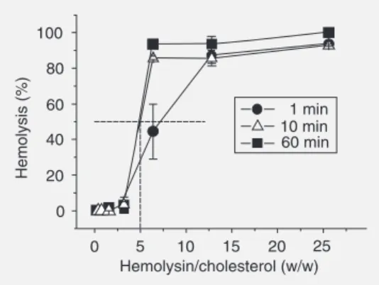
To estimate the concentration of the hemolytically active component in our hemo-lysin preparation, serial 2-fold dilutions of cholesterol-containing liposomes were sup-plemented with a fixed amount (equal to 0.1 mg dry weight) of the hemolysin. The reac-tion was started by erythrocyte addireac-tion and the extent of hemolysis was measured at 1, 10, and 60 min (Figure 6). Fifty percent inhibition of hemolysis occurred only when the dry weight of hemolysin exceeded the weight of cholesterol by 5-fold. The hemo-lysin-cholesterol complex appeared to be stable, with no conspicuous release of hemo-lysin from this complex for at least 60 min (data not shown).
Several lines of evidence suggest that the
P. quadrangularis hemolysin is a saponin. A 1:1 molar ratio of cholesterol/hemolysin was
2.0
2.0 2.5
1.5
1.5 1.0
1.0 0.5
0.5 0.0
Log hemolysin (µg/ml)
Log hemolysis
Figure 3. Concentration-de-pendent hemolysis by Passiflora quadrangularis hemolysin. He-molysis was quantified following incubation for 1 min. Data are reported as mean ± SEM per-cent hemolysis for 4 experi-ments in a double logarithmic plot. The SEM was <3%. Straight lines are least-squares fit. The slope value was 1.3 ± 0.1. All other conditions for the experiment are described in Ma-terial and Methods.
0.0 0 20 40 60 80 100
0.2 0.4 0.6 0.8 1.0
Hemolysin (mg/ml)
Hemolysis (%)
Control +PC +PC/Chol Figure 4. Influence of liposomes
on the hemolytic activity of the
Passiflora quadrangularis hemo-lysin. The hemolysin was pre-incubated with liposomes as de-scribed in Material and Methods. The hemolysin samples were serially double diluted with standard physiological solution and the erythrocyte suspension was then added. Hemolysis was quantified following incubation for 1 min. Data are reported as mean ± SD percent hemolysis for 4 experiments. Squares,
tri-angles, and inverted triangles show the effects of untreated hemolysin, and of hemolysin pre-incubated with phosphatidylcholine (PC) liposomes and PC/cholesterol (Chol) lipo-somes, respectively. All other conditions for the experiment are described in Material and Methods.
Figure 5. Typical recordings of transmembrane current under the influence of the hemolysin. Hemolysin (final concentration: 20 µg/ml) was added to the cis-compartment at the time indi-cated by the arrow. Planar bi-layers were formed from pure phosphatidylcholine (PC) (A) or from a PC/cholesterol mixture (1:1; w/w) (B) by the method of Mueller et al. (29). Current and time scales are given in the fig-ure. Other conditions are de-scribed in Material and Methods.
Dose dependence and estimation of the number of hemolysin molecules forming an elementary pore
To determine how many molecules of hemolysin comprise the transmembrane water pore, the dependence of erythrocyte
0 40 80
Current (nA)
60
20 100
Time (s) 8
6
4
2
0
Hemolysin 20 µg/ml
B
sufficient to prevent hemolysis, as was es-tablished for digitonin (19,20). The molecu-lar weight of saponin is comparable to that of cholesterol, which is accessible to the hemo-lysin only in the external leaflet of liposome membranes. On this basis, one can estimate that the crude hemolysin preparation may contain up to 10% (by weight) of the active hemolysin. Hence, dry P. quadrangularis
leaves may contain up to 0.8% active com-ponent. It is possible that this approach over-estimates the hemolysin content. The actual value could be still lower if a small percent-age of multilayer liposomes were present.
Selective saponin extraction and the
P. quadrangularis hemolysin
To further explore the possibility that the
P. quadrangularis hemolysin is a saponin, the traditional selective saponin extraction procedure was used. Four organic solvents (hexane, chloroform, ethyl acetate, and n-butanol) were successively employed. With this method, the last n-butanol fraction con-tains crude saponin. The highest specific hemolytic activity (660 HU/mg) was found in the n-butanol extract (Table 1). This value is ~10 times higher than those in the initial hydroethanol solution or in any other solu-tions/phases in the purification process. Ex-periments analogous to those in Figures 1-6 were done with this partially purified hemoly-sin (n-butanol fraction). The data obtained were identical except for the hemolysin con-centration, which was ~10 times less (data not shown). Moreover, the acid hydrolysis prod-ucts of this hemolysin gave a color reaction with vanillin, a classic test for saponins (32).
Discussion
The present study demonstrates for the first time that leaves of P. quadrangularis
possess a potent, heat-stable, non-proteina-ceous hemolysin(s) and that its activity is increased by trypsin treatment. This
activa-Table 1. Distribution of hemolytic activity among the fractions used for saponin extraction.
Dry weight Specific Total
(mg) activity activity
(units/mg) (units)
Hydroethanol 5400 64 345,600
extract
n-Hexane 1500 10 15,000
WaterH phase 3430 80 274,400
Chloroform 700 20 14,000
WaterC phase 2100 80 168,000
Ethyl acetate 420 20 8,400
WaterE phase 1480 80 118,400
n-Butanol 155 660 102,300
WaterB phase 1360 10 13,600
One hemolytic unit is the amount of hemolysin that is able to completely lyse 100 µl of a 1% erythrocyte suspension in 5 min at 25 ± 1ºC. Loss in total activity seen at each step of the purification procedure is the result of discarding the inter-phase zone material. Each value is the mean of three replicates with a standard deviation of not more than 20% of the mean.
1 min 10 min 60 min 100
80
60
40
20
0
Hemolysis (%)
0 5 10 15 20 25
Hemolysin/cholesterol (w/w)
Figure 6. Titration of the hemo-lytic effect of the Passiflora quadrangularis hemolysin with cholesterol. Phosphatidylcho-line/cholesterol liposomes were two-fold serially diluted on mi-croplates and mixed with a fixed amount (0.1 mg dry weight) of the hemolysin and the erythro-cyte suspension was then added. Hemolysis was quanti-fied following incubation for the time given in figure. The hemo-lysin/cholesterol ratio (w/w) is given on the abscissa. The ar-row indicates a condition in which 50% inhibition of hemolysis was observed. Data are reported as mean ± SD percent hemolysis for 4 experiments. Circles = 1 min; triangles = 10 min; squares = 60 min.
molecules participate in transmembrane pore formation. In an attempt to show the level of oligomerization achieved by the hemolysin during pore formation we analyzed the dose-effect relationship. We established that the slope of the dose dependence in double-logarithmic plots exceeded one. This method has been widely employed for establishing the number of molecules necessary for the formation of a pore in erythrocyte mem-branes and in planar lipid bilayers by differ-ent types of membrane active compondiffer-ents (30,31,33). From the slope we conclude that more than one molecule of the hemolysin participates in the formation of an elemen-tary pore in erythrocyte membranes.
To determine the nature of the membrane target responsible for the high sensitivity of erythrocyte membranes to the hemolysin, we employed lipid bilayers (spherical and planar). By using liposomes of different li-pid compositions we found that PC-lipo-somes did not prevent hemolysis. On the other hand, the ability of cholesterol-con-taining liposomes to prevent erythrocyte ly-sis, even in the presence of large hemolysin concentrations, was impressive. The most plausible explanation would be the prefer-ential binding of the hemolysin to choles-terol-containing liposomes, which protect erythrocytes from hemolysis. The difference between control hemolysin activity and ac-tivity in the presence of PC-liposomes was not statistically reliable at all hemolysin con-centrations. The data suggest that
choles-terol is the probable target for the hemolysin. Hemolysin-cholesterol complexes seem to be very stable, since we did not observe any release of hemolysin from this complex dur-ing a 60-min period.
Experiments with planar lipid bilayer membranes gave additional support to this apparent role of cholesterol. We established that BLMs formed from pure PC were insen-sitive to the hemolysin, whereas the conduc-tance of cholesterol-containing bilayers was significantly increased by the hemolysin in a time-dependent manner. These results indi-cate that cholesterol is a target for the action of this hemolysin against membranes.
In addition, characteristics such as resis-tance to heating and to trypsin treatment, specificity for and formation of a stable com-plex with cholesterol, as well as frothing ability and a positive vanillin color test, all suggest that the hemolysin is indeed a sapo-nin.
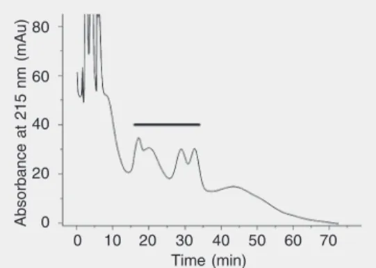
In order to confirm this point, the hemo-lysin was extracted by a procedure devel-oped for saponins, which involves succes-sive extractions of the crude hemolysin with n-hexane, chloroform, ethyl acetate, and n-butanol. A ~10-fold purification was achieved. The hemolytic activity, as expected (18,25-27), was recovered in the butanol fraction. Moreover, when subjected to HPLC analysis (Figure 7), the hemolysin’s profile resembled those of other saponin-contain-ing samples chromatographed under similar conditions (34), thus confirming its saponin nature.
Although saponins possess detergent-like properties (e.g., positive froth test) due to their amphiphilic character, and detergents are able to form pores in membranes (35), our data suggest that the P. quadrangularis
hemolysin does not induce membrane disor-der through a simple detergent action. It is well known (36,37) that membrane destabi-lization by detergents is mostly influenced by the membrane physical state, bilayers in the gel state being much more resistant to
Figure 7. HPLC chromatogram of Passiflora quadrangularis
hemolysin. HPLC conditions: C18Waters column, 3.8 mm in-ternal diameter x 300 mm, 15-20 µm operated at room tempera-ture (22ºC) with a mobile phase of CH3OH/H2O = 50/50 at a flow rate of 1 ml/min. The effluent was monitored at 215 nm using an AKTA™ chromatography system (GE-Amersham Biosciences, Piscataway, NJ, USA). The ap-proximate elution position of the hemolysin is indicated by the solid horizontal line.
Absorbance at 215 nm (mAu)
80
60
40
20
0
0 10 20 30 40 50 60 70
membrane-disruptive agents than bilayers in the liquid-crystalline state. Cholesterol enhances membrane resistance to detergents. In the present study, only bilayers contain-ing cholesterol were sensitive to the action of the hemolysin. Moreover, for a series of saponins (digitonin, aescine, tomatine, stevi-oside, and ginsenoside Rg1) it was demon-strated (38) that neither the surface proper-ties nor the interfacial tension-lowering prop-erties of saponins (in n-decane/water) could be correlated with their ability to induce hemolysis.
Our results showing that only choles-terol-containing BLMs are susceptible to the hemolysin are reminiscent of earlier studies (39) in which it was reported that digitonin induced a noticeable change in glucose per-meability of vesicles of egg yolk lecithin containing cholesterol, but not of choles-terol-free vesicles. Digitonin/cholesterol complex formation in the membranes has been established a long time ago (19,20), and these data suggest that complex forma-tion might be related to the hemolytic activ-ity. Moreover, although saponins possess detergent-like properties, they can increase the permeability of cell membranes without destroying them, and therefore are used for the detection of intracellular antigens on intact cells (22,23). Consistent with these studies, we propose that the P. quadrangula-ris hemolysin also acts by forming a
com-plex with cholesterol that creates transmem-brane pores in erythrocyte memtransmem-branes, re-sulting in water influx and finally leading to swelling and membrane rupture.
The name ‘saponin’ denotes a large and chemically heterogeneous group of sterol glycosides and triterpene glycosides linked to one or two polar oligosaccharides. The rates of hemolysis induced by sterol glyco-sides are generally much higher than those of triterpene glycosides (24). The high rate of erythrocyte lysis evoked by the P. qua-drangularis hemolysin and its prevention by cholesterol-containing liposomes suggest that this hemolysin may belong to the sterol glycoside group. Its exact chemical struc-ture will be the subject of further study.
Acknowledgments
We are indebted to Dr. Yara de Miranda Gomes (Centro de Pesquisas Aggeu Maga-lhães, FioCruz) for the use of a microplate reader and to Dr. José Luiz de Lima Filho (Laboratório de Imunopatologia Keizo Asami, LIKA, Federal University of Per-nambuco, Recife, PE, Brazil) for allowing us to use the AKTA™ purifier. We are grate-ful to Dr. Steven D. Aird (Department of Biology, Norfolk State University, Norfolk, VA, USA) for editing, that improved the clarity of the manuscript.
References
1. Dhawan K, Kumar S & Sharma A (2001). Comparative biological activity study on Passiflora incarnata and P. edulis. Fitoterapia, 72: 698-702.
2. Petry RD, Reginatto F, de-Paris F et al. (2001). Comparative phar-macological study of hydroethanol extracts of Passiflora alata and
Passiflora edulis leaves. Phytotherapy Research, 2: 162-164. 3. Mowrey DB (1993). Herbal Tonic Therapies. Keats Publishing, New
Canaan, CT, USA.
4. Nippon MKK (1993). Angiotensin converting enzyme and aldose-reductase inhibitory agent - comprises Passiflora quadrangularis
extract with organic solvent or water, or vitexin. Patent No. 1995-009562 [02].
5. Chandel RS & Rastogi RP (1980). Triterpenoid saponins and sapo-genins: 1973-1978. Phytochemistry, 19: 1889-1908.
6. Rao AV & Sung MK (1995). Saponins as anticarcinogens. Journal of Nutrition, 125 (Suppl): 717S-724S.
7. Shao Y, Chin CK, Ho CT et al. (1996). Anti-tumor activity of the crude saponins obtained from asparagus. Cancer Letters, 104: 31-36.
8. Birner J & Nicolls JM (1973). Passicol, an antibacterial and antifun-gal agent produced by Passiflora plant species: preparation and physicochemical characteristics. Antimicrobial Agents and Chemo-therapy, 3: 105-109.
9. Lutomski J & Malek B (1975). Pharmacochemical investigations on raw materials of the genus Passiflora. 3. Phytochemical investiga-tions on raw materials of Passiflora edulis forma Flavicarpa. Planta Medica, 27: 222-225.
4.4-Hydroxy-2-cyclopentenone an antipseudomonas and cytotoxic component from
Passiflora-tetrandra. Planta Medica, 57: 129-131.
11. Rao AV & Gurfinkel DM (2000). The bioactivity of saponins: triterpenoid and steroidal glycosides. Drug Metabolism and Drug Interactions, 17: 211-235.
12. Kinlen PJ, Menon VP & Pirakitikulr V (2001). Composition useful as a food additive for reducing serum cholesterol levels comprises a mixture of a phytostanol or its ester and a surfactant (Patent num-ber: WO200132036-A1; AU200114636-A). International Patent Classification: A23L-001/035; BNA2-3L001/30.
13. Ridoux O, Di Giorgio C, Delmas F et al. (2001). In vitro antileishma-nial activity of three saponins isolated from ivy, alpha-hederin, beta-hederin and hederacolchiside A1 in association with pentamidine and amphotericin B. Phytotherapy Research, 15: 298-301. 14. Feng XZ, Dong M & Xu SX (2001). A new triterpenoidal saponin
from Ixeris sonchifolia and its cytotoxic activity. Pharmazie, 56: 663-664.
15. Li XX, Davis B, Haridas V et al. (2005). Proapoptotic triterpene electrophiles (avicins) form channels in membranes: cholesterol dependence. Biophysical Journal, 88: 2577-2584.
16. Santos WR, Bernardo RR, Peçanha LM et al. (1997). Haemolytic activities of plant saponins and adjuvants. Effects of Periandra mediterranea saponin on the humoral response to the FML antigen of Leishmania donovani. Vaccine, 15: 1024-1029.
17. Kersten GF, Spiekstra A, Beuvery EC et al. (1991). On the structure of immune-stimulated saponin-lipid complexes (iscoms). Biochimi-ca et BiophysiBiochimi-ca Acta, 1062: 165-171.
18. Reginatto FH, Kauffmann C, Schripsema J et al. (2001). Steroidal and triterpenoidal glucosides from Passiflora alata. Journal of the Brazilian Chemical Society, 12: 32-36.
19. Akiyama T, Takagi S, Sankawa U et al. (1980). Saponin-cholesterol interaction in the multibilayers of egg yolk lecithin as studied by deuterium nuclear magnetic resonance: digitonin and its analogues.
Biochemistry, 19: 1904-1911.
20. Nishikawa M, Nojima S, Akiyama T et al. (1984). Interaction of digitonin and its analogs with membrane cholesterol. Journal of Biochemistry, 96: 1231-1239.
21. Lapetina EG, Watson SP & Cuatrecasas P (1984). Myo-inositol 1,4,5-trisphosphate stimulates protein phosphorylation in saponin-permeabilized human platelets. Proceedings of the National Acade-my of Sciences, USA, 81: 7431-7435.
22. Goldenthal KL, Hedman K, Chen JW et al. (1985). Postfixation detergent treatment for immunofluorescence suppresses localiza-tion of some integral membrane proteins. Journal of Histochemistry and Cytochemistry, 33: 813-820.
23. Jacob MC, Favre M & Bensa JC (1991). Membrane cell permeabili-zation with saponin and multiparametric analysis by flow cytometry.
Cytometry, 12: 550-558.
24. Takechi M & Tanaka Y (1995). Haemolytic time course differences
between steroid and triterpenoid saponins. Planta Medica, 61: 76-77.
25. Sotheeswaran S, Bokel M & Kraus W (1989). A hemolytic saponin, randianin, from Randia Dumetorum. Phytochemistry, 28: 1544-1546. 26. Mimaki Y, Kanmoto T, Sashida Y et al. (1996). Steroidal saponins from the underground parts of Chlorophytum comosum and their inhibitory activity on tumor promoter-induced phospholipids metabo-lism of HELA cells. Phytochemistry, 41: 1405-1410.
27. Itabashi M, Segawa K, Ikeda Y et al. (2000). A new bioactive steroidal saponin, furcreastatin, from the plant Furcraea foetida. Carbohydrate Research, 323: 57-62.
28. Krasilnikov OV, Capistrano MFP, Yuldasheva LN et al. (1997). Influence of Cys-130 S. aureus alpha-toxin on planar lipid bilayer and erythrocyte membranes. Journal of Membrane Biology, 156: 157-172.
29. Mueller P, Rudin DO, Tien HT et al. (1963). Methods for the forma-tion of single bimolecular lipid membranes in aqueous soluforma-tion.
Journal of Physical Chemistry, 67: 534-535.
30. Chernitskii EA, Sen’kovich OA & Kozlova NM (1996). Heterogeneity of pores formed in erythrocyte membrane by lipophilic hemolysins.
Biofizika, 41: 1270-1274.
31. Abramov AY, Zamaraeva MV, Hagelgans AI et al. (2001). Influence of plant terpenoids on the permeability of mitochondria and lipid bilayers. Biochimica et Biophysica Acta, 1512: 98-110.
32. Wu J, Lin L & Chau F (2001). Ultrasonic-assisted extraction of ginseng saponins from ginseng roots and cultured ginseng cells.
Ultrasonics Sonochemistry, 8: 347-352.
33. Krasilnikov OV, Muratkhodjaev JN & Zitzer AO (1992). The mode of action of Vibrio cholerae cytolysin. The influences on both erythro-cytes and planar lipid bilayers. Biochimica et Biophysica Acta, 1111: 7-16.
34. Yang D-J, Lu T-J & Hwang LS (2003). Simultaneous determination of furostanol and spirostanol glycosides in Taiwanese yam (Dioscorea spp) cultivars by high performance liquid chromatogra-phy. Journal of Food and Drug Analysis, 11: 271-276.
35. Senkovich OA & Chernitsky EA (1998). On the size of pores arising in erythrocytes under the action of detergents. Membrane and Cell Biology, 11: 679-689.
36. Rowland RN & Woodley JF (1980). The stability of liposomes in vitro
to pH, bile salts and pancreatic lipase. Biochimica et Biophysica Acta,620: 400-409.
37. Lasch J (1995). Interaction of detergents with lipid vesicles. Bio-chimica et Biophysica Acta, 1241: 269-292.
38. Steurer S, Wurglics M, Likussar W et al. (1999). Lack of correlation between surface and interfacial activities of saponins and their hemolytic properties. Pharmazie, 54: 766-767.