CLINICAL INVESTIGATION
www.bjournal.com.br
www.bjournal.com.br
Braz J Med Biol Res, July 2012, Volume 45(7) 665-675
doi:
10.1590/S0100-879X2012007500066
Vascular dysfunction by myofibroblast activation in patients with
idiopathic pulmonary fibrosis and prognostic significance
E.R. Parra, R. Falzoni and V.L. Capelozzi
Institutional Sponsors
The Brazilian Journal of Medical and Biological Research is partially financed by
Faculdade de Medicina de Ribeirão Preto Campus
Ribeirão Preto
Explore High - Performance MS Orbitrap Technology In Proteomics & Metabolomics
Vascular dysfunction by myofibroblast activation
in patients with idiopathic pulmonary fibrosis
and prognostic significance
E.R. Parra, R. Falzoni and V.L. Capelozzi
Departamento de Patologia, Faculdade de Medicina, Universidade de São Paulo, São Paulo, SP, Brasil
Abstract
In this study, we demonstrated the importance of telomerase protein expression and determined the relationships among
telomerase, endothelin-1 (ET-1) and myofibroblasts during early and late remodeling of parenchymal and vascular areas in usual interstitial pneumonia (UIP) using 27 surgical lung biopsies from patients with idiopathic pulmonary fibrosis (IPF). Telomerase+, myofibroblasts α-SMA+, smooth muscle cells caldesmon+, endothelium ET-1+ cellularity, and fibrosis severity were evaluated in 30 fields covering normal lung parenchyma, minimal fibrosis (fibroblastic foci), severe (mural) fibrosis, and vascular areas of UIP by the point-counting technique and a semiquantitative score. The impact of these
markers was determined in pulmonary functional tests and follow-up until death from IPF. Telomerase and ET-1
expres-sion was significantly increased in normal and vascular areas compared to areas of fibroblast foci. Telomerase and ET-1 expression was inversely correlated with minimal fibrosis in areas of fibroblast foci and directly associated with severe fibrosis in vascular areas. Telomerase activity in minimal fibrosis areas was directly associated with diffusing capacity of the lung for oxygen/alveolar volume and ET-1 expression and indirectly associated with diffusing capacity of the lungs for carbon monoxide and severe fibrosis in vascular areas. Cox proportional hazards regression revealed a low risk of death for females with minimal fibrosis displaying high telomerase and ET-1 expression in normal areas. Vascular dysfunction by telomerase/ET-1 expression was found earlier than vascular remodeling by myofibroblast activation in UIP with impact on IPF evolution, suggesting that strategies aimed at preventing the effect of these mediators may have
a greater impact on patient outcome.
Key words: Idiopathic pulmonary fibrosis; Vascular activity; Immunohistochemistry; Survival
Introduction
Correspondence: E.R. Parra, Departamento de Patologia, FM, USP, Av. Dr. Arnaldo, 455, 01246-903 São Paulo, SP, Brasil. Fax: +55-11-3064-2744. E-mail: erparra20003@ yahoo.com.br
Received November 14, 2011. Accepted April 9, 2012. Available online April 27, 2012. Published July 2, 2012.
Idiopathic pulmonary fibrosis (IPF) is a devastating chronic fibrosing interstitial pneumonia of unknown etiol
-ogy that typically increases in prevalence with advanced age, characterized by excessive collagen deposition and irreversible remodeling of the lung parenchyma (1,2). The
histologic pattern associated with IPF is the usual interstitial pneumonia (UIP), characterized by patchy and temporally
heterogeneous fibrosis with excessive extracellular matrix
(ECM) and honeycomb change, interspersed with normal
and collapsed areas (1). Although the etiology of IPF/UIP is not understood, many investigators hypothesize that
dysregulated communication between injured epithelial
cells and activated mesenchymal cells represents an im
-portant factor in its pathogenesis (3). Vascular changes
are another event frequently observed in IPF (4,5). In previous studies, we found a direct relationship between progressive vascular occlusion by collagen/elastic fiber
deposition and tissue remodeling in surgical lung biopsies
from patients with IPF (4,5). These findings probably are the consequence of persistent myofibroblast activation
by mesenchymal cells, resulting in a heterogeneous
-clusion or areas of fibroblastic foci, which are thought to be the sites of active ECM, collagen and elastic synthesis, and are considered to be the leading edge of fibrosis and vascular remodeling (8). In this regard, endothelin-1 (ET-1)
is one of the more potent inducers of collagen deposition,
with its fibrogenic effects being central in wound healing and tissue repair. A major role of ET-1 involves transition of quiescent fibroblasts to an “activated” phenotype, whereby
matrix production and wound contraction are important
consequences (9). Telomerase is a specialized polymerase
that adds telomere repeats to the ends of chromosomes by
using its intrinsic RNA component as a template, thereby
compensating for the telomere loss that normally occurs
with each cell division (10). Telomerase has been shown
to be essential for unlimited cell proliferation and has been linked to immortality (11). Bleomycin-induced lung injury
and fibrosis is also known to induce telomerase activity in the affected lung tissue and isolated lung fibroblasts (12). The fibroblasts isolated from such lungs undergoing fibro
-sis show increased intrinsic proliferative capacity and are able to differentiate into α-SMA expressing myofibroblasts, which are also a key source of cytokines with inflammatory and fibrogenic properties (13,14). An injured lung fibroblast
population may contain telomerase expressing cells with an
extended life span, which could contribute to the observed increased numbers of lung fibroblasts (12).
In view of these considerations suggesting an interac
-tion among telomerase, endothelium and myofibroblast activation, the aim of the present study was to evaluate 1) whether telomerase, ET-1 and myofibroblast α-SMA are expressed in vascular and parenchymal areas of UIP; 2) to verify if the measures of mediator expression correlate with some measure of fibrosis, and 3) to determine the impact of the resultant parenchymal/vascular remodeling on patient survival. It was hypothesized that in UIP, a devastating chronic fibrosing interstitial pneumonia, the mediators under study exert their effect by influencing parenchyma/vascular remodeling/fibrosis with impact on patient outcome.
Material and Methods
This research was approved by the Institutional Ethics and Scientific Committees (protocol No. 0960/08) and all patients gave written informed consent to participate in
the study.
Open lung biopsies
Pulmonary specimens were obtained by surgical lung
biopsy from 27 patients with IPF/UIP (14 males and 13 females; mean age 64 ± 1.4 years), according to criteria outlined in the American Thoracic Society/European Re -spiratory Society International Multidisciplinary Consensus
Classification of idiopathic interstitial pneumonias (1). Only specimens from patients who fulfilled these consensus
criteria were included. Three lung specimens obtained by
open surgical biopsy from each patient were collected ac-cording to the normal, intermediate and more affected areas in different parts of the lung determined by high-resolution computed tomography. The histologic UIP pattern of IPF was characterized by the patchy subpleural and paraseptal distribution of parenchymal injury. Temporal heterogeneity
was seen at low magnification, with alternating areas of normal lung parenchyma with septal fibro-myxoid tissue (fibroblastic foci), honeycomb and vascular thickening.
Baseline characteristics
The pulmonary function tests included vital capacity, forced expiratory volume in 1 s (FEV1), forced vital capac -ity (FVC), FEV1/FVC ratio 100X, total lung capac-ity (TLC),
residual volume (RV), and carbon monoxide transfer factor (diffusing capacity of the lung for carbon monoxide, DLCO).
The percentages of TLC, RV, and RV/TLC were measured by the helium dilution method using a Master Screen
ap-paratus (Erich Jaeger GmbH, Germany). The DLCO and DLCO/alveolar volume (VA) were assessed by the single
breath holding helium dilution method (15). Lung function results (Table 1) are reported as the percentage of patient
lung function compared to predicted values for a standard -ized healthy population. In all patients, the arterial partial
pressure of carbon dioxide (PaCO2) and partial pressure
of oxygen (PaO2) were also measured at rest.
Immunohistochemistry
A standard peroxidase technique was used, with Har -ris’s hematoxylin as the counterstain, to identify
telom-erase+ epithelial cells, ET-1+ endothelium and α-SMA+
Table 1. Clinical data of the patients.
Variables Idiopathic pulmonary fibrosis
Age at biopsy (years) 64.5 ± 7.4
Gender (males/females) 13/14 Spirometry
FEV1 (% predicted) 78 ± 4.4 FVC (% predicted) 69.5 ± 3.5
FEV1/FVC 20 ± 8.8
TLC (% predicted) 79 ± 5 RV (% predicted) 102 ± 15.9 TLC/RV (%predicted) 44.2 ± 3.4
DLCO (% predicted) 59.8 ± 8
DLCO/VA (% predicted) 59 ± 11.7
PaCO2 60.2 ± 3.7
PaO2 38.1 ± 1.3
Data are reported as means ± SEM. FEV1 = forced expiratory volume in 1 s; FVC = forced vital capacity; TLC = total lung ca -pacity; RV = residual volume; DLCO = diffusing capacity of the lung for carbon monoxide; VA = alveolar volume; PaCO2 = partial
myofibroblasts and caldesmon+ smooth muscle cells in normal tissue, fibroblastic foci and vascular areas of UIP. Since the α-SMA antibody stains myofibroblasts as well
as smooth muscle cells, which are a regular component of arteries, the caldesmon (C21) antibody sc-58700 was
specifically used to characterize these cells. All antibod -ies used were biotinylated rabbit polyclonal antibod-ies.
Anti-telomerase, anti-ET-1, anti-αSMA and anti-caldesmon polyclonal antibodies (Santa Cruz Biotechnology, Inc., USA)
were incubated with tissue sections at 1:100 dilution. The
Max Polymer Novolink amplification kit (Leica, Newcastle Inc., UK) was used for signal amplification and 3,3′-diamin
-obenzidine tetrachloride (0.25 mg dissolved in 1 mL 0.02%
hydrogen peroxide) was used as a precipitating substrate
for signal detection. The specificity of primary antibodies was confirmed by appropriate reagent controls (omitting
the primary antibody or substituting non-immune serum for the primary antibody in the staining protocol), which
revealed no staining.
Histomorphometry
Regional differences between samples from the same patient in the three lung specimens obtained by open
surgi-cal biopsy in each patient were evaluated for telomerase+ epithelium, ET-1+ endothelium, α-SMA+ myofibroblasts, caldesmon+ smooth muscle cells, and fibrosis severity in 30 fields by the point-counting technique (16) and a semi
-quantitative score (4).As previously described, the histologic
pattern of UIP was characterized by alternating areas of
normal lung parenchyma, minimal fibrosis (fibroblastic foci), severe (mural) fibrosis, honeycombing, and vascular thickening. At 400X magnification, 30 fields were chosen
from across multiple sites accounting/adjusting for cellularity
or as number of positively staining cells per mesenchymal
tissue. In other words, normal lung parenchyma, minimal
fibrosis (fibroblastic foci), severe (mural) fibrosis, and vas
-cular areas present in UIP in each field were determined
according to the number of points hitting mesenchymal tissue, as a proportion of the total grid area. The number
of positive cells within the normal lung parenchyma, mini
-mal fibrosis (fibroblastic foci), severe (mural) fibrosis, and vascular areas were then counted. The immunostaining cellularity was determined as the number of positive cells in each field divided by the normal lung parenchyma, mini
-mal fibrosis (fibroblastic foci), severe (mural) fibrosis, and vascular areas. The final results are reported as means ± SEM of 3 lung specimens from each patient in 30 random, non-coincident microscopic fields.
The severity of fibrosis was assessed by semiquantita
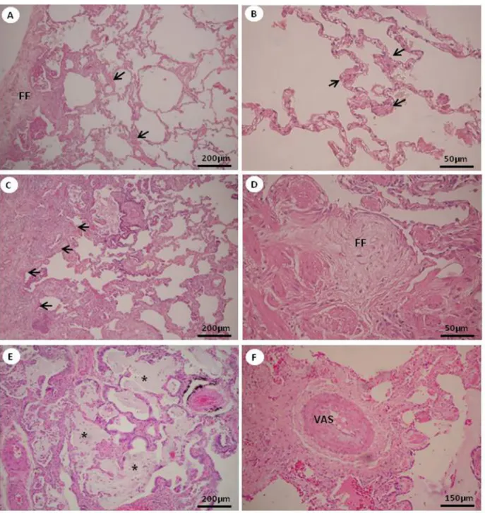
-tive analysis in UIP areas of 1) mild interstitial thickening by fibroblastic foci (minimal fibrosis, Figure 1A and B), and 2) severe mural-organizing fibrosis with honeycombing and foci of actively proliferating fibroblasts and myofibroblasts (severe fibrosis, Figure 1C-F).
Interobserver comparisons were performed for 20% of the
slides by 2 observers (E.R.P. and V.L.C.). The coefficient of variation for the interobserver error of cell count was <5%.
Statistical analysis
Data are reported as means ± SEM with 95% confidence intervals. ANOVA with the Bonferroni test were used to analyze the relationship between continuous variables and
the residuals were examined to ensure that their distribution was approximately normal. The relationship between the
immunostaining cellularity was evaluated by Pearson’s cor
-relation. Actuarial survival was estimated using the
Kaplan-Meier method and compared using the log-rank test. Cox proportional hazards regression was used to ascertain the
individual contribution of factors associated with survival and to compare adjusted survival between groups. The statistical program used was SPSS 18.0 (SPSS Inc., USA). The level of significance was set at 5% in all tests.
Results
Telomerase activation
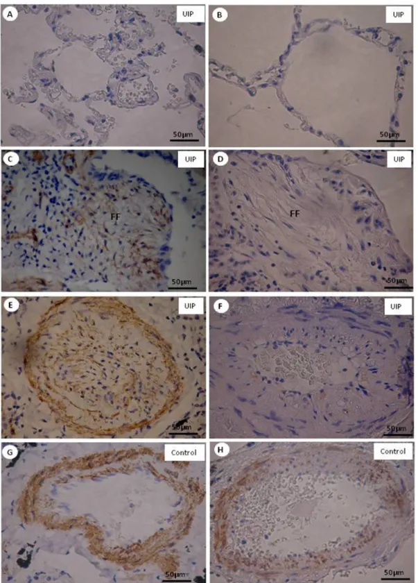
Immunochemical staining of epithelium telomerase+ in
normal (Figure 2B), minimal fibrosis (Figure 2C), severe fibrosis (Figure 2D), and vascular (Figure 2F) areas of UIP,
appears as brownish cells. Epithelium telomerase+
im-munostaining was more exuberant in normal and vascular areas than in fibrosing areas of the histologic pattern of UIP than in control areas (Figure 2A and E).
Quantitative analysis confirmed the morphologic dis -tribution of telomerase+ epithelium expression (Table 2).
Telomerase+ epithelium was significantly overexpressed in normal and vascular areas than in UIP fibrosing and
control areas.
Myofibroblasts and smooth muscle cell replication
α-SMA+ myofibroblasts in normal (Figure 3A), fibros
-ing (Figure 3C) and vascular (Figure 3E) areas of UIP and control areas (Figure 3G), when stained by immuno
-histochemistry, appear as brownish cells. α-SMA+ myofi -broblasts were more prominent than smooth muscle cells
caldesmon+ in vascular (Figure 3F) than fibrosing (Figure 3D) and control (Figure 3H) areas. Normal areas did not present α-SMA+ myofibroblasts or caldesmon+ smooth muscle cells (Figure 3B).
Quantitative analysis confirmed the morphologic distri
-bution of α-SMA+ myofibroblasts and caldesmon+ smooth muscle cells (Table 2). Interestingly, α-SMA+ myofibroblasts were significantly more increased than caldesmon+ smooth muscle cells in vascular areas of UIP (P < 0.001).
Endothelium activation
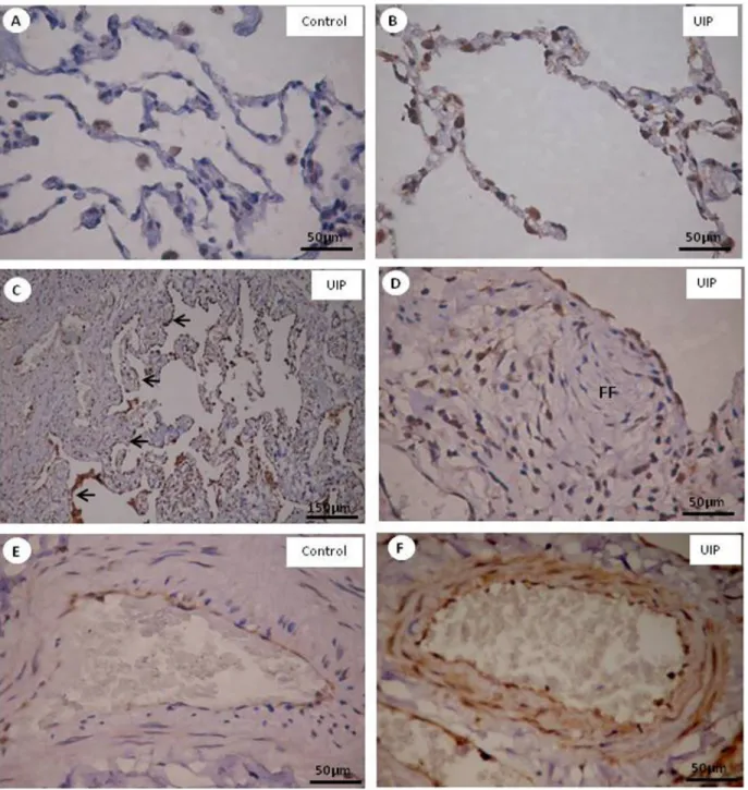
ET-1+ endothelium in normal (Figure 4B), minimal
cells in normal and vascular areas expressed ET-1 when compared to fibrosing areas of the UIP histologic pattern and control areas (Figure 4A and E).
Quantitative analysis confirmed the morphologic distri
-bution of ET-1 expression that was significantly increased
in normal and vascular areas of UIP when compared to fibrosing areas (Table 2).
Association between mediators
A direct association was found between telomerase and
Figure 1. Degree of fibrosis in the pattern of usual interstitial pneumonia (UIP). UIP minimal fibrosis is characterized by interstitial thickening (arrows) and by fibroblastic foci (FF) (A and B) and UIP severe fibrosis is characterized by severe mural-organizing fibrosis
(arrows) (C) with foci of actively proliferating fibroblasts and myofibroblasts (FF) (D) and honeycombing (stars) (E). Vascular (VAS)
ET-1 expression in vascular areas of UIP (r = 0.45; P = 0.03). An inverse association was found between telomerase and ET-1 expression in normal and minimal fibrosis areas of UIP (r = -0.42; P = 0.03). Telomerase and ET-1 expression were positively associated with vascular areas (r = 0.54; P = 0.004) and negatively associated with minimal fibrosis
and normal areas (r = -0.44; P = 0.02) of UIP.
Association between mediators and fibrosis severity
Telomerase and ET-1 expression were inversely cor
-related with minimal fibrosis in normal and fibroblast foci areas (r = -0.38; P = 0.04 and r = -0.39; P = 0.03, respec
tively) and directly associated with severe fibrosis in vascular areas (r = 0.41; P = 0.03 and r = 0.54; P = 0.004) of UIP.
Association between media-tors and functional tests
A direct association was
found between diffusing capac-ity of the lung for
oxygen/al-veolar volume (DLCO/VA) and telomerase activity (r = 0.49; P = 0.01) in minimal fibrosis areas. An indirect association was found between DLCO and severe fibrosis in vascular areas
displaying high ET-1 expression
(r = -0.74; P = 0.02).
Survival analysis
The median follow-up was 42.70 months. Ten patients
were still alive and 17 died
from causes related to IPF.
ROC curve analysis showed
that the optimum cut-off point of expression was 14.55% for telomerase and 12.54% for
ET-1. These cut-off points resulted in a significant differ
-ence in Kaplan-Meier curves. The 5-year survival rate of patients with minimal fibrosis in vascular areas displaying ET-1 ≤12.54% was 57.49 vs 31.13% in the group with
minimal fibrosis displaying higher ET-1 levels (P < 0.01). The 5-year survival rate of patients with minimal fibrosis in vascular areas displaying telomerase ≤14.55% was 59.55
vs 28.88% in the group with minimal fibrosis and higher
telomerase levels (P < 0.01).
Several combinations of clinical and morphological variables were analyzed to generate a mathematical model with an impact on IPF patient survival. The results of the
best combination, determined by Cox model analysis,
ap-pear in Table 3. The following variables showed a significant impact on patient survival: gender, age, fibrosis severity,
telomerase in UIP normal lung parenchyma, telomerase
in minimal (fibroblastic foci) fibrosis, ET-1 in normal lung parenchyma, and minimal (fibroblast foci) fibrosis. Multi
-variate analyses showed a low risk of death for females, UIP normal lung parenchyma or minimal (fibroblastic foci) fibrosis displaying high telomerase and ET-1 expression in
UIP normal lung parenchyma areas.
Discussion
We investigated the participation of telomerase and the factors responsible for its activation or suppression in the
fibrotic process found in pulmonary specimens obtained by surgical lung biopsy from patients with IPF. Specifically, we investigated telomerase and ET-1 expression in alveolar epithelium, endothelium, and myofibroblast cells present in normal lung parenchyma, minimal (fibroblast foci) fibrosis and vascular areas of UIP to verify the impact of these markers on the remodeling/fibrosis process. In the present study, telomerase and ET-1 were significantly increased in normal lung parenchyma and vascular areas compared to fibroblast foci areas of UIP. Unexpectedly, myofibroblast α-SMA+ expression was more prominent than smooth muscle caldesmon+ cells in vessels. Interestingly, we found a positive association between telomerase and ET-1 in UIP vascular areas, whereas a negative association was found
between telomerase and ET-1 in normal lung parenchyma.
A positive association was found between telomerase and myofibroblast α-SMA in UIP vascular areas. In addition, the
expression of these mediators correlated with the
measure-ment of fibrosis degree, suggesting that they exert their effect by influencing remodeling/fibrosis in UIP.
The current study revealed that telomerase increases myofibroblast activation in vascular areas of UIP. In fact,
telomerase can be expressed by different types of cells
and its presence indicates not only a persistent activity to
maintain a supply of activated cells (e.g., fibroblasts), but is also important in the regulation of cell proliferation (17,18),
apoptosis (19,20), and cell differentiation (21,22). Although Table 2. Histomorphometric markers and cellularity in non-fibrosing lung tissue (control) and different areas of fibrosing lung (usual interstitial pneumonia, UIP).
Lung tissue Telomerase α-SMA Caldesmon Endothelin-1
Non-fibrosing lung (control)
Septal areas 0.78 ± 0.42 0 0 0.01 ± 0.02 Vascular areas 0.01 ± 0.02 9.57 ± 2.90 5.2 ± 1.15 0.0003 ± 0.0003 Fibrosing lung (UIP)
Normal lung parenchyma 17.8 ± 6.7+ 0 0 17.2 ± 6.4#
Minimal fibrosis 7.4 ± 2.1 8.2 ± 3.7 0.002 ± 0.0005§ 5.7 ± 2.2
Severe (mural) fibrosis 6.4 ± 2.5 12.0 ± 6.6 0.06 ± 0.001§ 7.2 ± 2.9
Vascular areas 18.2 ± 7.2+ 51.7 ± 10.4* 0.45 ± 0.02§ 18.0 ± 4.3#
Data are reported as means ± SEM percent of markers for 3 lung specimens obtained by open surgical biopsy from each patient (10 random, non-coincident microscopic fields were analyzed in each specimen). All lung specimens were analyzed for epithelial, myofibroblast, endothelial and smooth muscle cells from normal lung parenchyma, and minimal fibrosis (fibroblastic foci), severe (mural) fibrosis and vascular areas of UIP for percentage of telomerase, α-SMA, calde -smon, and endothelin-1 fractional areas. +Telomerase expression values in UIP normal lung
parenchyma and vascular areas differed significantly from minimal (fibroblastic foci) and severe (mural) fibrosis (P < 0.01). *α-SMA expression values in UIP vascular areas were significantly different from normal lung parenchyma and fibrosis areas (P < 0.01). §Caldesmon expression
Figure 3. Alpha-smooth muscle actin (α-SMA) (A, C, E, G) and caldesmon (B, D, F, H) expression in normal (A, B), fibroblastic foci (FF) of fibrosing (C, D) and vascular (E, F) areas of usual interstitial pneumonia (UIP) pattern, and vascular control areas (G, H) stained by immunohistochemistry. α-SMA+/myofibroblasts are more prominent than
telomerase activity is a nonspecific signal of cell prolifera
-tion involving different cell types besides fibroblasts, this
last assumption does not disagree with our results, which
show an influence of telomerase expression on vessels and prognosis. Our findings agree with the study of Nozaki et al.
(12) who demonstrated that bleomycin-induced lung injury
and fibrosis are also known to induce telomerase activity in the affected lung tissue and isolated lung fibroblasts. The ac
-tivity, however, is expressed only during the period of active fibrosis, and appears to be localized primarily in fibroblasts
Figure 4. Endothelial ET-1+ expression in control areas (A, E) and in normal (B), minimal (C, arrows) and severe (D) fibrosing, showing
rather than myofibroblasts (23). The activity also declines
with increased passaging in vitro, which is associated
with increased differentiation into myofibroblasts. These studies suggest that the increased enzyme activity could be secondary to higher cell division rates and therefore the increase in telomerase activity would be secondary to the inflammatory and fibrotic process instead of its cause. The factors responsible for regulating telomerase activity
in these tissues and cells are mostly unknown. More recent
data from our group have shown that abnormal expression of telomerase/apoptosis limits type II alveolar epithelial cell
replication in the early remodeling of UIP (24). In fact, we found that epithelial telomerase expression in normal lung
parenchyma and fibroblastic foci areas was lower than in normal septal areas, probably reflecting the loss and shortening of telomeres activity. It is tempting to speculate about the existence of a specific checkpoint beyond which loss of telomerase activity due to injurious stimuli and/or
inherited mutations may lead to loss of epithelial cells and
promote genomic instability. Activation of telomerase after
the onset of genomic instability, coupled with a repeatedly
injurious microenvironment, may affect the physiological differentiation of normal alveolar epithelial progenitors
towards a transformed mesenchymal phenotype, resulting in abnormal reepithelization in normal parenchymal areas
and contributing to the fibrogenic process characterized by fibroblastic foci. Telomerase would inhibit the senescence
of many normal cells, such as epithelial and mesenchymal cells, and most probably would increase the incidence of
tumors and fibrosis in the patient. Thus, carcinogenesis and fibrogenesis may be similar processes respectively involving epithelial and mesenchymal (fibroblasts) neoplastic cells.
We also found that telomerase and ET-1
expres-sion were positively associated with vascular tissue and
negatively associated with normal lung parenchyma and fibroblast focus areas of UIP. ET-1 can also be synthesized by endothelial and epithelial cells, vascular smooth muscle
cells, airway epithelium, granulocytes, and macrophages
(25). Moreover, increased concentrations and biosynthesis
of ET-1 were demonstrated in the lungs of patients with pulmonary hypertension and systemic sclerosis (26).
Fur-thermore, preproendothelin-1 messenger RNA is elevated
in airway epithelial cells, proliferating type II pneumocytes,
endothelial cells, and inflammatory cells present in IPF
(27,28). The increased cell population expressing ET-1 and telomerase may indicate a continuous stimulation and transformation of these cells. In fact, we found a more
evident expression in epithelial cells of normal alveolar areas, reflecting not only the stress to which these cells
were submitted but also a probable attempt to maintain a
large population to avoid alveolar collapse, as previously
demonstrated by our group (24). We also found that many
epithelial cells and myofibroblasts present in fibroblast
focus areas expressed ET-1 and telomerase showing a
high activity of these areas. Actually, ET-1 plays a role in several stages of wound healing: stimulating fibroblast dif -ferentiation and collagen metabolism, promoting the wound healing response by the production of collagens IV, V and
VII, involvement in the reconstitution of the basement mem -brane and appropriate wound closure and other unknown
effects on different types of cells (29,30).
Our study has clinical consequences. In order to estab
-lish the relevance of these findings to the evolution of the patients, the mediators under study were evaluated as a function of fibrosis severity and physiological testing. We found a direct association between DLCO/VA and minimal fibrosis areas displaying high telomerase activity and be
-tween DLCO and severe fibrosis areas of UIP displaying Table 3. Cox proportional hazards regression to ascertain the individual contribution of clinical and morphological fac -tors associated with survival and to compare adjusted survival between groups [-2 log likelihood = 28.90; chi-square = 38.26; P < 0.0001].
β SE Wald test P value Exp (β) 95%CI for Exp (β)
Lower Upper
Gender -3.74 1.48 6.38 0.01 41.94 2.31 761.02
Age (years) 1.02 0.34 8.94 0.003 2.78 1.42 5.43
UIP fibrosis 9.26 0.01
UIP severe (mural) fibrosis -9.70 3.26 8.87 0.003 0.00 0.00 0.03
UIP minimal (fibroblastic foci) fibrosis -12.04 3.98 9.16 0.002 0.00 0.00 0.01
Telomerase in UIP minimal fibrosis -1.85 0.97 3.60 0.05 6.35 0.94 42.78
Telomerase in UIP normal lung parenchyma 6.05 2.35 6.62 0.01 0.002 0.00 0.23 Endothelin-1 in UIP normal lung parenchyma -5.13 1.81 8.00 0.005 0.006 0.00 0.20 Endothelin-1 in UIP minimal (fibroblast foci) fibrosis 5.72 2.13 7.24 0.007 0.003 0.00 0.21
high ET-1 expression. Although hypoxia is universal in IPF patients, decreases in DLCO caused by a defect in gas
transfer certainly present in the pulmonary parenchyma of UIP, also contribute to hypoxemia. These functional
changes observed are probably explained in part by the vascular involvement. The expression of these mediators
probably represents another step in the wound healing
process in IPF. We believe that high expression of these markers is crucial to maintain the activity of parenchymal injury by the regulation of compromised vascular tone. Many lines of evidence suggest that telomerase activation under
chronic hypoxia may result in enhanced and sustained
smooth muscle proliferation and vessel wall remodeling, findings characteristic of proliferative vascular disorders, such as pulmonary hypertension (31). On the other hand,
the continuous ET-1 stimulus from the endothelium and
smooth muscle of vascular cells activates the proliferation and the remodeling process of these vessels. Clearly, in our study poor patient survival was associated with high vascular expression of ET-1 and telomerase, suggesting
that a major damage to vessels leads to more parenchymal alterations during the development of pulmonary fibrosis
caused by maintained hypoxia.
In conclusion, telomerase activity increases endothelial and myofibroblast activation in early remodeling/fibrosis of IPF with impact on evolution, suggesting that strategies aimed at preventing the effect of these mediators may have
a greater impact on patient outcome.
Acknowledgments
We are grateful to the biologist Sandra de Morais Fernezlian from the Laboratory of Immunohistochemistry.
We would like to thank Dr. Ronaldo Adib Kairalla and Dr. Carlos Roberto Ribeiro de Carvalho from the Division of
Respiratory Diseases – Heart Institute (InCor), Faculdade
de Medicina, Universidade de São Paulo, for providing
many pulmonary function tests of these patients. Research
supported by CNPq and FAPESP (#2008/53022-3).
References
1. American Thoracic Society/European Respiratory Society International Multidisciplinary Consensus Classification of the Idiopathic Interstitial Pneumonias. This joint statement of the American Thoracic Society (ATS), and the European Respiratory Society (ERS) was adopted by the ATS board of directors, June 2001 and by the ERS Executive Com -mittee, June 2001. Am J Respir Crit Care Med 2002; 165:
277-304.
2. Gribbin J, Hubbard RB, Le Jeune I, Smith CJ, West J, Tata LJ. Incidence and mortality of idiopathic pulmonary fibrosis and sarcoidosis in the UK. Thorax 2006; 61: 980-985.
3. Selman M, King TE, Pardo A. Idiopathic pulmonary fibrosis: prevailing and evolving hypotheses about its pathogenesis and implications for therapy. Ann Intern Med 2001; 134:
136-151.
4. Parra ER, David YR, da Costa LR, Ab’Saber A, Sousa R, Kairalla RA, et al. Heterogeneous remodeling of lung ves -sels in idiopathic pulmonary fibrosis. Lung 2005; 183:
291-300.
5. Parra ER, Kairalla RA, de Carvalho CR, Capelozzi VL. Abnormal deposition of collagen/elastic vascular fibres and prognostic significance in idiopathic interstitial pneumonias.
Thorax 2007; 62: 428-437.
6. Tomasek JJ, Gabbiani G, Hinz B, Chaponnier C, Brown RA. Myofibroblasts and mechano-regulation of connective tissue remodelling. Nat Rev Mol Cell Biol 2002; 3: 349-363.
7. Serini G, Gabbiani G. Mechanisms of myofibroblast activity and phenotypic modulation. Exp Cell Res 1999; 250:
273-283.
8. Elias JA, Zitnik RJ, Ray P. Fibroblast immune-effector func -tion. In: Phipps RP (Editor), Pulmonary fibroblast heteroge -neity. Boca Raton: CRC Press; 1992. p 295-322.
9. Shichiri M, Adachi S, Sedivy JM, Marumo F, Hirata Y. Bi -phasic regulation of the preproendothelin-1 gene by c-myc.
Endocrinology 1997; 138: 4584-4590.
10. Greider CW. Telomere length regulation. Annu Rev Biochem
1996; 65: 337-365.
11. Counter CM, Hirte HW, Bacchetti S, Harley CB. Telomerase activity in human ovarian carcinoma. Proc Natl Acad Sci U S A 1994; 91: 2900-2904.
12. Nozaki Y, Liu T, Hatano K, Gharaee-Kermani M, Phan SH. Induction of telomerase activity in fibroblasts from bleomycin-injured lungs. Am J Respir Cell Mol Biol 2000;
23: 460-465.
13. Phan SH, Varani J, Smith D. Rat lung fibroblast collagen metabolism in bleomycin-induced pulmonary fibrosis. J Clin Invest 1985; 76: 241-247.
14. Zhang HY, Gharaee-Kermani M, Zhang K, Karmiol S, Phan SH. Lung fibroblast alpha-smooth muscle actin expression and contractile phenotype in bleomycin-induced pulmonary fibrosis. Am J Pathol 1996; 148: 527-537.
15. Quanjer PH, Tammeling GJ, Cotes JE, Pedersen OF, Peslin R, Yernault JC. Lung volumes and forced ventilatory flows. Report Working Party Standardization of Lung Function Tests, European Community for Steel and Coal. Official Statement of the European Respiratory Society. Eur Respir J Suppl 1993; 16: 5-40.
16. Hsia CC, Hyde DM, Ochs M, Weibel ER. An official research policy statement of the American Thoracic Society/European Respiratory Society: standards for quantitative assessment of lung structure. Am J Respir Crit Care Med 2010; 181:
394-418.
17. Greider CW. Telomerase activity, cell proliferation, and can -cer. Proc Natl Acad Sci U S A 1998; 95: 90-92.
18. Sarin KY, Cheung P, Gilison D, Lee E, Tennen RI, Wang E, et al. Conditional telomerase induction causes proliferation of hair follicle stem cells. Nature 2005; 436: 1048-1052.
S. Regulation of telomerase activity and anti-apoptotic func -tion by protein-protein interac-tion and phosphoryla-tion.
FEBS Lett 2003; 536: 180-186.
20. Zhang X, Mar V, Zhou W, Harrington L, Robinson MO. Telomere shortening and apoptosis in telomerase-inhibited human tumor cells. Genes Dev 1999; 13: 2388-2399.
21. Rosenberger S, Thorey IS, Werner S, Boukamp P. A novel regulator of telomerase. S100A8 mediates differentiation-dependent and calcium-induced inhibition of telomerase activity in the human epidermal keratinocyte line HaCaT. J Biol Chem 2007; 282: 6126-6135.
22. Savoysky E, Yoshida K, Ohtomo T, Yamaguchi Y, Akamatsu K, Yamazaki T, et al. Down-regulation of telomerase activity is an early event in the differentiation of HL60 cells. Biochem Biophys Res Commun 1996; 226: 329-334.
23. Liu T, Nozaki Y, Phan SH. Regulation of telomerase activity in rat lung fibroblasts. Am J Respir Cell Mol Biol 2002; 26:
534-540.
24. Waisberg DR, Barbas-Filho JV, Parra ER, Fernezlian S, de Carvalho CR, Kairalla RA, et al. Abnormal expression of telomerase/apoptosis limits type II alveolar epithelial cell replication in the early remodeling of usual interstitial pneu-monia/idiopathic pulmonary fibrosis. Hum Pathol 2010; 41:
385-391.
25. Battistini B, Dussault P. Biosynthesis, distribution and
metabolism of endothelins in the pulmonary system. Pulm Pharmacol Ther 1998; 11: 79-88.
26. Abraham DJ, Vancheeswaran R, Dashwood MR, Rajkumar VS, Pantelides P, Xu SW, et al. Increased levels of en -dothelin-1 and differential endothelin type A and B receptor expression in scleroderma-associated fibrotic lung disease.
Am J Pathol 1997; 151: 831-841.
27. Giaid A, Polak JM, Gaitonde V, Hamid QA, Moscoso G, Le -gon S, et al. Distribution of endothelin-like immunoreactivity and mRNA in the developing and adult human lung. Am J Respir Cell Mol Biol 1991; 4: 50-58.
28. Saleh D, Furukawa K, Tsao MS, Maghazachi A, Corrin B, Yanagisawa M, et al. Elevated expression of endothelin-1 and endothelin-converting enzyme-1 in idiopathic pulmonary fibrosis: possible involvement of proinflammatory cytokines.
Am J Respir Cell Mol Biol 1997; 16: 187-193.
29. Guidry C, Hook M. Endothelins produced by endothelial cells promote collagen gel contraction by fibroblasts. J Cell Biol
1991; 115: 873-880.
30. Desmouliere A. Factors influencing myofibroblast differentia -tion during wound healing and fibrosis. Cell Biol Int 1995; 19:
471-476.