Cadasil
Pathogenesis, clinical and radiological
indings and treatment
Charles André
ABSTRACT
Cerebral autosomal dominant arteriopathy with subcortical infarcts and leukoencephalopathy (CADASIL) is the most common genetic cause of ischemic strokes and a most important model for the study of subcortical vascular dementia. This unrelentlessly progressive disease affects many hundreds of families all over the world but is not well studied in Brazil. This manuscript reviews pathogenetic, clinical, radiological and therapeutic features of CADASIL. The causal mutations are now very well known, but the same can not be said about its intimate pathogenetic mechanisms. The variable clinical presentation should lead physicians to actively pursue the diagnosis in many settings and to more thouroughly investigate family history in first degree relatives. A rational approach to genetic testing is however needed. Treatment of CADASIL is still largely empiric. High-quality therapeutic studies involving medications and cognitive interventions are strongly needed in CADASIL.
Key words: CADASIL, etiology, genetics, diagnosis, therapeutics.
CADASIL: patogênese, achados clínicos e radiológicos e tratamento
RESUMO
CADASIL é a causa genética mais freqüente de infartos cerebrais e constitui modelo importante de estudo de demências vasculares subcorticais. De natureza inexoravelmente progressiva, afeta milhares de pessoas em todo o mundo. Sua importância é pouco reconhecida entre nós, o que nos levou à presente revisão dos principais aspectos patogenéticos, clínicos, neuroradiológicos e terapêuticos da doença. As mutações causais são hoje bem conhecidas, mas os mecanismos patogenéticos íntimos ainda permanecem misteriosos. A apresentação clínica variável deve fazer com que os médicos considerem o diagnóstico em vários contextos clínicos e investiguem de forma mais extensa que o usual a história familial deparentes de primeiro grau. Além disso, uma abordagem racional é necessária para reduzir custos e aumentar a eficiência do diagnóstico genético. O tratamento atual de pacientes com CADASIL é basicamente empírico. Estudos clínicos sobre medicamentos e intervenções cognitivas de alto nível metodológico constituem uma necessidade urgente.
Palavras-chave: CADASIL, etiologia, genética, diagnóstico, terapêutica.
Correspondence Charles André
Av Rodolpho Paulo Rocco 255 Serviço de Neurologia 10E36 Cidade Universitária
21941-913 Rio de Janeiro RJ - Brasil E-mail: [email protected]
Received 4 November 2009
Accepted 18 November 2009 Associate Professor of Neurology, School of Medicine: Federal University of Rio de Janeiro, Rio de Janeiro RJ, Brazil. CADASIL (cerebral autosomal
domi-nant arteriopathy with subcortical infarcts and leukoencephalopathy) is a hereditary disease with high penetrance in which oc-clusion of small arteries in the brain of adults results in small deep brain infarcts and progressive accumulation of demy-elination areas in the brain. Its
manifes-tations are diverse and in most individu-als include recurrent headache of migraine pattern, focal deicits secondary to brain infarction (more rarely bleeding) and, in later stages, progressive neuropsychiatric disorders including dementia.
the main genetic cause of stroke. Emphasis is given to cognitive and neuropsychiatric symptoms.
deinitions
CADASIL is an autosomal dominant disease result-ing from mutations of the gene encodresult-ing the transmem-brane receptor Notch 3, located on chromosome 19. Re-garded as a prototype of subcortical vascular dementia re-lated to subcortical microangiopathy, it also results in ad-ditional psychiatric disorders, particularly mood chang-es, usually in association with the development of cogni-tive impairment.
he disease is reported in several countries from all continents. Its frequency is probably underestimated and CADASIL may be one of the most common inherited neu-rological conditions. he prevalence of Notch 3 gene muta-tions has been estimated as more than 4 per 100,000 adults.
he disease appears in adult life. Its manifestations are virtually restricted to the central nervous system, espe-cially the brain, and are caused by the progressive devel-opment of disseminated white matter lesions in associ-ation with small infarcts - lacunes - in subcortical areas. Migraine with aura and focal neurologic deicits caused by these lacunar infarctions are characteristic forms of presentation in young or middle age. Over the years, mood disorders, diverse neurological deicits and cogni-tive disturbances add up. To the extent that the total vol-ume of lesions increases and cerebral atrophy develops, the frequency and severity of motor diiculties and cog-nitive dysfunction also increase.
he cognitive disorder is progressive, and typically re-lects accumulating injury resulting from subcortical mi-crovascular disease. Initially, diiculties in information processing and slowing of cognitive processes are most evident; later, changes in memory and other high order cognitive functions lead to the development of dementia. Depression is the commonest mood disorder, occurring in 20% of individuals. In the inal stages, individuals are bed-driden, apathetic and totally dependent. Death usually re-sults from medical complications, especially malnutrition and infectious diseases such as aspiration pneumonia.
Genetics and pathogenesis
he genetics of CADASIL is directly linked to that of the Notch receptor family. hese surface receptors medi-ate signal transduction with ligands (such as Jagged [Jag] and Delta [D]) in neighboring cells, which are also type I transmembrane receptors. Notch 3 mutations are respon-sible for CADASIL, whose main characteristic is a vascu-lar degeneration, indicating that this system plays an im-portant role in maintaining structural stability and func-tion of vascular smooth muscle cells (VSMC).
Studying two large French families in 1993,
Tourni-er-Lasserve and cols. found that the gene responsible for CADASIL should be located on chromosome 19 in a 14-cM centimorgans) segment between D19S221 and D19S2221. As two other autosomal dominant diseases -
familial hemiplegic migraine and episodic hereditary cer-ebellar ataxia - were also mapped on chromosome 19 in close proximity with CADASIL, the question of allelism in these three conditions soon arised2,3.
he 14-cM segment on chromosome 19q12 was pro-gressively reduced in successive studies, until Joutel et al. were able to reduce it to a critical 800 kb interval that would probably contain the gene for CADASIL, now in 19p13.14. Among several genes in this region, there was
one that exhibited strong homology with Notch 3 gene encoding region 5 in mice. Further studies showed that the human homologue of the Notch 3 gene actually was located in that critical 800 Kb region5.
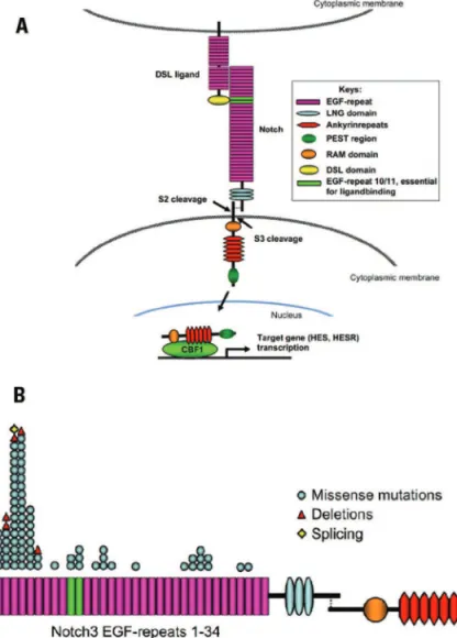
In the last decade, more than 80 Notch 3 mutations were identiied in more than 400 families with CADASIL. hese mutations are distributed across 34 repetitions of the epidermal growth factor (EGFR) that comprise the ex-tracellular domain of Notch 36. All mutations responsible
for CADASIL lead to an odd number of cysteine residues. he Notch 3 gene encodes a single pass transmem-brane receptor comprising 2,321 amino acids with an ex-tracellular domain containing 34 epidermal growth factor (EGF)-like repeats (with 6 cysteine residues in each) and 3 Lin-12 repeats related to the transmembrane and intracel-lular domains . It consists of 33 exons (23 extracelintracel-lular)5,7,8
(Fig 1A). All mutations occur in extracellular domains, more speciically the epidermal growth-like factor-re-peats (EGF-refactor-re-peats), from a non-paired cysteine4,7. Most
mutations occur in codons of exon 4, followed by exons 3, 5, 6 and 117,9,10 (Fig 1B). he exon 3 seems to be the
sec-ond most commonly afected site in French, English and German families7,9; exon 11 could, however, be the
sec-ond most prevalent in Dutch families11.
he Notch proteins constitute a family of surface re-ceptors that promote transduction of signals between neighboring cells. heir interaction with ligands leads to cleavage of its intracellular portion, which translocates to the nucleus and activates transcription of speciic factors, thus regulating subsequent gene expression (Fig 1).
This signaling pathway has been highly preserved throughout phylogeny, and seems to exert a central role in determining cell fate during embryonic development, including various aspects of vascular development such as vasculogenesis, angiogenesis, vascular remodeling, and diferentiation of VSMC12,13. In humans, mutations
In mammals, there are four Notch receptors. While other genes of the Notch group are ubiquitous, the Notch 3 gene is restricted to VSMC of arterial walls14.
he Notch 3 protein, when activated, undergoes se-rial cleavage by proteolysis, producing extracellular and transmembrane domain spliting. After cleavage, these two fragments form a heterodimer on the cell surface of VSCM. Mutations and deletions of the Notch 3 gene re-sponsible for CADASIL promote accumulation of the Notch receptor ectodomain 3 in the vascular wall, prob-ably by reducing their clearance eiciency15. his
accu-mulation occurs near but not within granules of osmeo-phylic material characteristic of the disease in electron microscopy studies.
he mechanisms by which the accumulation of recep-tor fragments and osmeophylic material in the adventitia and medial layer of brain arterioles correlates with vascu-lar functional changes and the development of brain le-sions in CADASIL are discussed. he molecular mech-anisms may include changes in the activation of Notch 3, but the classic Notch activation is not changed, hence lesions can not be solely explained by an increase or re-duction of the Notch 3-related signaling classical pathway
(mediated by CBF1/JBP-Jk). Alternative signaling path-ways mediated by Notch 3, or a cross-regulation of Notch 3 with other signaling pathways may thus play a central role in the survival of VSCM. Alternatively, the Notch 3 mutation can gain new functions not yet studied on VSCM, including toxic ones8.
As mentioned, the Notch3 receptors are found mainly in adult VSMC. heir main function seems to be related to the maintenance of vascular structural and functional stability. he pathogenesis of CADASIL is most likely re-lated to a disturbance in vascular mechanotransduction, with reduced low-induced vasodilation and increased vascular myogenic tone induced by pressure16. Skin
vas-oreactivity is altered in disease. For instance, the kinetics of reactive hyperemia after cuf occlusion displays a de-layed and slow curve17. Endothelium-dependent
vasodi-lation is impaired in resistance arteries (not conductive ones) in the forearm of patients18. Transgenic mice
ex-pressing mutant Notch 3 develop CADASIL typical vas-cular alterations19. hese mice exhibit altered
autoregu-lation of cerebral blood low (CBF): the mutation appears to reduce the relaxation ability or to increase vascular re-sistance of rere-sistance vessels20.
his dynamic process is also associated with deposi-tion of granular osmeophylic material (GOM) and degen-eration of the middle layer of cerebral arteries (and oth-er organs and toth-erritories), and results in reduced CBF in brain white matter. Cerebral and leptomeningeal arteries and arterioles become thickened, with stenosis of pene-trating arteries in the white matter and cortex21-23.
Grad-ually, difuse myelin pallor and rarefaction of hemispher-ic white matter (sparing the U ibers), deep focal lesions, particularly in periventricular areas and centrum semio-vale, and lacunar infarctions in the white matter, basal ganglia and pons add up21,24. Virchow-Robin spaces tend
to become dilated, perhaps by changes related to age or the progression of degeneration of penetrating cerebral arteries25. Further changes were demonstrated by MR
spectroscopy: metabolic reductions in the relationship between mean NAA/Cr, NAA/Cho and Cho/Cr become obvious, especially in anterior (VS. posterior) regions of the centrum semiovale26. hese changes are more severe
in symptomatic individuals, suggesting a correlation be-tween them and the also increasing pathological indings. he dense osmeophylic deposits form granules (10 to 15 nm in diameter) and occupy the middle layer of the vessel, extending to its adventitial layer, but sparing the vascular endothelium21,27,28. hey are located near the cell
membrane of VSMC, and stain positively for PAS (period-ic acid Schif ) - suggesting the presence of glycoproteins - and negatively for elastin and amyloid. here is no
evi-dence of deposition of immunoglobulins. he VSMC, sep-arated by granular material, are swollen and degenerated, and sometimes disappear and are replaced by collagen29.
Vascular abnormalities characteristic of the brain of patients with CADASIL, like the GOM around the smooth muscle cells, are also common in the medial lay-er of artlay-eries of othlay-er organs such as livlay-er, spleen, heart, kidney, retina, muscles and skin, and even in large arter-ies such as aorta and carotid arterarter-ies27,28. Vascular lesions
and accumulation of GOM may be detected in biopsies of muscle and nerve, and skin biopsies are commonly used for diagnostic purposes. Nevertheless, the clinical man-ifestations of the disease are almost always restricted to the central nervous system, speciically the brain.
diagnosis of Cadasil
he diagnosis of CADASIL should be particularly sus-pected in the presence of (adapted from30):
1. Typical clinical picture: one or more subcortical infarcts, especially early (up to 60 years) or with famil-ial history, migraine, usually with aura (including atypi-cal or prolonged); progressive cognitive changes or sub-cortical-type dementia;
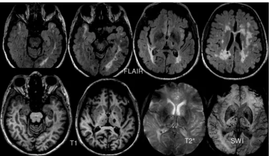
2. Typical neuroradiological indings on magnetic res-onance imaging (MRI): multifocal and bilateral FLAIR/ T2 hyperintensities in the periventricular and deep white matter, with lesions mainly afecting the anterior tempo-ral pole, frontal and parietal lobes, external capsule, pons
and basal ganglia; focal hypointensities on T1 (lacunar infarcts) and lesions suggestive of microhemorrhages in SWI or gradient-echo T2* (Fig 2);
3. Positive familial history: autosomal dominant pat-tern for one or more of the typical clinical features.
CADASIL is sometimes not even considered in dif-ferential diagnoisis for several reasons, especially lack of MRI availability and apparently negative family history. Computed tomography (CT) clearly shows the subcor-tical infarcts - typically lacunar, but also larger lesions31 -
and also shows white matter lesions, especially in more advanced cases. In a study speciically designed to an-swer the second question, 30% of patients with CADA-SIL apparently had a documented negative family histo-ry32. his is mainly related to a restricted search only for
early cerebrovascular disease in the family, and neglect-ing other clinical presentations, such as mood disorders, cognitive impairment and headache.
Deinitive diagnosis is conirmed in patients with clin-ical and radiologclin-ical features (see below) by the inding of mutations in the Notch 3 gene, located on chromosome 19 - 19p13.1. Mutations that promote orientation errors (mis-sense mutations) are the most common, followed by those that promote simple gene deletions. Pathogenic muta-tions remove or insert cysteine residues in EGF-repeats in the extracellular domain of Notch 3 receptor (N3ECD)8,33.
he molecular diagnosis of CADASIL can be, how-ever, tiring and fruitless, especially if it does not follow a rational routine. Most mutations are found in gene exon 4: this site should be investigated irst in suspected cases. In a British study prospectively evaluating diferent diag-nostic strategies, 15 diferent mutations were identiied in 48 families, 73% of them in exon 4 (less than 10% each in exons 3, 5 and 6)9.
A case in which the molecular diagnosis was made in a fetus whose father was afected by CADASIL has been published34. he possibility of early intra-uterus diagnosis
in a progressive disease with high, but incomplete, pen-etrance raises new ethical questions to the medical pro-fession.
In the study of Markus et al.9, the diagnostic value of
characteristic MRI changes was also evaluated. Moder-ate to severe white matter changes in the anterior tem-poral regions exhibited slightly lower sensitivity (89% vs. 93%), but much higher speciicity than those in the exter-nal capsule (86% vs. 45%).
Skin biopsy is considered useful in diagnosis. Spec-iicity is high, approaching 100%, but sensitivity is low (less than 50%); the involvement is often focal, and thour-ough assessment of the material is necessary to make the diagnosis9,35.
Clinical and radiological manifestations
CADASIL may be seen as a pleomorphic disease: the dominant manifestations may vary in diferent families; and the clinical picture and functional course also difer in individuals of the same family. Important phenotypic diferences between families could be attributed in part to the various causal mutations36. As in other “pure”
domi-nant diseases, homozygotes do not seem to do worse than heterozygotes, even if they exhibit increased burden of GOM in tissues37.
he possible role of additional genetic inluences, like the presence of speciic alleles of the Apolipoprotein E, on disease progression is debated38. he white matter
le-sion load, the amount and speciic location of ischemic lesions, the presence and number of microhemorrhages, the degree of cerebral atrophy, and functional (diasqui-sis) or anatomical changes of critical intra-and interhemi-spheric connecting pathways are other factors that may inluence the nature and severity of clinical manifesta-tions (see section on cognitive changes) .
Cardinal clinical manifestations however include mi-graine with aura, transient ischemic attacks (TIAs) and
ixed focal neurologic deicits caused by lacunar infarc-tions, and cognitive decline of subcortical type. In some patients, mood disorders, especially depression, may pre-dominate. In the long run the disease always worsens, but in shorter periods - two years, for example - evolu-tion is much variable: there may be periods of rapid dete-rioration, clinical stability or even occasionally improve-ment39. Time to death is also highly variable - 10 to 30
years. Death occurs around 65 (men) 70 years (women) on average, from accumulation of morbidities and clinical complications related to infection and immobility40.
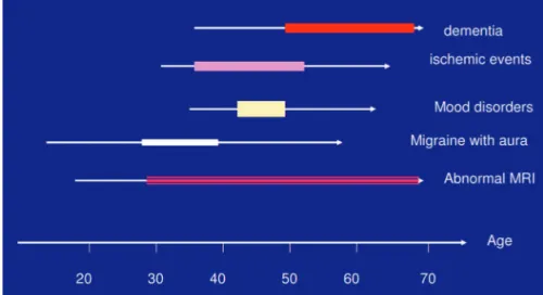
he dominant clinical indings vary according to the disease stage (Fig 3). Neuroradiological abnormalities, which may be present from childhood41, are often already
detected at symptom onset and are almost universally present in symptomatic patients (possible exception in in-dividuals with migraine only) or around 35 years42,43.
CADASIL may be classiied in three clinical/radio-logical stages44:
I: generally between 20 to 40 years of age, with mi-graine and MRI showing white matter-deined lesions42.
II: age between 40 and 60, characterized by transient or permanent ischemic insults leading to motor disor-ders, sensory, sensory and cognitive impairments, with or without psychiatric disorders. MRI shows basal gan-glia and conluent white matter lesions;
III: usually after 60 years, with subcortical demen-tia associated with pseudobulbar signs - dysarthria, dys-phagia and emotional lability; and in some cases apathy, mutism and akinesia. Pyramidal signs are almost always marked. MRI shows difuse leukoencephalopaty and mul-tiple infarcts of basal ganglia.
Some individuals with the mutation may remain asymp-tomatic for long periods, even with well-deined lesions on MRI45,46, while others exhibit only one major disease
char-acteristic, such as migraine with aura, TIA or lacunar in-farctions, depression or anxiety, or slow evolution of cog-nitive impairment42,47. Diagnosis is usually made around
age 45, but greater attention to relatives of afected patients to detect neurological and psychiatric manifestations may lead to an earlier diagnosis. The average age for symp-tom onset is 37 years48, and is independent of gender49,50.
Migraine is common between 20 and 40 years, occur-ring in about one quarter of patients, but can already be present in the early teens. Ischemic events, occurring in at least 60 to 80% of individuals, arise mainly between 30 to 50 years, accumulating over two or three decades, with increasing functional limitation. Mood disorders, primar-ily depression, occur in 10 to 20% of subjects, appearing at any age, but mostly around 40 to 50 years, a period in which cognitive disorders, initially subtle, may lead to the slow development of advanced dementia in the sixth and seventh decades.
Migraine
he most characteristic initial symptom is migraine, usually with aura. he International Headache Society considers migraine as an inappropriate diagnosis in the presence of an obvious vascular cause, but there is a trend to keep the name migraine in patients with CADASIL.
Its frequency varies considerably among affected families49,51. Among 45 patients in 7 CADASIL families,
migraine with aura was present in 22%49. Desmond et al
found migraine in 42 of 105 individuals (40%). On the other hand, none of the members of an Italian family pre-sented symptoms52. here are other families in which
mi-graine and later insidiously progressive cognitive disorders are the sole manifestations of the disease, but the inal di-agnosis of CADASIL in these cases is still uncertain53.
The first migraine attacks occur before 20 years of age44,54 and may precede or be linked to early
neuroradio-logical changes - punctate hyperintensities in deep hemi-sphere or periventricular white matter - that may also be present in some individuals with primary migraine30.
Women with CADASIL and migraine with aura tend to have symptom onset earlier than afected men43.
In the study by Dichgans et al.50, migraine was present
in 30% of men and 44% of women, and 95% of patients af-fected by migraine headaches presented uo to 38 years of age; also, 14% reported increased frequency or severity of migraine near the irst ischemic event. his clinical dete-rioration could therefore have predictive value.
The relationship between migraine and CADASIL may be justiied by the identiication of genetic abnor-malities in the same chromosome 19 in familial hemi-plegic migraine and episodic hereditary cerebellar atax-ia. As in primary migraine with aura, visual or sensory symptoms predominate. However, more than 50% of in-dividuals may exhibit atypical, hemiplegic or prolonged auras43, and even rarer presentations, with meningism,
mental confusion, fever and coma55. he frequency of
at-tacks varies between afected individuals, from less than one to several attacks a month56.
In addition to the therapeutic and prophylactic measures in the primary migraine, patients with CA-DASIL may show benefit with the prophylactic use of acetazolamide57,58.
Lacunar infarcts and other cerebrovascular injuries
isch-emic insults. he average age of their initial occurrence is 42 years, with extremes of 20 to 65 years48-50. he
post-partum period may be of high risk, but this is still poorly studied59. Transient vascular events may initially be
con-fused with migraine with atypical or prolonged aura. he onset and severity of ischemic events may vary greatly within a family. he progressive accumulation of isch-emic lesions probably contributes to the emergence of cognitive manifestations, particularly executive dysfunc-tion, slowly progressing to dementia.
Most lesions have a diameter smaller than 15 mm and are typically rounded or oval lacunar infarctions (Fig 2). Larger infarctions and atypical - elongated, complex lesions are not, however, rare - 17% of injuries according to one series60. hese larger lesions could be due to the
involve-ment of small arteries of larger dimensions, the conluence of smaller lesions, or secondary tissue degeneration. Al-though the disease afects predominantly smaller arteries and long penetrating arterioles up to 100-120 µm, steno-sis of intracranial major arteries is occasionally detected61.
Classical lacunar syndromes such as isolated unilater-al motor or sensory deicits and ataxic hemiparesis-dys-arthria are typical49. Other presentations such as aphasia,
hemianopia and other cortical disorders are observed less frequently. Vascular events usually occur in the early stag-es of the disease and as they accumulate, are associated with other clinical manifestations such as mood disorders and cognitive deicits51. In later stages, varied
neurologi-cal sequelae, inability to walk, urinary incontinence, and pseudobulbar manifestations are almost universally pres-ent in dempres-ented patipres-ents.
Small asymptomatic ischemic lesions are not un-common, occurring in 10% of patients evaluated prospectively62,63. Multiple infarcts can also occur
con-currently64.
Cerebral hemorrhage is traditionally considered rare, constituting the subject of literature reports65,66. his
vi-sion has been, however, challenged, with case series re-ferring up to 25% of patients with CADASIL with symp-tomatic hemorrhage67. Asymptomatic microbleeds
(pre-dominantly in the thalamus, basal ganglia and brainstem) are even more common, and seem to increase in inci-dence with advancing age of the patients67-69 (Fig 2).
Pre-disposing factors for cerebral bleeding are poorly under-stood but may include, in addition to microhemorrhag-es, the extent of white matter changes and ischemic le-sion burden, the use of antiplatelet agents, disturbances in glucose metabolism and hypertension68,70.
Epilepsy
Although little discussed, seizures have significant relevance: 5 to 10% of patients with CADASIL develop epilepsy. he mean age of onset is 50 years50. It usually
appears after the onset of ischemic strokes or cognitive problems. Clinical worsening after the occurrence of sei-zures was reported in six of 10 patients who had grand mal seizures50. Most cases are well controlled with
com-monly used anticonvulsants. Some patients, however, de-velop status epilepticus, including non-convulsive71. his
can even be the explanation (alternative to atypical mi-graine crisis) to some of the reported cases of acute en-cephalopathy and rapid onset coma55,72,73, although other
explanations have also been proposed74.
Psychiatric disorders
At least 20 to 40% of patients with CADASIL exhib-it psychiatric disorders. In a report of 454 patients, 106 (24%) had mood disorders75. Depression is the most
com-mon manifestation - at least half of the cases. he inci-dence varies widely in diferent families, and the sever-ity can also vary in members of one family, with some episodes being especially diicult to treat39. Manic and
depressive episodes may alternate in a few cases76.
Isch-emic lesions in speciic locations - such as basal ganglia and frontal white matter - may facilitate the emergence of mood disorders77,78.
Mood changes and other psychiatric manifestations - adjustment disorders and anxiety, psychotic events and rarely schizophrenia79, usually appear after diagnosis,
from 40 years on, in patients with previous ischemic epi-sodes or cognitive disordersl . Exceptions may lead to
di-agnostic errors80,81.
Cognitive decline and dementia
Cognitive impairment and dementia are the second most common manifestation of CADASIL, after cerebro-vascular injury82. he emergence of neuropsychological
changes may however occur late, even in patients with well-established manifestations of the disease. In a study done in Colombia, young individuals with typical mu-tation maintained their normal cognitive performance (when compared to non-carrier members of the same family) over 4 years83. In another Italian study,
asymp-tomatic mutation carriers with or without visible white matter lesions on MRI showed no decline in cognitive performance over 2 years of follow-up82 .
Not infrequently, on the other hand, subtle and insid-iously worsening cognitive symptoms may appear years before TIA and cerebral infarctions. Symptomatic (for vascular lesions) individuals without cognitive complaints already show changes in tests of executive function84. In
addition, about 10% of individuals with CADASIL show relatively pure cognitive dysfunction85.
De-mentia is an ominous prognostic inding, present in about 80% of patients at the time of death49.
In young people, manifestations of executive dysfunc-tion (almost 100% between 35 and 50 years of age) and attentional deicits (69%) are among the earliest cogni-tive changes86. Working memory is also hampered87.
In-sight may be impaired, leading the patient to deny any speciic complaint. Worried family members not infre-quently bring patients to assessment, with the vague im-pression of innatention, behavioural changes or indifer-ence. he initial signs denote essentially underlying dys-function of frontal-subcortical circuits86,88. In this initial
stage, verbal episodic memory and visuospatial functions - which tend to predominate in standard screening tests commonly performed in clinical practice - are usually preserved, which can lead to the false impression of de-pression and normal cognitition. A characteristic pattern of mnestic problems mainly afecting free recall, may be present in 70% of patients (see below).
Neuropsychological tests sensitive to unveil these changes in the early stages of “subcortical” cognitive im-pairment include: Digit span (forward and reverse order); Trail making B; Stroop Test; Digit-symbols Correlation; Wiskonsin Cards Test; Rey-Osterreith Figure Memory Test.
he digit-span in direct order essentially tests the abil-ity to hold increasing amounts of information (sequences of increasing numbers) for a very short period, and is es-sentially a test of working memory. In reverse order (back-ward repetition of number sequences), the test evaluates, moreover, the ability to process information quickly while it is retained in memory, typically an executive function. Many of the other tests mentioned here are mea-sured against the clock, ie, speed-dependent tests89.
Pa-tients with CADASIL exhibit markedly slow performance in this type of test. hese tests also assess the ability of the individual to switch concepts and work with sequential tasks, create strategies and planning, and to both moni-tor and solve problems in their own performance. Errors in planning and monitoring of responses are also frequent in this context. Patients with CADASIL may also display impaired performance on tests of verbal luency and ide-ational praxis in relatively early stages of the disease88,90.
he error proile on memory tests is characteristic and represents preservation of the mesiotemporal cortex cir-cuits responsible for the processes of encoding informa-tion (usually damaged in Alzheimer’s disease and related conditions). In trials evaluating various aspects of memo-ry, such as the Grober and Buschke test (16 words of dif-ferent semantic categories)91 and other similar tests,
pa-tients with CADASIL have relative preservation of the stages of encoding information and recognition of the saved information but in contrast exhibit marked
distur-bance on free recall - early and late - of this information (often with intrusions), although its performance improve partially with the presentation of hints and tips86,92.
his same pattern of mnestic change may still be pres-ent in advanced disease stages in up to two thirds of pa-tients86. In many cases, however, as age advances and the
burden of white matter lesions and subcortical infarcts in-creases, a general decline in cognitive function tends to manifest with progressive involvement of new cognitive domains, such as instrumental activities, reasoning, lan-guage, and visuospatial functions86,87,93. his general
de-cline indicates increasing cortical dysfunction combined with the initial subcortical aggression, and is associated with the development of more or less difuse brain at-rophy and extensive spread of white matter aggression. here can be rapid deterioration on occasions, possibly (but not exclusively) related to new ischemic events, but progression is usually insidious, often with long periods of apparent stability (and even some improvement)39, in
a pattern similar to neurodegenerative disorders. Gait problems (90%), urinary incontinence (80 to 90%) and pseudobulbar symptoms and signs (50%) are extremely frequent. In advanced stages, patients are un-able to walk40 and frequently apathetic/abulic, but rarely
exhibit typical ‘cortical’ changes such as aphasia, aprax-ia and agnosaprax-ia86. he dissociated pattern of memory loss
(with relative preservation of recognition and some im-provement with clues and hints) often remains.
Systematic analysis of several groups of diagnostic cri-teria for vascular dementia indicated that the NINDS-AIREN criteria exhibit 90% sensitivity for correct classii-cation of CADASIL patients with dementia94. he missed
cases are mainly those in which there are no focal neuro-logical signs. Concomitant use of neuroradioneuro-logical infor-mation virtually eliminates doubt in these cases: all de-mentia patients show MRI abnormalities.
Many authors tried to deine the main determinants of onset and progression of cognitive impairment. he most consistently found predictor is age39,95,96. he
sever-ity of MRI structural changes, extensively evaluated in volumetric, regional blood volume and anisotropy (dif-fusion tensor imaging) studies is also correlated to cog-nitive changes. Speciically, variables such as the speciic location of white matter lesions (eg the cingulate pathway and frontal regions), the number and total volume of isch-emic lesions, the degree of difuse cortical atrophy or the presence of atrophy in speciic areas such as hippocam-pus and thalamus, correlate (with marked variations be-tween studies) to the evolution of cognitive impairment and progression of secondary functional limitations95-100.
Other manifestations of the disease
Unusual presentations appearing in the international literature will be only briely mentioned.
Regarding the central nervous system, there are sev-eral reports of clinical epileptic syndromes and complex and acute encephalopathies, including the rapid devel-opment of coma (see the epilepsy section for referenc-es). Parkinsonism and other extrapyramidal manifesta-tions such as dystonias are occasionally reported101-103, as
well as disconnection syndromes from involvement of the corpus callosum104,105. Also isolated cases of
thrombophil-ia, primary vasculitis and vascular malformations and ce-rebral aneurysms associated with CADASIL have been reported106-108.
Visual impairment from retinal migraine has been de-scribed109. Possible involvement of the optic nerve and
retina, including arteriolar narrowing and possible mi-croinfarctions or bleeding, is much discussed in the lit-erature. Although pathological, electrophysiological and vascular reactivity changes are frequent and relatively well studied, clinically relevant impairment seems rare110-116.
Injury of other cranial nerves, especially the eighth cra-nial nerve is eventually described, and may be related to ischemia in the pontine nuclei117-119 .
Abnormal electromyographic indings are probably common. Polyneuropathy and myopathy (related to mi-tochondrial dysfunction or vascular insuiciency) are oc-casionally reported120-122. Myelopathy, even related to
co-existing tumor, has also been described123.
Vascular involvement in other areas, such as the coro-nary arteries, has also been reported. Prospective studies suggest, however, that this type of event must be rare11,124.
Alopecia (a common feature in CARASIL) or prominent lesions of the skin or other organs and systems also ap-pear to be rare125-127. Venous insuiciency was a frequent
manifestation in one oriental family128.
Treatment of Cadasil
Present treatment of CADASIL is basically empiri-cal, mainly because only a few patients are usually in-cluded in diferent therapeutic studies. hus, the man-agement of acute complications of stroke, migraine (ex-cept for the use of acetazolamide), epilepsy, and psychiat-ric disorders follows trends in other patient groups. Occa-sional reports suggest that traditional approaches to med-ical and surgmed-ical problems may also be appropriate in pa-tients with CADASIL129,130. Also, attempts to rehabilitate
patients with dementia or more subtle cognitive changes are based on the presumprion that the natural process of motor cortical reorganization that occurs in CADASIL as axonal injury increases131 may be inluenced, without any
conirmatory evidence.
Ignorance is almost complete in some areas. he use
of thrombolytics in patients with CADASIL and ischemic brain has not so far been reported. heoretically, it should be associated with an increased risk of bleeding, espe-cially in patients with many microbleeds in brain MRI. he same increased risk could be present in aspirin us-ers in the prevention of further lacunar infarctions: there are no reports of cerebral hemorrhage associated with the use of antiplatelet agents, and little is known about platelet function in individuals with CADASIL, in which the pathological attack concentrates on the middle layer of arteries. A clinical trial should probably monitor over 600 patients in order to demonstrate a relative reduction of 40% in the number of new infarcts over 2 years in pa-tients with CADASIL39.
In the future, neurobiological markers of early inju-ry will probably be used in studies tinju-rying to inluence the onset and progression of clinical changes; this should help reducing the number of patients needed in therapeutic studies. he search for these markers in MR studies in-volving analysis of the patterns of appearance and pro-gression of atrophy and accumulation of white matter le-sions and other typical structural changes is under way, as well as analyses of difusion histograms and metabolic relations as assessed by spectroscopy89,132-135.
he main marker of disease progression is, unfortu-nately, increasing age, which can be seen as a non mod-iiable risk factor. Additional genetic factors have been studied as determinants of clinical variability, but up to now did not lead to new therapeutic strategies. he main modiiable vascular risk factors are usually absent in pa-tients with CADASIL.
Strict control of blood pressure (BP) is usually consid-ered mandatory in patients at high risk of developing vas-cular complications. A study of ambulatory blood pres-sure monitoring in Japanese patients with CADASIL and controls matched for age and gender showed that the for-mer group tend to exhibit impaired reduction of average blood pressure at night136. he authors suggested that this
could have pathogenic implication in the development of cerebral ischemic complications. he development of mi-crobleeds (but not of ischemic lesions or the volume of white matter changes) in CADASIL seems to be correlat-ed with a history of hypertension and the levels of systolic blood pressure (and glicated hemoglobin)70 .
corre-lation between low levels of mean daytime BP (MBP) and Mini-Mental State Examination (MMSE) scores (but not with the estimated volume of white matter lesions). As BP instability is now considered a risk factor for the wors-ening or development of white matter changes in spo-radic subcortical vascular dementia, reduction of already low average BP levels could contribute to reduction of re-gional CBF also in individuals with CADASIL, with po-tential deterioration of cognitive disorders due to further reduction of low and further damage in areas, mainly of the white matter, in such patients. Hence, until proved otherwise, great care should be taken to avoid exces-sive or repeated reductions in BP in CADASIL patients.
he induction of endothelial production of nitric oxide by HMG CoA reductase inhibitors (statins) and its poten-tial therapeutic beneit in individuals with CADASIL has been evaluated in a small study with transcranial Dop-pler in 24 patients using atorvastatin (40 and then 80 mg/ day)138. he mean low velocity and vasoreactivity after
in-duction of hypercapnia and after intravenous infusion of l-arginine (the natural substrate of endothelial synthetase of nitric acid) measured before and along 8 weeks of ator-vastatin use remained unchanged with the drug use.
Prophylactic manipulation of serum homocysteine - with by its pro-inlammatory and atherogenic efects - has also been proposed. his strategy has not yet been formally tested in clinical trials, but research at the Mayo Clinic showed that patients with CADASIL and cerebro-vascular events have higher baseline and post-stimulation (6 hours after oral methionine, 100 mg/kg) homocysteine levels than control subjects also with cerebral ischemia139.
While attempts to modify the natural course of CA-DASIL - or to prevent its onset in future studies using pre-clinical markers as outcomes - are still in very early stages, another strategy has been studied in patients already with signiicant cognitive deicit. his is the use of centrally acting cholinesterase inhibitors, similar to what occurs in degenerative diseases like Alzheimer’s disease and cer-tain groups of sporadic subcortical vascular dementia140.
In 2003, Mesulam et al. studied the brain of a 36 year-old patient and showed that cortical cholinergic deicit exclusively attributable to small subcortical infarcts was present even in the absence of pathology suggestive of Al-zheimer-type dementia141.
In vitro study of the brains of patients with CADA-SIL detected a large reduction of both the activity of ace-tylcholine transferase in frontal and temporal neocortex and its distribution, as assessed by immunocytochemis-try, of this enzyme and of the Neurotropin p75 receptor in Meynert’s nucleus basalis142. In a diferent approach,
Italian researchers evaluated cortical cholinergic innerva-tion through an electrophysiological technique - observa-tion of a phenomenon called short latency aferent
inhi-bition (SAI). his phenomenon is observed in the analy-sis of evoked motor potentials when transcranial magnet-ic stimulation is induced with some delay from the time needed to an aferent input to reach the somatosensory cortex, and is a function of cortical cholinergic activity in that region143 In this study of 10 patients with CADASIL
and 10 age-matched controls, the amount of SAI was sig-niicantly lower in individuals with CADASIL, again indi-cating the presence of a cholinergic deicit.
Together, therefore, several research lines point to the presence of a cholinergic deicit in patients with CADA-SIL, justifying the planning of a clinical trial speciical-ly evaluating the use of a centralspeciical-ly acting anticholinest-erase - donepezil. CADASIL seems particularly suitable for therapeutic studies in vascular dementia, due to ho-mogeneity of the underlying pathology, although the dis-ease actually have greatly variable its clinical presentation and progression varies greatly.
his multicentric study, involving researchers in 10 countries, was published in 2008144. It evaluated 168
pa-tients, using evolutive changes in a cognitive subscore adapted for vascular dementia - the Vascular-Alzheimer’s Disease Assessment Scale-cognitive subscale (V-ADAS-cog) - as the primary endpoint and evaluating a number of secondary outcomes. In summary, the study failed to demonstrate beneicial efects of the use of donepezil - 10 mg/day - on the V-ADAS-cog scale (p=0.956) or MMSE after 18 weeks of use. Ten of 84 patients discontinued the drug because of side efects. he use of donepezil was as-sociated with improved performance in some tests that assess executive functions - such as trail making A and B (TMT-A and TMT-B) and EXIT-25 (a structured inter-view145). However, the actual clinical implications of these
efects detected are unknown, and treatment did not in-luence positively the ability to perform activities of daily living or a number of other secondary outcomes.
A series of critics regarding the study methodology appeared following its publication146. One relates to the
use of the V-ADAS Cog as an evaluation tool. his in-strument supplements ADAS-cog (maximum score - 70 points) with a maze test and a number cancellation test for better assessment of attention and executive function (10 additional points)147 but possibly has, as the original
instrument, low sensitivity to detect evolutive changes in patients who traditionally do not show major problems in orientation, memory and understanding.
likely beneit from the use of cholinesterase inhibitors, since only a minority of included subjects met criteria for dementia - for example, more than two thirds scored nor-mally (27 or more points) in the MMSE. It should be re-membered that clinical trials using cholinesterase inhib-itors or memantine in patients with dementia of vascular origin only included patients fulilling strict clinical cri-teria for dementia140 .
ConClusion
Despite recent developments in understanding CA-DASIL, the disease is still little known, especially in its most intimate pathogenetic mechanisms and hence the primary determinants of clinical course, which is quite variable in diferent families and even in individuals of the same family.
In the near future the development and critical eval-uation of protocols for pharmacological intervention and cognitive rehabilitation should be priorized. hese treat-ment protocols need to better address the cognitive char-acteristics of the disease and include measures of func-tional impact of interventions.
aCknowledGmenTs- he author gratefully acknowledges Dr. Julio Cesar Vasconcelos da Silva, Science Mastership student at the Federal University of Rio de Janeiro, for the opportunity to meet and work with individuals afected by CADASIL and the sharing of knowledge and expe-riences; and to Prof. Dr. Lidia Cardoso, who also worked in that project.
RefeRenCes
1. Tournier-Lasserve E, Joutel A, Melki J, et al. Cerebral autosomal dominant ar-teriopathy with subcortical infarcts and leukoencephalopathy maps to chro-mosome 19q12. Nat Genet 1993;3:256-259.
2. Joutel A, Bousser MG, Biousse V, et al. A gene for familial hemiplegic migraine maps to chromosome 19. Nat Genet 1993;5:40-45.
3. Rajput V, Kramer ED. Adult onset familial hemiplegic migraine. Headache 1995;35:423-427.
4. Joutel A, Ducros A, Alamowitch S, et al. A human homolog of bacterial ace-tolactate synthase genes maps within the CADASIL critical region. Genom-ics 1996;38:192-198.
5. Joutel A, Corpechot C, Ducros A, et al. Notch-3 mutations in CADASIL, a hereditary adult-onset condition causing stroke and dementia. Nature 1996;383:707-710.
6. Monet M, Domenga V, Lemaire B, et al. The archetypal R90C CADASIL-NOTCH3 mutation retains CADASIL-NOTCH3 function in vivo. Hum Mol Genet 2007;16:982-992.
7. Joutel A, Corpechot C, Ducros A, et al. Notch3 mutations in cerebral auto-somal dominant arteriopathy with subcortical infarcts and leukoencephal-opathy (CADASIL), a mendelian condition causing stroke and vascular de-mentia. Ann N Y Acad Sci 1997;826:213-217.
8. Wang T, Baron B, Trump D. An overview of Notch3 function in vascular smooth muscle cells. Progr Biophys Molec Biol 2008;96:499-509.
9. Markus HS, Martin RJ, Simpson MA, et al. Diagnostic strategies in CADASIL. Neurology 2002;59:1134-1138.
10. Peters N, Opherk C, Bergmann T, Castro M, Herzog J, Dichgans M. Spectrum of mutations in biopsy-proven CADASIL: implications for diagnostic strate-gies. Arch Neurol 2005; 62:1091-1094.
11. Lesnik Oberstein SA, Jukema JW, van Duinen SG, et al. Myocardial infarction in cerebral autosomal dominant arteriopathy with subcortical infarcts and leukoencephalopathy (CADASIL). Medicine 2003; 82:251-256.
12. Iso T, Hamamori Y, Kedes L. Notch signaling in vascular development. Arte-rioscler Thromb Vasc Biol 2003; 23:543-553.
13. Alva JA, Iruela-Arispe ML. Notch signaling in vascular morphogenesis. Curr Opin Hematol 2004; 11:278-283.
14. Villa N, Walker L, Lindsell CE, Gasson J, Iruela-Arispe ML, Weinmaster G. Vas-cular expression of Notch pathway receptors and ligands is restricted to ar-terial vessels. Mech Dev 2001;108:161-164.
15. Ishiko A, Shimizu A, Nagata E, Takahashi K, Tabira T, Suzuki N.Notch3 ectodo-main is a major component of granular osmiophilic material (GOM) in CA-DASIL. Acta Neuropathol 2006;112:333-339.
16. Dubroca C, Lacombe P, Domenga V, et al. Impaired vascular mechanotrans-duction in a transgenic mouse model of CADASIL arteriopathy. Stroke 2005; 36:113-117.
17. Gobron C, Vahedi K, Vicaut E, et al. Characteristic features of in vivo skin micro-vascular reactivity in CADASIL. J Cereb Blood Flow Metab 2007; 27:250-257. 18. Stenborg A, Kalimo H, Viitanen M, Terent A, Lind L. Impaired endothelial function
of forearm resistance arteries in CADASIL patients. Stroke 2007; 38:2692-2697. 19. Ruchoux MM, Domenga V, Brulin P, et al. Transgenic mice expressing mu-tant Notch3 develop vascular alterations characteristic of cerebral autosom-al dominant arteriopathy with subcorticautosom-al infarcts and leukoencephautosom-alopathy. Am J Pathol 2003; 162:329-342.
20. Lacombe P, Oligo C, Domenga V, Tournier-Lasserve E, Joutel A. Impaired cere-bral vasoreactivity in a transgenic mouse model of cerecere-bral autosomal dom-inant arteriopathy with subcortical infarcts and leukoencephalopathy arteri-opathy. Stroke 2005; 36:1053-1058.
21. Baudrimont M, Dubas F, Joutel A, Tournier-Lasserve E, Bousser MG. Autosom-al dominant leukoencephAutosom-alopathy and subcorticAutosom-al ischemic stroke. A clini-copathological study. Stroke 1993; 24:122-125.
22. Okeda R, Arima K, Kawai M. Arterial changes in cerebral autosomal dominant arteriopathy with subcortical infarcts and leukoencephalopathy (CADASIL) in relation to pathogenesis of difuse myelin loss of cerebral white matter: ex-amination of cerebral medullary arteries by reconstruction of serial sections of an autopsy case. Stroke 2002;33:2565-2569.
23. Miao Q, Paloneva T, Tuominen S, et al. Fibrosis and stenosis of the long pen-etrating cerebral arteries: the cause of the white matter pathology in cere-bral autosomal dominant arteriopathy with subcortical infarcts and leuko-encephalopathy. Brain Pathol 2004;14:358-364.
24. Davous P, Fallet-Bianco C. Familial subcortical dementia with arteriopathic leu-koencephalopathy. A clinico-pathological case. Rev Neurol 1991;147:376-384. 25. Cumurciuc R, Guichard JP, Reizine D, Gray F, Bousser MG, Chabriat H. Dilation
of Virchow-Robin spaces in CADASIL. Eur J Neurol 2006;13:187-190. 26. Macrı MA, Colonnese C, Garrefa G, et al. A chemical shift imaging study on
regional metabolite distribution in a CADASIL family. Magn Reson Imaging 2006; 24:443-447.
27. Ruchoux MM, Guerouaou D, Vandenhaute B, Pruvo JP, Vermersch P, Leys D. Systemic vascular smooth muscle cell impairment in cerebral autosomal dominant arteriopathy with subcortical infarcts and leukoencephalopathy. Acta Neuropathol 1995; 89:500-512.
28. Ruchoux MM, Maurage CA. CADASIL: cerebral autosomal dominant arteriop-athy with subcortical infarcts and leukoencephaloparteriop-athy. J Neuropathol Exp Neurol 1997;56:947-964.
29. Zhang WW, Ma KC, Andersen O, Sourander P, Tollesson PO, Olsson Y. The mi-crovascular changes in cases of hereditary multi-infarct disease of the brain. Acta Neuropathol 1994; 87:317-324.
30. Gladstone JP, Dodick DW. Migraine and cerebral white matter lesions: when to suspect cerebral autosomal dominant arteriopathy with subcortical in-farcts and leukoencephalopathy (CADASIL). Neurologist 2005;11:19-29. 31. Hervé D, Mangin JF, Molko N, Bousser MG, Chabriat H. Shape and volume of
lacunar infarcts: a 3D MRI study in cerebral autosomal dominant arteriopathy with subcortical infarcts and leukoencephalopathy. Stroke 2005; 36:2384-2388. 32. Razvi SS, Davidson R, Bone I, Muir KW. Is inadequate family history a barrier
to diagnosis in CADASIL? Acta Neurol Scand 2005;112:323-326.
33. Peters N, Opherk C, Zacherle S, Capell A, Gempel P, Dichgans M. CADASIL-associated Notch3 mutations have diferential efects both on ligand bind-ing and ligand-induced Notch3 receptor signalbind-ing through RBP-Jk. Exp Cell Res 2004;299:454-464.
34. Milunsky A, Konialis C, Shim SH, et al.The prenatal diagnosis of cerebral au-tosomal dominant arteriopathy with subcortical infarcts and leukoenceph-alopathy (CADASIL) by mutation analysis. Prenat Diagn 2005;25:1057-1058. 35. Schultz A, Santoianni R, Hewan-Lowe K. Vasculopathic changes of CADASIL
can be focal in skin biopsies. Ultrastruct Pathol 1999;23:241-247. 36. Joutel A, Monet M, Domenga V, Riant F, Tournier-Lasserve E. Pathogenic
37. Tuominen S, Juvonen V, Amberla K, et al. Phenotype of a homozygous CA-DASIL patient in comparison to 9 age-matched heterozygous patients with the same R133C Notch3 mutation. Stroke 2001;32:1767-1774.
38. van den Boom R, Lesnick Oberstein SA, van den Berg-Huysmans AA, Fer-rari MD, van Buchem MA, Haan J. Cerebral autosomal dominant arteriop-athy with subcortical infarcts and leukoencephaloparteriop-athy: structural MR im-aging changes and apolipoprotein E genotype. AJNR Am J Neuroradiol 2006;27:359-362.
39. Peters N, Herzog J, Opherk C, Dichgans M. A two-year clinical follow-up study in 80 CADASIL subjects: progression patterns and implications for clinical tri-als. Stroke 2004;35:1603-1608.
40. Opherk C, Peters N, Herzog J, Luedtke R, Dichgans M. Long-term prognosis and causes of death in CADASIL: a retrospective study in 411 patients. Brain 2004;127:2533-2539.
41. Fattapposta F, Restuccia R, Pirro C, et al. Early diagnosis in cerebral autosom-al dominant arteriopathy with subcorticautosom-al infarcts and leukoencephautosom-alopa- leukoencephalopa-thy (CADASIL): the role of MRI. Funct Neurol 2004;19:239-242.
42. Chabriat H, Levy C, Taillia H, Iba-Zizen MT, Vahedi K, et al. Patterns of MRI le-sions in CADASIL. Neurology 1998;51:452-457.
43. Vahedi K, Chabriat H, Levy C, Joutel A, Tournier-Lasserve E, Bousser MG. Mi-graine with aura and brain magnetic resonance imaging abnormalities in patients with CADASIL. Arch Neurol 2004;61:1237-1240.
44. Vérin M, Rolland Y, Landgraf F, et al. New phenotype of the cerebral auto-somal dominant arteriopathy mapped to chromosome 19: migraine as the prominent clinical feature. J Neurol Neurosurg Psychiatry 1995;59:579-585. 45. Mourad A, Levasseur M, Bousser MG, Chabriat H. CADASIL with minimal
symptoms after 60 years. Rev Neurol 2006;16:827-831.
46. Pescini F, Bianchi S, Salvadori E, et al. A pathogenic mutation on exon 21 of the NOTCH3 gene causing CADASIL in an octogenarian paucisymptomatic patient. J Neurol Sci 2008;267:170-173.
47. Pradotto L, Azan G, Doriguzzi C, Valentini C, Mauro A. Sporadic vascular de-mentia as clinical presentation of a new missense mutation within exon 7 of NOTCH3 gene. Neurol Sci 2008;271:207-210.
48. Desmond DW, Moroney JT, Lynch T, Chan S, Chin SS, Mohr JP. The natural history of CADASIL: a pooled analysis of previously published cases. Stroke 1999;30:1230-1233.
49. Chabriat H, Vahedi K, Iba-Zizen MT, et al. Clinical spectrum of CADASIL: a study of 7 families. Cerebral autosomal dominant arteriopathy with subcor-tical infarcts and leukoencephalopathy. Lancet 1995;346:934-939. 50. Dichgans M, Mayer M, Uttner I, et al. The phenotypic spectrum of CADASIL:
clinical indings in 102 cases. Ann Neurol 1998;44:731-739.
51. Vahedy K, Chabriat H, Ducros A, et al. Analysis of CADASIL clinical natural his-tory in a series of 134 patients belonging to 17 families linked to chromo-some 19. Neurology 1996;46:A211 [abstract].
52. Sabbadini G, Francia A, Calandriello L, et al. Cerebral autosomal dominant arteriopathy with subcortical infarcts and leukoencephalopathy (CADASIL). Clinical, neuroimaging, pathological and genetic study of a large Italian fam-ily. Brain 1995;118:207-215.
53. Mellies JK, Bäumer T, Müller JA, et al. SPECT study of a German CADASIL fam-ily: a phenotype with migraine and progressive dementia only. Neurology 1998;50:1715-1721.
54. Hutchinson M, O’Riordan J, Javed M, et al. Familial hemiplegic migraine and autosomal dominant arteriopathy with leukoencephalopathy (CADASIL). Ann Neurol 1995;38:817-824.
55. Le Ber I, Carluer L, Derache N, Lalevée C, Ledoze F, Defer GL. Unusual presentation of CADASIL with reversible coma and confusion. Neurology 2002;59: 1115-1116. 56. Chabriat H, Tournier-Lasserve E, Vahedi K, et al. Autosomal dominant mi-graine with MRI white-matter abnormalities mapping to the CADASIL locus. Neurology 1995;45:1086-1091.
57. Weller M, Dichgans J, Klockgether T. Acetazolamide-responsive migraine in CADASIL. Neurology 1998;50:1505.
58. Forteza AM, Brozman B, Rabinstein AA, Romano JG, Bradley WG. Acetazol-amide for the treatment of migraine with aura in CADASIL. Neurology 2001; 57:2144-2145.
59. Roine S, Pöyhönen M, Timonen S, et al. Neurologic symptoms are common during gestation and puerperium in CADASIL. Neurology 2005;64:1441-1443. 60. Hervé D, Mangin JF, Molko N, Bousser MG, Chabriat H.Shape and volume of lacunar infarcts: a 3D MRI study in cerebral autosomal dominant arteriopathy with subcortical infarcts and leukoencephalopathy. Stroke 2005;36:2384-2388. 61. Choi EJ, Choi CG, Kim JS. Large cerebral artery involvement in CADASIL.
Neu-rology 2005;65:1322-1324.
62. Moon SY, Ki CS, Kim JW, Suh YL, Kwon JC, Na DL. Silent infarcts demonstrat-ed by difusion-weightdemonstrat-ed MRI in CADASIL. Eur Neurol 2003;49:178-180.
63. O’Sullivan M, Rich PM, Barrick TR, Clark CA, Markus HS. Frequency of subclin-ical lacunar infarcts in ischemic leukoaraiosis and cerebral autosomal domi-nant arteriopathy with subcortical infarcts and leukoencephalopathy. AJNR Am J Neuroradiol 2003;24:1348-1354.
64. Gobron C, Viswanathan A, Bousser MG, Chabriat H. Multiple simultaneous ce-rebral infarctions in cece-rebral autosomal dominant arteriopathy with subcor-tical infarcts and leukoencephalopathy. Cerebrovasc Dis 2006;22:445-446. 65. Maclean AV, Woods R, Alderson LM, et al. Spontaneous lobar haemorrhage
in CADASIL. J Neurol Neurosurg Psychiatry 2005;76:456-457.
66. Oh JH, Lee JS, Kang SY, Kang JH, Choi JC. Aspirin-associated intracerebral hem-orrhage in a patient with CADASIL. Clin Neurol Neurosurg 2008;110:384-386. 67. Choi JC, Kang SY, Kang JH, Park JK. Intracerebral hemorrhages in CADASIL.
Neurology 2006;67:2042-2044.
68. Lesnik Oberstein SA, van den Boom R, van Buchem MA, et al. Dutch CADASIL Re-search Group. Cerebral microbleeds in CADASIL. Neurology 2001;57:1066-1070. 69. Dichgans M, Holtmannspötter M, Herzog J, Peters N, Bergmann M, Yousry
TA. Cerebral microbleeds in CADASIL: a gradient-echo magnetic resonance imaging and autopsy study. Stroke 2002;33:67-71.
70. Viswanathan A, Guichard JP, Gschwendtner A, et al. Blood pressure and hae-moglobin A1c are associated with microhaemorrhage in CADASIL: a two-centre cohort study. Brain 2006;129:2375-2383.
71. Valko PO, Siccoli MM, Schiller A, Wieser HG, Jung HH.Non-convulsive status epilepticus causing focal neurological deicits in CADASIL. J Neurol Neuro-surg Psychiatry 2007;78:1287-1289.
72. Schon F, Martin RJ, Prevett M, Clough C, Enevoldson TP, Markus HS. CADA-SIL coma”: an underdiagnosed acute encephalopathy. J Neurol Neurosurg Psychiatry 2003;74:249-252.
73. Kleinig TJ, Kimber T, Thompson PD. Acute encephalopathy as the initial symptom of CADASIL. Intern Med J 2007;37:786-787.
74. Feuerhake F, Volk B, Ostertag CB, et al. Reversible coma with raised intracrani-al pressure: an unusuintracrani-al clinicintracrani-al manifestation of CADASIL. Acta Neuropathol 2002;103:188-192.
75. Valenti R, Poggesi A, Pescini F, Inzitari D, Pantoni L. Psychiatric disturbances in CADASIL: a brief review. Acta Neurol Scand 2008;118:291-295.
76. Kumar SK, Mahr G. CADASIL presenting as bipolar disorder. Psychosomatics 1997;38:397-398.
77. Aylward EH, Roberts-Twillie JV, Barta PE, et al. Basal ganglia volumes and white matter hyperintensities in patients with bipolar disorder. Am J Psychi-atry 1994;151:687-693.
78. Bhatia KP, Marsden CD. The behavioural and motor consequences of focal lesions of the basal ganglia in man. Brain 1994;117:859-876.
79. Lågas PA, Juvonen V. Schizophrenia in a patient with cerebral autosomally dominant arteriopathy with subcortical infarcts and leucoencephalopathy (CADASIL disease). Nord J Psychiatry 2001;55:41-42.
80. Pantoni L, Pescini F, Inzitari D, Dotti MT. Postpartum psychiatric disturbances as an unrecognized onset of CADASIL. Acta Psychiatr Scand 2005;112:241. 81. Leyhe T, Wiendl H, Buchkremer G, Wormstall H. CADASIL: underdiagnosed
in psychiatric patients? Acta Psychiatr Scand 2005;111:392-396.
82. Trojano L, Ragno M, Manca A, Caruso G. A kindred affected by cerebral autosomal dominant arteriopathy with subcortical infarcts and leukoen-cephalopathy (CADASIL). A 2-year neuropsychological follow-up. J Neurol 1998;245:217-222.
83. Henao-Arboleda E, Aguirre-Acevedo DC, Pacheco C, Yamile-Bocanegra O, Lopera F. Monitoring cognitive characteristics in a population with hereditary cerebrovascular disease (CADASIL) in Colombia. Rev Neurol 2007;45:729-733. 84. Taillia H, Chabriat H, Kurtz A, et al. Cognitive alterations in non-demented
CA-DASIL patients. Cerebrovasc Dis 1998;8:97-101.
85. Joutel A, Tournier-Lasserve E. Molecular basis and physiopathogenic mech-anisms of CADASIL: a model of small vessel diseases of the brain. J Soc Biol 2002;196:109-115.
86. Bufon F, Porcher R, Hernandez K, et al. Cognitive proile in CADASIL. J Neu-rol Neurosurg Psychiatry 2006;77:155-180.
87. Amberla K, Wäljas M, Tuominen S, et al. Insidious cognitive decline in CADA-SIL. Stroke 2004;35:1598-1602.
88. Peters N, Opherk C, Danek A, Ballard C, Herzog J, Dichgans M. The pattern of cognitive performance in CADASIL: a monogenic condition leading to sub-cortical ischemic vascular dementia. Am J Psychiatry 2005;162:2078-2085. 89. Holtmannspötter M, Peters N, Opherk C, et al. Difusion magnetic resonance
histograms as a surrogate marker and predictor of disease progression in CA-DASIL: a two-year follow-up study. Stroke 2005;36:2559-2565.
90. Charlton RA, Morris RG, Nitkunan A, Markus HS. The cognitive proiles of CA-DASIL and sporadic small vessel disease. Neurology 2006;66:1523-1526. 91. Grober E, Buschke H, Crystal H, Bang S, Dresner R. Screening for dementia by
92. Chabriat H, Bousser M-G. Neuropsychiatric manifestations in CADASIL. Dia-logues Clin Neurosc 2007;9:199-208.
93. Almhvist O. Cognitive syndrome(s) in preclinical and clinical vascular demen-tia. Int Psychogeriatr 2003;15 (Suppl 1):S127-S131.
94. Benisty S, Hernandez K, Viswanathan A, et al. Diagnostic criteria of vascular dementia in CADASIL. Stroke 2008;39:838-844.
95. O’Sullivan M, Ngo E, Viswanathan A, et al. Hippocampal volume is an inde-pendent predictor of cognitive performance in CADASIL. Neurobiol Aging 2007 Oct 24. [Epub ahead of print- January 2009]
96. Viswanathan A, Gschwendtner A, Guichard JP, et al. Lacunar lesions are in-dependently associated with disability and cognitive impairment in CADA-SIL. Neurology 2007;69:172-179.
97. Liem MK, van der Grond J, Haan J, et al. Lacunar infarcts are the main corre-late with cognitive dysfunction in CADASIL. Stroke 2007;38:923-928. 98. O’Sullivan M, Barrick TR, Morris RG, Clark CA, Markus HS. Damage within a
network of white matter regions underlies executive dysfunction in CADA-SIL. Neurology 2005;65:1584-1590.
99. Bruening R, Dichgans M, Berchtenbreiter C, et al. Cerebral autosomal dom-inant arteriopathy with subcortical infarcts and leukoencephalopathy: de-crease in regional cerebral blood volume in hyperintense subcortical lesions inversely correlates with disability and cognitive performance. AJNR Am J Neuroradiol 2001;22:1268-1274.
100. O’Sullivan M, Singhal S, Charlton R, Markus HS. Difusion tensor imaging of thalamus correlates with cognition in CADASIL without dementia. Neurolo-gy 2004 Mar 9;62:702-707.
101. Van Gerpen JA, Ahlskog JE, Petty GW. Progressive supranuclear palsy pheno-type secondary to CADASIL. Parkinsonism Relat Disord 2003;9:367-369. 102. Miranda M, Dichgans M, Slachevsky A, et al. CADASIL presenting with a
move-ment disorder: a clinical study of a Chilean kindred. Mov Disord 2006;21: 1008-1012.
103. Wegner F, Strecker K, Schwarz J, Wagner A, Heinritz W, Sommerer F, Thal DR, Schneider JP, Kendziorra K, Sabri O. Vascular parkinsonism in a CADASIL case with an intact nigrostriatal dopaminergic system. J Neurol 2007;254:1743-1745. 104. Iwatsuki K, Murakami T, Manabe Y, et al. Two cases of Japanese CADASIL with
corpus callosum lesion. Tohoku J Exp Med 2001;195:135-140.
105. Felician O, Barbeau E, Gavaret M, et al. A case of late-onset CADASIL with in-terhemispheric disconnection features. J Neurol 2003;250:1242-1244. 106. Pantoni L, Sarti C, Pescini F, et al. Thrombophilic risk factors and unusual
clini-cal features in three Italian CADASIL patients. Eur J Neurol 2004;11:782-787. 107. Schmidley JW, Beadle BA, Trigg L. Co-occurrence of CADASIL and isolated
CNS angiitis. Cerebrovasc Dis 2005;19:352-354.
108. Pescini F, Sarti C, Pantoni L, et al. Cerebellar arteriovenous malformation and vertebral artery aneurysm in a CADASIL patient. Acta Neurol Scand 2006; 113:62-53.
109. Ravaglia S, Costa A, Santorelli FM, Nappi G, Moglia A. Retinal migraine as un-usual feature of cerebral autosomal dominant arteriopathy with subcortical infarcts and leukoencephalopathy (CADASIL). Cephalalgia 2004;24:74-77. 110. Parisi V, Pierelli F, Malandrini A, et al. Visual electrophysiological responses in
subjects with cerebral autosomal arteriopathy with subcortical infarcts and leukoencephalopathy (CADASIL). Clin Neurophysiol 2000;111:1582-1588. 111. Parisi V, Pierelli F, Fattapposta F, et al. Early visual function impairment in
CA-DASIL. Neurology 2003;60:2008-2010.
112. Haritoglou C, Rudolph G, Hoops JP, Opherk C, Kampik A, Dichgans M. Reti-nal vascular abnormalities in CADASIL. Neurology 2004;62:1202-1205. 113. Rufa A, De Stefano N, Dotti MT, et al. Acute unilateral visual loss as the irst
symptom of cerebral autosomal dominant arteriopathy with subcortical in-farcts and leukoencephalopathy. Arch Neurol 2004;61:577-580.
114. Cumurciuc R, Massin P, Pâques M, et al. Retinal abnormalities in CADASIL: a retro-spective study of 18 patients. J Neurol Neurosurg Psychiatry 2004; 75:1058-1060. 115. Rufa A, Malandrini A, Dotti MT, Berti G, Salvadori C, Federico A. Typical patho-logical changes of CADASIL in the optic nerve. Neurol Sci 2005;26:271-274. 116. Roine S, Harju M, Kivelä TT, et al. Ophthalmologic indings in cerebral
auto-somal dominant arteriopathy with subcortical infarcts and leukoencephal-opathy: a cross-sectional study. Ophthalmology 2006;113:1411-1417. 117. Matsumoto H, Tsumoto M, Yamamoto T, et al. A case of early stage CADASIL
showing only dizziness and vertigo with a novel mutation of Notch 3 gene. Rinsho Shinkeigaku 2005;45:27-31.
118. Phillips JS, King JA, Chandran S, Prinsley PR, Dick D. Cerebral autosomal dom-inant arteriopathy with subcortical infarcts and leukoencephalopathy (CA-DASIL) presenting with sudden sensorineural hearing loss. J Laryngol Otol 2005;119:148-151.
119. Rufa A, Cerase A, Monti L, et al. Acute vestibular syndrome in a patient with cerebral autosomal dominant leukoencephalopathy with subcortical infarcts and leukoencephalopathy (CADASIL). J Neurol Sci 2008;271:211-213. 120. Sicurelli F, Dotti MT, De Stefano N, et al. Peripheral neuropathy in CADASIL. J
Neurol 2005;252:1206-1209.
121. Schröder JM, Züchner S, Dichgans M, Nagy Z, Molnar MJ. Peripheral nerve and skeletal muscle involvement in CADASIL. Acta Neuropathol 2005;110:587-599. 122. Finsterer J. Neuromuscular implications in CADASIL. Cerebrovasc Dis 2007;24:
401-404.
123. Skowrońska M, Lewandowska E, Członkowska A. Co-existing spinal canal tumours and CADASIL: a diagnostic challenge. Neurol Neurochir Pol 2006; 40:526-529.
124. Cumurciuc R, Henry P, Gobron C, et al. Electrocardiogram in cerebral auto-somal dominant arteriopathy with subcortical infarcts and leukoencephal-opathy patients without any clinical evidence of coronary artery disease: a case-control study. Stroke 2006;37:1100-1102.
125. Yamada H, Yasuda T, Kotorii S, Takahashi K, Tabira T, Sunada Y. Report of a pa-tient with CADASIL having a novel missense mutation of the Notch 3 gene-association with alopecia and lumbar herniated disk. Rinsho Shinkeigaku 2001;41:144-146.
126. Ratzinger G, Ransmayr G, Romani N, Zelger B. CADASIL-an unusual manifesta-tion with prominent cutaneous involvement. Br J Dermatol 2005;152:346-349. 127. Kusaba T, Hatta T, Kimura T, et al. Renal involvement in cerebral autosomal dominant arteriopathy with subcortical infarcts and leukoencephalopathy (CADASIL). Clin Nephrol 2007;67:182-187.
128. Saiki S, Sakai K, Saiki M, et al. Varicose veins associated with CADASIL result from a novel mutation in the Notch3 gene. Neurology. 2006;67:337-339. 129. Raghu C, Loubeyre C, Obadia E, Morice MC. Primary angioplasty in CADASIL.
Catheter Cardiovasc Interv 2003;59:235-237.
130. Dieu JH, Veyckemans F. Perioperative management of a CADASIL type arte-riopathy patient. Br J Anaesth 2003;91:442-444.
131. Reddy H, De Stefano N, Mortilla M, Federico A, Matthews PM. Functional re-organization of motor cortex increases with greater axonal injury from CA-DASIL. Stroke 2002;33:502-508.
132. Molko N, Pappata S, Mangin JF, et al. Monitoring disease progression in CA-DASIL with difusion magnetic resonance imaging: a study with whole brain histogram analysis. Stroke 2002;33:2902-2908.
133. van den Boom R, Lesnik Oberstein SA, Ferrari MD, Haan J, van Buchem MA. Cerebral autosomal dominant arteriopathy with subcortical infarcts and leu-koencephalopathy: MR imaging indings at diferent ages--3rd-6th decades. Radiology 2003;229:683-690.
134. Singhal S, Rich P, Markus HS. The spatial distribution of MR imaging abnor-malities in cerebral autosomal dominant arteriopathy with subcortical in-farcts and leukoencephalopathy and their relationship to age and clinical features. AJNR Am J Neuroradiol 2005;26:2481-2487.
135. Peters N, Holtmannspötter M, Opherk C, et al. Brain volume changes in CA-DASIL: a serial MRI study in pure subcortical ischemic vascular disease. Neu-rology 2006;66:1517-1522.
136. Manabe Y, Murakami T, Iwatsuki K, et al. Nocturnal blood pressure dip in CA-DASIL. J Neurol Sci 2001;193:13-16.
137. Rufa A, Dotti MT, Franchi M, et al. Systemic blood pressure proile in cere-bral autosomal dominant arteriopathy with subcortical infarcts and leuko-encephalopathy. Stroke 2005;36:2554-2558.
138. Peters N, Freilinger T, Opherk C, Pfeferkorn T, Dichgans M. Efects of short term atorvastatin treatment on cerebral hemodynamics in CADASIL. J Neu-rol Sci 2007;260:100-105.
139. Flemming KD, Nguyen TT, Abu-Lebdeh HS, et al. Hyperhomocysteinemia in patients with cerebral autosomal dominant arteriopathy with subcortical in-farcts and leukoencephalopathy (CADASIL). Mayo Clin Proc 2001;76:1213-1218. 140. Kavirajan H, Schneider LS. Eicacy and adverse efects of cholinesterase in-hibitors and memantine in vascular dementia: a meta-analysis of randomised controlled trials. Lancet Neurol 2007;6:782-792.
141. Mesulam M, Siddique T, Cohen B. Cholinergic denervation in a pure multi-infarct state: observations on CADASIL. Neurology 2003;60:1183-1185. 142. Keverne JS, Low WC, Ziabreva I, Court JA, Oakley AE, Kalaria RN. Cholinergic
neuronal deicits in CADASIL. Stroke 2007;38:188-191.
143. Manganelli F, Ragno M, Cacchiò G, et al. Motor cortex cholinergic dysfunc-tion in CADASIL: a transcranial magnetic demonstradysfunc-tion. Clin Neurophysiol 2008;119:351-355.
144. Dichgans M, Markus HS, Salloway S, et al. Donepezil in patients with subcor-tical vascular cognitive impairment: a randomised double-blind trial in CA-DASIL. Lancet Neurol 2008;7:310-318.
145. Royall DR, Mahurin RK, Gray KF. Bedside assessment of executive cognitive impairment: the executive interview. J Am Geriatr Soc 1992;40:1221-1226. 146. Schneider LS. Does donepezil improve executive function in patients with
CADASIL? Lancet Neurol 2008;7:287-289.