Valentim MH et al. / Primary neuroendocrine breast carcinoma
Radiol Bras. 2014 Mar/Abr;47(2):125–127 125
0100-3984 © Colégio Brasileiro de Radiologia e Diagnóstico por Imagem
Case Report
Primary neuroendocrine breast carcinoma: a case report
and literature review
*
Carcinoma neuroendócrino primário da mama: relato de caso e revisão da literatura
Valentim MH, Monteiro V, Marques JC. Primary neuroendocrine breast carcinoma: a case report and literature review. Radiol Bras. 2014 Mar/ Abr;47(2):125–127.
Abstract
R e s u m o
The authors present a case of a neuroendocrine carcinoma in an asymptomatic 75-year-old woman, detected in routine breast screening. The lesion was visible at mammography as a well circumscribed, medium density nodule, with no associated microcalcifications, and at ultrasonography as a hypoechoic nodule, with irregular shape and ill-defined margins. Magnetic resonance imaging revealed findings consistent with malignancy.
Keywords: Carcinoma; Neuroendocrine; Breast; Imaging.
Os autores apresentam um caso de carcinoma neuroendócrino numa mulher de 75 anos assintomática detectado em estudo imagino-lógico mamário de rotina. A lesão apresentava-se na mamografia como nódulo bem circunscrito de média densidade, sem microcalci-ficações associadas e ecograficamente como nódulo hipoecogênico de morfologia irregular e limites mal definidos. Na ressonância magnética mamária as características da lesão eram concordantes com malignidade.
Unitermos: Carcinoma; Neuroendócrino; Mama; Imaginologia.
* Study developed at the Radiology Department of Instituto Português de Onco-logia de Lisboa Francisco Gentil, Lisbon, Portugal.
1. MD, Radiology Resident, Centro Hospitalar de Lisboa Ocidental, Lisbon, Por-tugal.
2. MD, Specialist of Radiology, Hospital Beatriz Ângelo, Loures, Portugal. 3. MD, Specialist of Radiology, Graduate Assistant, Instituto Português de Onco-logia de Lisboa Francisco Gentil, Lisbon, Portugal.
Mailing Address: Dra. Maria Helena Valentim. Alameda Fernão Lopes, 29, 3° Dto. Lisbon, Portugal. E-mail: [email protected].
Received October 16, 2012. Accepted after revision July 18, 2013.
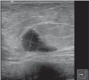
teration of posterior echoes, measuring 19 mm in its great-est diameter (Figure 2).
Considering the imaging characteristics of the mass, the patient’s age range, and the fact that it was a new finding, ultrasonography-guided percutaneous biopsy was performed. Anatomopathological analysis revealed a solid, neuroendo-crine carcinoma with a carcinoid-like pattern (Figure 3a), and positive for neuron specific enolase, chromogranin and synaptophysin markers (Figure 3b).
Maria Helena Valentim1, Vanessa Monteiro2, José Carlos Marques3
INTRODUCTION
Neuroendocrine carcinomas are rare malignant tumors which, in most of cases, are located in the lungs and gas-trointestinal tract. Primary location of such tumors in the breast is extremely rare, corresponding to less than 0.1% of all breast tumors(1,2).
Most published studies about neuroendocrine breast car-cinoma emphasize anatomopathological findings, with scarce reference to imaging findings(3,4).
The present report describes mammographic, sonographic and magnetic resonance imaging (MRI) findings of a case of asymptomatic, primary neuroendocrine breast carcinoma in a 75-year-old woman.
CASE REPORT
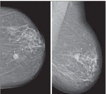
Mammography detected an ovoid, well defined mass with medium density in the upper-inner quadrant of the left breast in an asymptomatic 75-year-old woman. No associated microcalcifications were observed (Figure 1). At ultrasonog-raphy, the mass corresponded to a hypoechogenic, morpho-logically irregular and ill defined, solid nodule, with no
Valentim MH et al. / Primary neuroendocrine breast carcinoma
Radiol Bras. 2014 Mar/Abr;47(2):125–127 126
Figure 2. Ultrasonography demonstrated a solid hypoechogenic mass with ill defined contours, without any alteration of posterior echoes, measuring 19 mm in its largest diameter.
The patient underwent preoperative breast MRI (Fig-ure 4) which confirmed the presence of a hypointense, ir-regular lesion with 20 mm in diameter on T2-weighted se-quences, with peripheral ring-enhancement after intravenous gadolinium injection, and kinetic curve demonstrating early intense contrast uptake, followed by washout. Additionally, another adjacent, small nodular focus measuring 5 mm was observed, with comparable features.
In order to rule out a possible extramammary location of neuroendocrine carcinoma, thoracic, abdominal and pel-vic computed tomography (CT) was performed, but no other lesion was detected.
The patient underwent conservative surgery and the anatomopathological analysis of the surgical specimen con-firmed the diagnosis of a moderately differentiated, multi-focal neuroendocrine breast carcinoma.
DISCUSSION
Neuroendocrine carcinoma primarily located in the breast is an extremely rare entity. Although exact data on its incidence is not available yet, a series developed by
Günhan-Figure 3. Neuroendocrine tumor with a carcinoid-like pattern. Tumor cells with a rosette pattern of distribution and some cells showing granular eosinophilic cytoplasm (a). Immunohistochemical staining positive for synaptophysin (b).
Valentim MH et al. / Primary neuroendocrine breast carcinoma
Radiol Bras. 2014 Mar/Abr;47(2):125–127 127
Bilgen et al.(5) with 1,845 proved cases of breast tumors re-ports an incidence as low as 0.27%. Such a tumor presents specific morphological characteristics, expressing neuroen-docrine markers on a high percentage (> 50%) of the cells(3,6). Neuron specific enolase, synaptohysin and chromogranin are generally utilized as markers; the first one is considered as less specific(7). In 2003, the World Health Organization clas-sified neuroendocrine breast carcinomas into three subtypes, as follows: solid, small cell and large cell carcinomas(2,7). However, in a recent revision undertaken in 2012(8), neu-roendocrine tumors, included in the class of rare epithelial tumors, were subdivided into well differentiated neuroendo-crine tumors, poorly differentiated neuroendoneuroendo-crine carcino-mas (small cell carcinocarcino-mas), and carcinocarcino-mas with neuroen-docrine differentiation. In the present case, the tumor was positive for the three neuroendocrine markers, with solid histological subtype and carcinoid-like pattern (the assess-ment was made prior to the 2012 revision).
The cases of primary neuroendocrine breast carcinoma described in the literature refer mostly to women in the age range between 40 and 70 years(7), and there are some few cases described in male individuals(5).
The diagnosis of primary neuroendocrine breast carci-noma can only be made if an extramammary location is ruled out or an in situ component is demonstrated(3,7,9). In the present case, although an in situ component has not been demonstrated, other locations, namely pulmonary or intes-tinal, were ruled out at CT.
Neuroendocrine carcinomas do not present any particu-lar imaging finding and, in many cases, the findings are com-parable to the ones of other types of breast tumors(10). On mammography, as described by Ogawa et al.(1), such tumors may present as well circumscribed lesions, with no associ-ated microcalcifications, mimicking benign lesions. Such findings are in agreement with the ones observed in the present case, where the lesion corresponded to a well defined mass of medium density and with no associated microcalci-fications. On ultrasonography, such tumors may present as either morphologically irregular solid lesions or lesions with
a cystic component, with ill defined margins and increased vascularization(1,4,5). Also, in the present case, ultrasonog-raphy revealed the presence of a hypoechogenic mass with irregular morphology and ill defined contours, with no cys-tic component. MRI demonstrated, like in other cases de-scribed in the literature(4,5), the presence of an irregular le-sion with early, intense, ring-enhancement, with morphologi-cal and kinetic characteristics of contrast uptake consistent with malignancy.
Thus, despite the rarity of neuroendocrine carcinomas, with nonspecific imaging findings, such tumors should be included in the differential diagnosis of a nodular lesion with no associated microcalcifications on mammography and sonographically corresponding to a hypoechogenic mass with microlobulated or irregular contours.
REFERENCES
1. Ogawa H, Nishio A, Satake H, et al. Neuroendocrine tumor in the breast. Radiat Med. 2008;26:28–32.
2. Menéndez P, García E, Rabadán L, et al. Primary neuroendocrine breast carcinoma. Clin Breast Cancer. 2012;12:300–3.
3. Kinoshita S, Hirano A, Komine K, et al. Primary small-cell neu-roendocrine carcinoma of the breast: report of a case. Surg Today. 2008;38:734–8.
4. Fujimoto Y, Yagyu R, Murase K, et al. A case of solid neuroendo-crine carcinoma of the breast in a 40-year-old woman. Breast Can-cer. 2007;14:250–3.
5. Günhan-Bilgen I, Zekioglu O, Ustün EE, et al. Neuroendocrine dif-ferentiated breast carcinoma: imaging features correlated with clini-cal and histopathologiclini-cal findings. Eur Radiol. 2003;13:788–93. 6. Nozoe T, Sueishi K, Mori E, et al. Primary neuroendocrine carci-noma of the breast: report of a case. Surg Today. 2011;41:829–31. 7. Kim JW, Woo OH, Cho KR, et al. Primary large cell neuroendo-crine carcinoma of the breast: radiologic and pathologic findings. J Korean Med Sci. 2008;23:1118–20.
8. Gobbi H. Classificação dos tumores da mama: atualização baseada na nova classificação da Organização Mundial da Saúde de 2012. J Bras Patol Med Lab. 2012;48:463–74.
9. Graça S, Esteves J, Costa S, et al. Neuroendocrine breast cancer. BMJ Case Rep. 2012 Aug 13;2012.