This is the pre-review version of the paper published in
J. Phys. Chem. B, 114, 8994–9001
The final version may be obtained from the publisher, at the following link
A tale of two acids: when arginine is a more appropriate
acid than H
3
O
+
.
Pedro J. Silvaa‡* , Claudia Schulzb‡, Dieter Jahnb, Martina Jahnb and Maria João Ramosc
a
REQUIMTE, Fac. de Ciências da Saúde, Univ. Fernando Pessoa, Rua Carlos da Maia, 296, 4200-150 Porto-Portugal
b
Department of Microbiology,Technische Universitaet Braunschweig, Spielmannstr. 7, 38106 Braunschweig - Germany
c
REQUIMTE, Faculdade de Ciências do Porto, Rua do Campo Alegre, 687, 4169-007 Porto – Portugal
‡
Abstract:
Uroporphyrinogen III decarboxylase catalyzes the fifth step in heme biosynthesis: the elimination of carboxyl groups from the four acetate side chains of uroporphyrinogen-III to yield coproporphyrinogen-III. We have previously found that the rate-limiting step is substrate protonation, rather than decarboxylation itself, and that this protonation can be effected by a nearby arginine residue (Arg37). In this report, we have studied the reasons for the unusual choice of arginine as a general acid catalyst. Our density functional calculations show that, although substrate protonation by H3O+ is both exergonic and very fast, in the presence of a protonated Arg37 substrate decarboxylation becomes rate-limiting and the substrate spontaneously breaks upon protonation. These results suggest that the active site must be shielded from solvent protons, and that therefore H3O+ should be excluded from a role in both protonations present in this mechanism. A second Arg residue (Arg41) is uniquely positioned to act as donor of the second proton, with an activation barrier below 2 kcal mol-1. Additional site-directed mutagenesis experiments confirmed that no coproporphyrinogen is formed in the absence of any of these these Arg residues. This counter-intuitive use of two basic residues as general acids in two different proton donation steps by uroporphyrinogen decarboxylase may have arisen as an elegant solution to the problem of simultaneously binding the very negative uroporphyrinogen (which requires a positively charged active site), and selectively protonating it while preventing excessive carboxylate stabilization by positive charges.
KEYWORDS: uroporphyrinogen decarboxylase; reaction mechanism; decarboxylation; density-functional theory; binding/unbinding events
*
I. Introduction
Fig. 1: Proposed reaction mechanism for UroD (adapted from Silva & Ramos, 2005). Since all four pyrrole rings present in the substrate are decarboxylated by UroD, the reaction cycle depicted must be repeated three more times.
With substrate protonation as the rate-limiting, stronger acids should yield faster reaction rates. Such reaction rates do not occur in the enzyme, and it is well known that in strongly acidic conditions H3O+ catalyzes uroporphyrinogen decarboxylation at much slower rates than the enzyme-catalyzed reaction7. These paradoxes, together with the counter-intuitive use of arginine as an acidic residue, prompted the present study on potential evolutionary shortcomings of the use of the much stronger acid, H3O+, as proton donor in the enzymatic reaction.
II. Methods
Quantum chemistry
All calculations were performed at the Becke3LYP level of theory8,9,10. In geometry optimizations a medium-sized basis set, 6-31G(d), was used, since it is well known that larger basis sets give very small additional corrections to the geometries, and their use for this end is hence considered unnecessary from a computational point of view11,12,13. More accurate energies of the optimized geometries were calculated with the triple-quality basis set 6-311+G(2d,2p). As in our previous work, our calculations
focused on the decarboxylation of pyrrole ring D of uroporphyrinogen III, since this is believed to be the first ring to undergo decarboxylation in physiological conditions14. Asp86, Arg37 and H3O+ were incorporated into the model. Two water molecules were also included, in order to provide some charge stabilization and for added realism. In order to prevent unrealistic movements of the modeled aminoacid sidechains, aminoacid C and C carbon atoms were constrained to their crystallographic positions3
Likewise, the intervening substrate pyrrole ring D was also anchored to its initial coordinates by freezing two of its carbon atoms. Constraints imposed on the geometries prevented frequency analyses: zero point (ZPE) and thermal effects (T=310.15 K, P=1 bar) were estimated from calculations on smaller unconstrained models: contributions for the proton transfer between H3O+ and the substrate were estimated from the proton transfer between solvated H3O+ and unsubstituted pyrrole; an estimate for ZPE and thermal effects for the decarboxylation of the protonated substrate was taken from our previous work4. A scaling factor of 0.9804 was used for the frequencies.
The polarizable conductor model15,16, as implemented in Gaussian0317 was used in order to account for the effects of the protein environment. The dielectric constant was chosen equal to 4, as commonly used for the active site of proteins18. Atomic charge and spin density distributions were calculated with a Mulliken population analysis19, using the larger basis set. All calculations were performed with the Gaussian03 suite of programs.
Continuum electrostatic calculations
and 47.5 Å. With the exception of Asp86 in the substrate-bound subunit, all Asp, Glu, His, Arg, Lys, Tyr and Cys residues, as well as the substrate acetate and propionate substituents, were allowed to titrate. The sampling of proton-binding states was done using the MCRP program (Monte Carlo for Reduction and Protonation), which implements a Monte Carlo method described by Baptista et al.27,28. pH was sampled at 0.2 pH units intervals in the 0-15 range. Occupation states and correlations were computed using 106 Monte Carlo steps.
Molecular dynamics
Site-directed mutagenesis of the human hemE gene
To exchange nucleotides in the human hemE gene, the QuikChange®II Site-Directed Mutagenesis Kit (Stratagene, Heidelberg, Germany) was used according to the manufacturer’s instructions by using the expression vector pHT#77 as template35. Oligonucleotides carrying nucleotide exchanges compared with the hemE gene sequence in the underlined positions were used to generate mutants: R37A: 5´-GTTTGGTGCATGGCCCAGGCAGGCCG-3´; R37K: GTTTGGTGCATGAAACAGGCAGGCCG-3´; R41A: CGCCAGGCAGGCGCCTACTTACCAGAG-GTTTGGTGCATGAAACAGGCAGGCCG-3´; R41K: 5´-CGCCAGGCAGGCAAATACTTACCAGAG-3´. All mutated genes were verified for gene integrity by DNA sequence determination.
Production and purification of Uroporphyrinogen-III-decarboxylase (UroD)
Protein Determination (Sigma – Aldrich, Munich, Germany) according to manufacturer’s instructions and concentrated by ultrafiltration (vivaspin 15, Vivascience, Hanover, Germany) and stored at 4 °C.
Enzyme activity assay
The UroD activity assay was carried out under strict anaerobic conditions. The reaction components were mixed in an anaerobic chamber. All solutions used were saturated with N2 prior to use. The standard assay mixture contained UroD (0.1 µM) and uroporphyrinogen-III (0,025 – 20 µM) in potassium phosphate buffer in a total volume of 240 µL after a modified procedure as described earlier37. After incubation at 37 °C in the dark for 30 min the reaction was stopped by the addition of 10 µL concentrated HCl and formed coproporphyrinogen-III was oxidised for 15 min to coproporphyrin-III by addition of 10 µL 30 % H2O2. The mixture was centrifuged for protein removal for 15 min at 15.000 x g. The obtained supernatant was analysed by High Performance Liquid Chromatography (HPLC). The HPLC system was Jasco 1500 series (Jasco, Gross-Umstadt, Germany). Therefore, 100 µL of the supernatant was mixed with 100 µL acetone/HCl (9:10). Twenty µL were loaded onto a 250 x 4,6 mm Equisil ODS C18 reversed phase column (Dr. Maisch GmbH, Ammerbuch, Germany). Separation was performed at a flow rate of 0.3 mL/min using 40 % (v/v) ammoniumacetate (pH 5.2) and 10 % (v/v) acetonitrile in methanole as mobile phase. Tetrapyrroles were detected by fluorescence measurement using an excitation wavelength of 409 nm and an emission wavelength of 618 nm. Uroporphyrin-III and Coproporphyrin-III (Frontier Scientific, Germany, European Union) were used as standards.
III. Results
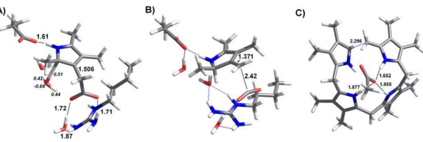
for proton transfer from H3O+ to the pyrrole ring was therefore performed subject to these constraints. We found that initially the reactive proton in H3O+ lies 1.92 Å away from the pyrrole C2 atom. Charge stabilization of H3O+ is provided by the pyrrole acetate substituent (1.53 Å away) and by a H2O molecule positioned approximately midway between H3O+ and Asp86. In this model, the protonated Arg37 stabilizes the acetate substituent, with one of its Nη1 guanidinium protons positioned 1.70 Å from one of the carboxylate oxygens, and a water molecule providing a bridge between the remaining carboxylate oxygen and the Nη2 guanidinium hydrogens.
Proton transfer from H3O+ to the pyrrole ring requires a very small (0.23 Å) movement of the H3O+ center of mass. In the transition state (Figure 2a), the O distance has increased to 1.26 Å , and the H-pyrrole distance has decreased to 1.37 Å. The original positive charge has partially spread from the H3O+ (which retains only +0.68 charge) to the pyrrole (which has acquired +0.32 charge). This step occurs with a very small energetic barrier (3.3 kcal.mol-1 electronic energy difference, ΔG=0.3 kcal.mol -1
). At pH=6.2, the optimum pH for the human enzyme reaction rate6, the initial energy of the system should be decreased by a factor of RT ln (H+), yielding an effective barrier of 8.8 kcal.mol-1. This reaction step is moderately exergonic, as the product lies 11.9 kcal mol-1 below the transition state. The protonated pyrrole ring formed in this step is stabilized through interaction with Asp86, and its acetic acid substituent strongly interacts with the protonated Arg37 residue. We computed the energetic contribution of these amino acids by repeating the calculations in their absence, while keeping their orbitals in order to avoid basis step superposition errors. These calculations show that Asp86 and Arg37 together lower the reaction product energy by 9 kcal mol-1.
barrier for decarboxylation enables other reaction pathways to become competitive with CO2 evolution. In particular, breaking of the protonated tetrapyrrole ring now becomes possible: elongation of the C-C bond between C2 of ring D and the methylene bridging rings D and A allowed us to identify a low-lying transition state 12.8 kcal.mol-1 above the protonated uroporphyrinogen (Figure 2c). The energy and geometry of this transition state are very similar to those observed in a related reaction performed by uroporphyrinogen III synthase38, which catalyzes the previous step in heme biosynthesis. It is clear from these calculations that in the presence of a protonated Arg37 substrate degradation occurs faster than decarboxylation, and that the correct reaction product cannot be formed quantitatively. These results show that H3O+ entry to the active site is disadvantageous for the enzyme-catalyzed reaction, and provide an interesting explanation for the unusual choice of an arginine residue as general acid in this reaction mechanism.
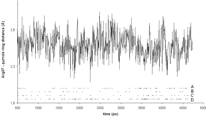
Figure 3: Minimum distance between Arg37 guanidinium protons and pyrrole C2 atoms of UroD-bound uroporphyrinogen III, as a function of simulation time. Snapshots with Arg37-C2 distances below 2.5 Å are highlighted (for each of the fours rings present in the substrate) in the lower portion of the graph. For ease of presentation, the graph only includes the first half of this simulation. Full data on this simulation, as well as simulations of Arg37 approach to the pyrrole rings in uroporphyrinogen III after sequential decarboxylation of rings D, A and B are available in the Supporting Information.
low substrate concentrations the first decarboxylation occurs on ring D. Simulations on D-ring-decarboxylated uroporphyrinogen III showed that without the electrostatic attraction provided by the ring D acetate group the guanidium group from Arg37 adopts an approximately equidistant position between rings A and B, so that the following decarboxylation is about as likely to occur on ring A (Arg37 approaches 80 % of the time) as on ring B (Arg37 approaches 66 % of the time). Ring C is shown to be harder to decarboxylate: after decarboxylation of rings D and A, Arg 37 approaches ring B 29.8 % of the time , vs. only 0.4 % of the time for ring C. Even after rings D, A and B are decarboxylated the approach of Arg37 to ring C still occurs quite intermittently (2 % of the time).
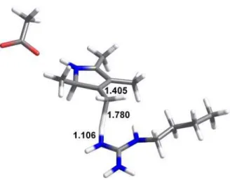
Figure 4: Structure of the transition state for proton transfer from Arg41 to the methylene substituent arising from substrate decarboxylation. All distances in Å.
Figure 5: Titration behavior of the acetic acid (open symbols) and propionic acid (filled symbols) substituents present in uroporphyrinogen III bound to uroporphyrinogen III decarboxylase. Substituents present on ring A are depicted as squares, those on ring B as diamonds, those on ring C as triangles and those on ring D as circles.
Figure 6: Activity profile of wild type (●) and R37A (○) uroporphyrinogen III decarboxylase.
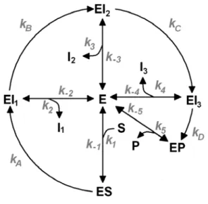
an N-terminal histidine tag in Escherichia coli and purified in a single step by nickel – NTA chromatography as described in materials and methods. The yield of protein (from 2 to 10 mg/ml) was measured by Bicinchoninic Acid Protein Assay. Their catalytic competence was studied with 0.1 µM UroD and a large range of substrate concentrations (from 0.025 to 35 µM) in potassium phosphate buffer for 30 min at 37 °C. The amount of formed coproporphyrinogen III was analysed by HPLC chromatography (data not shown). Kinetic measurements on the wild type protein (Figure 6) showed that its reaction rate law does not fit a straightforward Michaelis-Menten form: instead of approaching a defined maximum rate as the substrate concentration is increased to infinity, we found that a maximum rate was attained at a relatively modest substrate amount (5 μM), and decreased at higher concentrations. Such an odd behavior may be explained by a kinetic model where partially decarboxylated intermediates may escape the binding site at rates competitive with the decarboxylation rate. At low substrate concentrations, these intermediates may get back inside and quickly finish conversion into coproporphyrinogen III. At higher concentrations, fresh uroporphyrinogen III competes with these intermediates for entry into the active site and prevents them from finishing conversion into coproporphyrinogen III (Figure 7). Numerical simulations of this reaction scheme confirm its ability to generate activity profiles consistent with the experimental data (Figure 8).
Reaction rates of binding/unbinding events are labeled numerically (positive labels for binding events, negative labels for unbinding events)
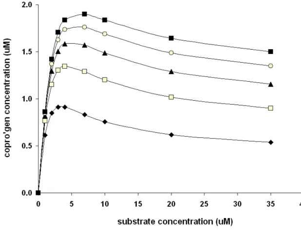
Figure 8: Kinetic profiles predicted by the reaction model depicted in Figure 7. Simulation parameters: Enzyme concentration=0.1 μmol.dm-3 ; Simulation time=2000 s; k1=k2=k3=k4=k5=105 M-1s-1;
k-1=k-2=k-3=k-4=k-5=0.1 s-1; kA=kB=kC=0.1 s-1; kD= 0.02 s-1 (♦) ; 0.04 s-1 (□); 0.06 s-1 (▲); 0.08 s-1 (○); 0.1 s-1 (■). Additional kinetic profiles under different parameters are available as Supporting Information
substrate concentration, and much smaller than observed with the wild-type enzyme. The amount of unreacted uroporphyrinogen remaining at the end of the reaction was virtually indistinguishable from the amount added initially, showing that catalysis by this mutant is not significant. In the R41K mutant, we could not detect the coproporphyrinogen III product, but non-negligible amounts of several other fluorescent products were observed.
IV Conclusions
From our computational study, the disadvantages of H3O+ as a general acid in this reaction become clear: although it quickly generates the protonated form of the substrate needed as an electron sink for the decarboxylation, the slow rate of decarboxylation in the presence of protonated Arg37 allows the breaking of protonated uroporphyrinogen III to become competitive with the main reaction. The use of Arg37 as a proton donor allows substrate protonation to occur, while at the same time removing the stabilizing effect of protonated Arg37 on the leaving carboxylate. Site-directed mutagenesis of this residue confirm its important role in the catalytic mechanism. The observed kinetic profile of the enzyme shows very peculiar features, consistent the release of partially decarboxylated intermediates from the acive site. The inherent flexibility of Arg37, as well as its central position in the active site, allow it to donate the catalytic proton to every pyrrole ring in uroporphyrinogen and therefore to catalyze the sequential decarboxylation of the substrate acetic acid substituents on the intact substrate and on the partially decarboxylated intermediates without the need for different binding modes for each of these species.
uroporphyrinogen (which requires a positively charged active site), and selectively protonating it while preventing excessive carboxylate stabilization by positive charges.
V. References
1. Wyckoff , E.E. & Kushner, J. P. (1994) Heme biosynthesis, the porphyrins and the liver. In Arias, I.m., Boyer, J.L., Fausto, N., Jakoby, W.B. Schachter, D.A. & Shafritz, D.A. (eds) The Liver: Biology and Pathobiology. Raven Press Ltd, New York, pp. 505-527
2. Sassa, S. & Kappas, A. (2000) J. Intern. Med.247: 169-178
3. Phillips J.D., Whitby F.G., Kushner J.P. & Hill C.P. (2003) EMBO J. 22, 6225-6233
4. Silva, P.J. & Ramos, M.J. (2005) J. Phys. Chem. B 109: 18195-18200.
5. Jones, R.M. & Jordan, P.M. (1993) Biochem. J. , 293: 703-712
6. de Verneuil, H,. Sassa, S. & Kappas, A.(1983) J. Biol. Chem.258:2454-2460
7. Koskelo P. & Toivonen I. (1970) Clin. Chem.16, 459-461
8. Becke, A. D. J. Chem. Phys.I, 98 (1993), 5648.
9. Lee, C.; Yang, W.; Parr, R. J. Phys. Rev. B, 37 (1988), 785.
10. Hertwig, R. W.; Koch, W.; J. Comp. Chem., 16 (1995), 576.
11. Siegbahn, P. E. M; Eriksson, L.; Himo, F.; Pavlov, M. J. Phys. Chem. B., 102 (1998), 10622.
12. Fernandes, P. A.; Ramos, M. J. (2003) J. Am. Chem. Soc., 125: 6311-6322
13. Riley K.E., Op’t Holt, B.T., Merz Jr., K.M. (2007) J. Chem. Theory Comput.,3, 407-433
14. Jackson AH, Sancovich HA, Ferramola AM, Evans N, Games DE, Matlin SA, Elder GH, Smith SG. (1976) Philos. Trans. R. Soc. Lond. B Biol. Sci. , 273: 191-206.
15. Barone, V.;Cossi, M. (1998) J. Phys. Chem. A, 102: 1995-2001
17. M. J. Frisch, G. W. Trucks, H. B. Schlegel, G. E. Scuseria, M. A. Robb, J. R. Cheeseman, J. A. Montgomery, Jr., T. Vreven, K. N. Kudin, J. C. Burant, J. M. Millam, S. S. Iyengar, J. Tomasi, V. Barone, B. Mennucci, M. Cossi, G. Scalmani, N. Rega, G. A. Petersson, H. Nakatsuji, M. Hada, M. Ehara, K. Toyota, R. Fukuda, J. Hasegawa, M. Ishida, T. Nakajima, Y. Honda, O. Kitao, H. Nakai, M. Klene, X. Li, J. E. Knox, H. P. Hratchian, J. B. Cross, C. Adamo, J. Jaramillo, R. Gomperts, R. E. Stratmann, O. Yazyev, A. J. Austin, R. Cammi, C. Pomelli, J. W. Ochterski, P. Y. Ayala, K. Morokuma, G. A. Voth, P. Salvador, J. J. Dannenberg, V. G. Zakrzewski, S. Dapprich, A. D. Daniels, M. C. Strain, O. Farkas, D. K. Malick, A. D. Rabuck, K. Raghavachari, J. B. Foresman, J. V. Ortiz, Q. Cui, A. G. Baboul, S. Clifford, J. Cioslowski, B. B. Stefanov, G. Liu, A. Liashenko, P. Piskorz, I. Komaromi, R. L. Martin, D. J. Fox, T. Keith, M. A. Al-Laham, C. Y. Peng, A. Nanayakkara, M. Challacombe, P. M. W. Gill, B. Johnson, W. Chen, M. W. Wong, C. Gonzalez, and J. A. Pople, Gaussian 03, Revision B.04, Gaussian, Inc., Pittsburgh PA, 2003.
18. Blomberg, M. R. A.; Siegbahn, P. E. M.; Babcock, G. T. (1998) J. Am. Chem. Soc., 120,8812.
19. Mulliken, R. S. (1955) J. Chem. Phys.23, 1833
20. Bashford, D.; Gerwert. K. (1992) J. Mol. Biol.224, 473-486
21. Sitkoff, D; Sharp, K.A.; Honig, B (1994) J. Phys. Chem., 98, 1978-1988
22. Whitby FG, Phillips JD, Kushner JP, Hill CP (1998) EMBO J., 17, 2463-2471
23. Dolinsky TJ, Nielsen JE, McCammon JA, Baker NA. (2004) Nucl. Acids Res., 32, W665-W667
24. Martel, P.J.; Soares, C.M.; Baptista, A.M.; Fuxreiter, M.; Náray-Szabó, G.; Louro, R.O.; Carrondo, M.A. (1999) J. Biol. Inorg. Chem., 4, 73-86
25. Antosiewicz, J.; McCammon, J.A.; Gilson, M.K. (1994) J. Mol. Biol., 238, 415-436
27. Baptista, A.M.; Martel, P.J., Soares, C.M. (1999) Biophys. J., 76, 2978-2998
28. Teixeira, V.H.; Soares, C.M.; Baptista, A.M. (2002) J. Biol. Inorg. Chem., 7, 200-216
29. Krieger E, Darden T, Nabuurs S, Finkelstein A, Vriend G (2004) Proteins57,678-683
30. Wang J, Cieplak P, Kollman PA (2000) J. Comput. Chem.21,1049-1074
31. Essmann U, Perera L, Berkowitz ML, Darden T, Lee H, Pedersen LG (1995) J Chem Phys 1995;
103, 8577-8593.
32. Berendsen HJC, Postma JPM, van Gunsteren WF, DiNola A, Haak JR. (1984) J. Chem. Phys. 81, 3684-3690.
33. Jakalian A, Jack DB and Bayly CI (2002) J. Comput. Chem. 23,1623-1641
34. Krieger E, Nielsen JE, Spronk CA, Vriend G (2006) J. Mol. Graph. Model.25, 481-486
35. Phillips, J. D., Whitby, F. G., Kushner, J. P. & Hill, C. P. (1997) Protein Sci.6, 1343-1346.
36. Romeis, B. (1968) Mikroskopische Technik (16. Aufl.) p. 593 (R. Oldenbourg Verlag, München).
37. Phillips, J. D. & Kushner, J. P. (1999) Current Protocols in Toxicology, 8.4.1-8.4.13.
38. Silva, P.J; Ramos, M.J. (2008) J. Phys. Chem. B, 112, 3144-3148
39. Lash, T.D.; Mani, U.N.; Lyons, E.A.; Thientanavanich, P.; Jones, M.A.(1999) J. Org. Chem.,